Ankara Üniv Vet Fak Derg, 58, 1-4, 2011
Magnetic resonance imaging of the guttural pouch (diverticulum tubae
auditivae) and its related structures in donkey (Equus asinus)
Çağdaş OTO, Reşide Merih HAZIROGLU
Ankara University, Faculty of Veterinary Medicine, Department of Anatomy, 06110 - Ankara, Turkey.
Summary: Anatomy of the guttural pouches and their associated structures such as bones, muscles, salivary glands, vessels and nerves were examined in detail in 4 adult donkey heads by magnetic resonance imaging (MRI). The protocol was performed using a superconducting magnet operating at a field strenght of 1,5 Tesla in 5 mm thickness transversal sections in human body coil. The obtained images provided excellent detail, so the relevant anatomic structures were described in whole slices. The pouch and cortical bone were scanned very low signal intensity, the muscles were slightly hypointense and the salivary glands were slightly hyperintense signals. Presented informations in this study will serve as a reference data to review MRI of the guttural pouch and its associated structures in clinical applications especially in the diagnosis of the guttural pouch diseases.
Key Words: Donkey, guttural pouch, magnetic resonance imaging
Merkepte (Equus asinus) hava keseleri (diverticulum tubae auditivae) ve ilişkili yapıların manyetik rezonans ile görüntülenmesi
Özet: Çalışmada 4 adet yetişkin merkep başında hava keseleri ve ilişkili oldukları yapıların (kemikler, kaslar, tükrük bezleri, damarlar ve sinirler gibi…) anatomisi detaylı olarak incelendi. Bu amaçla insan vücut koili içerisinde, 1,5 tesla gücündeki magnet aracılığıyla alınmış, 5 mm kalınlığındaki transversal kesitler kullanıldı. Elde edilen görüntüler mükemmel düzeyde detay sağladı ve tüm slaytlardaki ilişkili yapılar belirlendi. Hava keseleri ve kemik korteksi çok düşük sinyal yoğunluğuyla taranırken, kaslar hafif hipointens, tükrük bezleri ise hafif hiperintens özellikte tarandı. Bu çalışma ile sunulan verilerin, klinik uygulamalarda başta hava kesesi hastalıklarının tanısı olmak üzere, hava keseleri ve ilişkili oldukları yapılar için, MR görüntülerinin değerlendirilmesinde referans olarak kullanılacağı düşünülmektedir.
Anahtar sözcükler: Hava kesesi, manyetik rezonans görüntüleme, merkep.
Introduction
The guttural pouches are a pair of mucosal sacs that are formed by ventro-lateral dilatation of the auditory tube which connects the pharynx and the middle ear in perissodactyls such as horse, donkey, tapir (10, 21). The pouches are symmetrically seperated by a septum, and they extend from the pharynx to the atlanto-occipital joint in both sides (8, 15, 17, 22). In addition to this, each pouch is divided into wider medial and narrower lateral compartments by the stylohyoid bone with a capacity ratio of 2:1 (8).
It is well known that the air-filled diverticuli have an important function of brain cooling (7, 8, 10, 13) and also intimately involved in the physiology of swallowing (13, 20). Because of their localization, the guttural pouches have unique and indicate relationship with several cranial nerves and blood vessels (10, 15, 17). So, diseases of the guttural pouch such as empyema, mycosis, tympany, neoplasia, cysts and muscle ruptures
make the pouch susceptible to serious and sometimes fatal clinical conditions (8).
The investigation of the equine guttural pouch has been performed by anatomical approach including dissection (3, 6), mold preparation (15, 17), radiography (12, 14, 16), endoscopy (8, 10) and computed tomography (21). On the other hand, there is no published material describing the magnetic resonance imaging (MRI) of the guttural pouch of donkey head. Donkeys occupy the second order in the world equine population (11, 23). They have been generally used for transporting, working and as a guarding animal (23). Today, MRI that is known as a better imaging technique for displaying the soft tissues, is commonly being used for observation of anatomical features of soft tissues in the head region (2).
The aim of this study is to provide an overview of the detailed anatomy and imaging features of the guttural pouch and its associated structures using transversal MR images in donkey.
Çağdaş Oto - Reşide Merih Hazıroğlu 2
Materials and Methods
The four adult donkey heads that were used in this study were obtained from donkeys slaughtered in order to feed the wild animals kept at Ankara city zoo. The heads were examined in detail by MRI that was performed using a superconducting magnet operating at a field strenght of 1,5 Tesla in T1 and T2-weighted transversal sections. The human body coil was used for scanning of the donkey heads. T1-weighted images were acquired by using following parameters; Repetition time (TR) = 660 msec, echo time (TE) = 14 msec, 177x256 matrix, (NEX) = 2, field of view (FOV) = 20 cm. And for T2-weighted images; Repetition time (TR) = 4224 msec, echo time (TE) = 99 msec, 242x256 matrix, (NEX) = 2, field of view (FOV) = 20 cm was standardized. The slice thickness was 5 mm and interslice spacing was 3 mm.
Definition of the anatomical structures are based on the atlases regarding equine anatomy (3, 9, 19) and MRI (4). Nomina Anatomica Veterinaria was used for the nomenculature (18).
Results
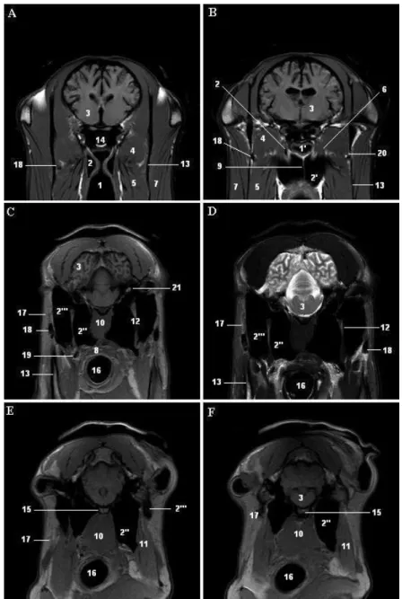
The guttural pouches were observed in the transversal MR sections of the head of donkey. Five T1-weighted and one T2-T1-weighted MR images were presented in a rostral to caudal series from the level of auditory tube orifice to the level of atlanto-occipital joint (Figure 1). Because of its air-filled structure, the auditory tube and its diverticulum gave negligible signal as completely hypointens and were imaged in dark black color in both T1-weighted and T2-weighted slices (Figure 2C), by this way it would be distinguished easily from the surrounding tissues such as bones, muscles and salivary glands.
In transversal sections, it was described that the right and left sacs were symmetrically separated by a thin septum in the rostral side (Figure 2B) and by rectus capitis ventralis muscle and longus capitis muscle in the caudal side (Figure 2E). The septum and muscles of the head were scanned with intermediate signal intensity and appeared in grey color in T1-weighted images (Figure 2C) and were scanned hypointense dark grey color with low signal intensity in T2-weighted images (Figure 2D). And it was observed that T1-weighted images depicted more anatomic detail than T2-weighted images.
Border of the pouches extended from the rostral side of mandibular ramus to the atlanto-occipital joint in rostral-caudal direction. They were covered with lateral and medial pterygoid muscles in the rostral portion (Figure 2B), and digastric muscle and parotid gland in the caudal portion laterally (Figures 2E, 2F), tensor and levator veli palatini muscles, sphenoid bone and occipital bone dorsally (Figures 2B, 2C, 2E) and pharynx muscles ventrally (Figures 1, 2). It was observed that they were also in close relationship with external carotid artery
(Figures 2C, 2D, 2E), maxillary vein (Figures 2A, 2B) and the nerve package of glossopharyngeal, vagal and accessory nerves (Figures 2C, 2D). On the other hand, the internal carotid artery could not be tagged on sections without using contrast medium.
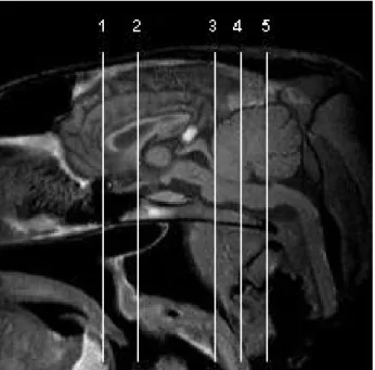
Figure 1. Midsagittal T1-weighted magnetic resonance image of donkey head showing the levels of transverse planes. 1 = level of autidory tube ostium, 2 = level of sphenoid bone, 3 = level of styloid bone, 4 = level of occipital bone, 5 = level of parotid gland.
Şekil 1. Merkep başında transversal düzlem seviyelerini gösteren T1-ağırlıklı sagittal manyetik rezonans görüntüsü. 1 = Ostium pharyngeum tubae auditiva seviyesi, 2 = Os sphenoidale seviyesi, 3 = Sytlohyoideum seviyesi, 4 = Os occipitale seviyesi, 5 = Glandula parotis seviyesi.
The auditory tube orifice was scanned as a triangle in shape at the level of rostral border of mandibular ramus (Figure 2A). The pouches started to appear on both sides of the pharyngeal recess and expanded ventro-laterally towards the caudal. At the level of the caudal border of mandibular angle, it was seen that the sacs were in the widest position and they reached to the inner surface of the mandible. The pouches were divided into two compartments by the stylohyoid bone at this level (Figures 2C, 2D). The compartments were observed more clearly in the caudal, and the medial part was larger than the lateral part in all slides of the whole heads. Cortical part of the mandibula was observed hipointens and black color appearing, but the medullar part was scanned high signal intensity because of the fat tissue. Stylohyoid bone was also seen hiperintens with light grey color (Figure 2C). Towards the caudal of the head, the sacs were getting smaller. At the levels of thympanic bulla and atlanto-occipital joint, firstly the lateral compartment and then the medial one disappeared in the transversal sections sequentially (Figures 2E, 2F).
Ankara Üniv Vet Fak Derg, 58, 2011 3
Discussion and Conclusion
Manglai et al. (17) reported that the pouches are divided two compartments as wider lateral and narrower medial. On the contrary, Carmalt (8) emphasized that the medial compartment was two times larger than the lateral. We clearly observed parallel data to Carmalt (8).
Manglai et al. (17) described the opening of auditory tube as a funnel form canal, but we observed
that in the transversal slides the entrance of the pharyngeal part of the pouches was like a triangle shape orifice.
Dorsal side of the pouches consisted of hard tissue (sphenoid bone and occipital bone) and except for the region connected to the stylohyoid bone, the other walls were in contact with soft tissue such as pharynx, esophagus and many muscles (17), as we clearly observed and exhibited in MRI. Baptiste et al. (5) pointed
Figure 2. T1-weighted (A, B, C, E, F) and T2-weighted (D) transversal magnetic resonance images that it levels are described in midsagittal section of the figure 1. 1 = Pharynx, 1’ = Recessus pharyngis, 2 = Auditory tube, 2’ = Guttural pouch, 2’’ = its medial compartment, 2’’’ = its lateral compartment, 3 = Brain, 4 = Lateral pterygoid muscle, 5 = Medial pterygoid muscle, 6 = Tensor and levator veli palatini muscles, 7 = Massater muscle, 8 = Palatopharyngeus muscle, 9 = Septum diverticuli, 10 = Longus capitis and rectus ventralis muscles, 11 = Digastric muscle, 12 = Sytloid bone, 13 = Mandibula, 14 = Sphenoid bone, 15 = Occipital bone, 16 = Cavum laryngis, 17 = Parotid gland, 18 = Maxillary vein, 19 = external carotid artery, 20 = Lingual nerve, 21 = IX., X., XI. cranial nerves. Şekil 2. Şekil 1’deki sagittal kesitte tanımlanan seviyelerden geçen T1-ağırlıklı (A, B, C, E, F) ve T2-ağırlıklı (D) transversal manyetik rezonans görüntüleri. 1 = Pharynx, 1’ = Recessus pharyngis, 2 = Tuba auditiva, 2’ = Diverticulum tubae auditiva, 2’’ = pars medialis, 2’’’ = Pars lateralis, 3 = Beyin, 4 = Musculus pterygoideus lateralis, 5 = Musculus pterygoideus medialis, 6 = Musculus tensor et levator veli palatini, 7 = Musculus massater, 8 = Musculus palatopharyngeus, 9 = Septum diverticuli, 10 = Musculus longus capitis et musculus rectus ventralis, 11 = Musculus digastricus, 12 = Sytlohyoideum, 13 = Mandibula, 14 = Os sphenoidale, 15 = Os occipitale, 16 = Cavum laryngis, 17 = Glandula parotis, 18 = vena maxillaris, 19 = Arteria carotis externa, 20 = Nervus lingualis, 21 = IX., X., XI. Nervi craniales.
Çağdaş Oto - Reşide Merih Hazıroğlu 4
out that movement of the head and contraction of the mastication muscles may affect the shape and size of the pouches. We observed that the size and shape of the pouches were slightly changed during the performance depending on the angle between the head and the neck. So we considered that it was not necessary to give a volumetric measurement.
Excellent discrimination of soft and mineralized tissues was evident in the T1-weighted images (1, 2, 24). Because the outline of the guttural pouches were presented more clearly in T1-weighted images, delineation of the anatomy of the auditory tube and its associated structures in T1-weighted images were superior to T2-weighted images (2). We have agreed strongly with the authors who emphasized the superiority of T1-weighted scanners.
In conclusion, the results of the examination suggested that donkey guttural pouch was almost the same as horse’s and utilization of the MRI provided detailed anatomic depiction. We believe that the tagged images presented in this study will serve as a reference data to review MR images of the guttural pouch and its associate structures in clinical applications.
References
1. Arencibia A, Vazquez JM, Jaber R, Gil F, Ramirez JA, Rivero M, Gonzales N, Wisner ER (2000): Magnetic
resonance imaging and cross sectional anatomy of the normal equine sinuses and nasal passages. Vet Radiol Ultrasound, 41, 313-319.
2. Arencibia A, Vazquez JM, Ramirez JA, Ramirez G, Vilar JM, Rivero A, Alayon S, Gil F (2001): Magnetic
resonance imaging of the normal equine brain. Vet Radiol Ultrasound, 42, 405-408.
3. Ashdown RR, Done SH (1987): Color Atlas of Veterinary
Anatomy Vol. II-The Horse, Part I-Head. 1-50. Gower Medical Publishing, London, NewYork.
4. Assheuer J, Sager M (1997): MRI and CT Atlas of the
Dog. 44-81. Blackwell Wissenschafts, Berlin.
5. Baptiste KE, Holladay SD, Freeman LE (1996):
Alterations in equine guttural pouch morphology with head position: observations using a new technique for producing accurate casts. Anat Rec, 246, 579-584.
6. Baptiste KE (1997): Functional anatomy observations of
the pharyngeal orifice of the equine guttural pouch (auditory tube diverticulum). Vet J, 153, 311-319.
7. Baptiste KE (1998): A preliminary study on the role of the
equine guttural pouches in selective brain cooling. Vet J,
155, 139-148.
8. Carmalt J (2002): Guttural pouch diseases in the horse.
Large Animal Veterinary Rounds Vol. 2.
Erişim: [http://www.drounds.ca/crus/laveng_0202.pdf], Erişim tarihi: 15.09.2009
9. Clayton HM, Flood PF (1996): Color Atlas of Large
Animal Applied Anatomy. 7-27. Mosby-Wolfe, Barcelona.
10. Dyce KM, Sack WO, Wensing CJG (2002): Textbook of
Veterinary Anatomy, 3rd ed., Chapter 18, The Head and Ventral Neck of the Horse. 496-501. Saunders-Elsevier Science, Philadelphia.
11. FAO (1997): FAO statistical database website. Food and Agriculture Organisation, Italy.
Erişim: [http://www.fao.org], [FAOSTATS: http://apps.fao.org/cgi-bin/nph-db.pl?subset=agriculture],
Erişim tarihi: 15.09.2009
12. Hance SR, Robertson JT, Bukowiecki CF (1992): Cystic
structures in the guttural pouch (auditory tube diverticulum) of two horses. J Am Vet Med Assoc, 200, 1981-1983.
13. Hodgson DR (1998): What is the function of the guttural
pouches: Selective brain cooling? Augmentation of swallowing? Still to be defined. Vet J, 155, 115-117.
14. Jones DL (1994): Squamous cell carcinoma of the larynx
and pharynx in horses. Cornell Vet, 84, 15-24.
15. Liebich HG, König HE (2004): Veterinary Anatomy of
the Domestic Mammals, Part 17, 577-579. In: HE König and HG Liebich (eds), Vestibulocochlear Organ, Schattauer, Stuttgart.
16. Macdonald DG, Fretz PB, Baptiste KE, Hamilton DL (1999): Anatomic, radiographic and physiologic comparisons of the internal carotid and maxillary artery in the horse. Vet J, 158, 182-189.
17. Manglai D, Wada R, Endo H, Kurohmaru M, Yoshihara T, Sasaki M, Oikawa M, Hayashi Y (2000):
Macroscopic anatomy of the auditory tube diverticulum (guttural pouch) in the thoroughbred equine – a slicon mold approach. Okajimas Folia Anat Jpn, 76, 335-346.
18. Nomina Anatomica Veterinaria (2005): Prepared by The International Commitee on The Veterinary Gross Anatomical Nomenclature (I.C.V.G.A.N.), Hannover. 19. Popesko P (1979): Atlas of topografical anatomy of the
domestic animals. Vol. 1, Head and Neck. 150-151. Ferdinand Enke Verlag, Stuttgart.
20. Rooney JR (1997): Why guttural pouches? The Equine
Disease Quarterly 6th. 3-4. University of Kentucky, College of Agriculture, U.K.
21. Sasaki M, Hayashi Y, Koie H, Yamaya Y, Kimura J, Manglai D, Kawashima S, Endo H, Yamamato M (1999): CT examination of the guttural pouch (auditory tube diverticulum) in Przewalski’s horse (equus przewalskii). J Vet Med Sci, 61, 1019-1022.
22. Sisson S (1975): Sisson’s and Grossman’s The Anatomy of
the Domestic Animals, Vol I. 5th ed. 723-725. In: R Getty (ed), The Ear, WB Saunders Company, Philadelphia. 23. Starkey P, Starkey M (2009): Regional and World
Trends in Donkey Populations. In Starkey P., Fielding D. (editors), Donkeys, People and Development. Animal Traction Network for Eastern and Southern Africa. Erişim:
[http://www.atnesa.org/donkeyspeopledevelopment.htm], Erişim tarihi: 15.08.2009
24. Vazquez JM, Rivero M, Gil F, Ramirez JA, Ramirez G, Vilar JM, Arencibia A (2001): Magnetic resonance
imaging of two normal equine brains and their associated structures. Vet Rec, 148, 229-232.
Geliş tarihi: 11.01.2010 / Kabul tarihi: 19.03.2010
Address for correspondance
Çağdaş Oto, DVM, PhD
Ankara University, Faculty of Veterinary Medicine Department of Anatomy
06110, Ankara, Turkey