Ankara Üniversitesi Tıp Fakültesi Mecmuası 2008, 61(4)
CERRAHİ BİLİMLER / SURGICAL SCIENCES Olgu Sunumu / Case Report
Primary Malignant Fibrous Histiocytoma of Bone in Distal
Radius
Radius Alt Uç Yerleşimli Kemiğin Primer Malign Fibröz Histiyositomu
Bülent Özkurt
1, Yusuf Yıldız
2, Sinan Bilgin
2, Yener Sağlık
21Ankara Numune Hastanesi, 5. Ortopedi Kliniği 2Ankara Üniversitesi Tıp Fakültesi, Ortopedi ve Travmatoloji
Anabilim Dalı
244
Received: 22.12.2008 • Accepted: 25.03.2009 Corresponding author
Uzm. Dr. Bülent Özkurt
Ankara Numune Hastenesi, 5. Ortopedi Kliniği Phone : +90 (505) 293 43 83 E-mail address : [email protected]
Primary Malignant Fibrous Histiocytoma of Bone in Distal Radius A case of primary malignant fibrous histiocytoma of bone (MFH-B) located in the distal region of the radius is presented. It is very rare for primary MFH-B to be located in upper extremity particularly in wrist region. The mainstay of treatment for MFH-B is wide resection combined with chemotherapy. This report demonstrates a case of MFH-B of distal radius, treated successfully with surgery combined with chemotherapy.
Key Words: primary, radius, malignant fibrous histiocytoma, bone
Kemiğin primer malin fibröz histiyositomu (KPMFH) üst ekstremite özellikle de bilek çevresinde çok çok nadir yerleşim gösterir. Kemiğin primer malin fibröz histiyositomunun tedavisinin te-melini lezyonun geniş olarak rezeksiyonu ile kemoterapi combine edilmesi oluşturur. Bu yazıda, radius alt kesiminde KPMFH tespit edilen ve ceraahi ile kemoterapi combine edilerek başarı ile tedavi edilen bir hasta sunuldu. Lezyon ekstraartiküler olarak kürete edildi. Yüz on dört aylık takip ve kontrolü sonrasında hastada hiçbir lokal nüks veya metastaza ait klinik ve radyolojik bulgu saptanmadı.
Anahtar Kelimeler: primer, radius, malin fibröz histiyositom, kemik
Malignant fibrous histiocytoma of bone was first described as a dis-tinct bone tumor in 1972 by Nor-man and FeldNor-man as malignant histiocytoma (malignant fibroxan-thoma) (1). Although many cases of malignant fibrous histiocytoma in somatic soft tissue have been re-ported, the description of its bony counterpart are relatively rare, representing 1 to 8% of all prima-ry malignant bone tumors (2,3). Although this tumor may occur at any site, the most common locali-zation site is knee (3). Reports of bony localizations involving the distal forearm and hand, in the lit-erature, are rare (3-5). We present a case with primary MFH-B of dis-tal radius.
Case Report
A 47-year-old man presented with pain in dorsal region of his right wrist. Duration of the symptoms was six months with increasing swelling in the past three months and wrist stiffness in the past 45 days. There was no history of pvious trauma, malignancy or re-cent illness. Clinical examination revealed swelling on distal radius with localized tenderness on the dorsal side. There was swelling on the dorsal and volar region of the distal radius. Active and passive pronation and supination of the forearm and total range of motion of the wrist were limited because of pain. No palpable lymph node was determined. Laboratory tests including complete blood cell count, biochemical values,
C-reac-Journal of Ankara University Faculty of Medicine 2008, 61(4)
245
Bülent Özkurt, Yusuf Yıldız, Sinan Bilgin, Yener Sağlık tive protein, erythrocyte sedimen-tation rate were normal. A radio-graph of the right wrist showed an expanding osteolytic lesion of the distal radius with cortical destruc-tion, extending up to the articular surfaces of both radiocarpal and distal radioulnar joints, with di-mensions of 30×35×65 mm (Fig-ure 1). There was no evidence of pathological fracture. Magnetic resonance imaging of the distal ra-dius showed volar and radial corti-cal destruction with direct spread into the adjacent soft tissue (M. pronator quadratus). There was
bone marrow edema within the adjacent diaphyseal region. Corti-cal destruction of the radioulnar and radiocarpal articular surfaces was evident (Figure 2, 3).
Whole-body scintigraphy and radio-graphic bone survey disclosed no additional lesions (Figure 4). The radiograph and computerized to-mography scans of the chest and abdomen were normal.
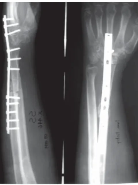
Wide resection of the tumor was performed on January 1999. The radius was cut one inch proximal to the most proximal tumor mar-gin. Wrist extensors, wrist flexors and m. brachioradialis were ex-cised at the same level as the ra-dius. Pronator quadratus, trian-gular fibrocartilage and distal ra-dioulnar joint capsule were ex-cised. The remaining tendons, distal ulna and carpal bones were preserved. Non-vascularized free fibular graft was obtained from the contra-lateral leg. The fibula was fixed to the radius shaft and the middle ray metacarpal shaft with 14 holes 3.5-mm DCP plate. Cancellous autograft, obtained from iliac crest, was inserted be-tween the carpal bones and fibu-lar head, proximal fibula and radi-us shaft junctions after excision of the articular cartilages of the fibu-lar head, carpal bones. Midcarpal arthrodesis was performed (Fig-ure 5).
Histological examination showed a high-grade, pleomorphic scoma. The tumor cells being ar-ranged in a storiform pattern and producing varying amounts of col-lagen. Numerous giant cells were distributed throughout the speci-mens. The appearances were typi-cal of a MFH-B (Figure 6).
Adjuvant chemotherapy was started three weeks after surgery. Chem-otherapy protocol consisted of high-dose methotrexate (8-12 g/ m2), adriamycin (60-75 mg/m2,8-h infusion), cisplatinum (120 mg/ m2) and citrovarum factor res-cue. Chemotherapy was well tol-erated and never life-threatening. No cardiac or hepatic toxicity was
Figure 1. Preoperative X-Ray of the patient
Figure 2. Preoperative MRI of the patient
Figure 3. Preoperative coronal MRI of the
patient
Figure 4. Preoperative scintigraphy of the
patient
Figure 6. Histopathological features of the
patient
Figure 5. Postoperative posteroanterior
and lateral x-ray of the patient after non-vascularized fibular grafting
Ankara Üniversitesi Tıp Fakültesi Mecmuası 2008, 61(4)
246 Primary Malignant Fibrous Histiocytoma of Bone in Distal Radius
seen. Mucositis requiring mouth care was encountered.
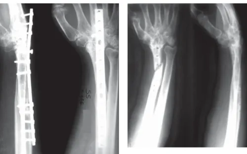
After a sixth year follow-up with-out any complication, on Janu-ary 2006, the plate was removed (Figure 7). The patient had satis-factory outcome. He had no pain and complete union at both wrist and proximal fibula-radius junc-tions was obtained. One month after removal of the plate, a frac-ture at the distal region of the graft occurred owing to a low en-ergy trauma (Figure 8). After a five months period of conservative treatment with a short arm cast, union could not be obtained. On July 2006, the patient underwent surgery and treated with vascu-larized fibula and internal fixa-tion (Figure 9). The patient is still under follow-up at 114th month, with no evidence of recurrence or metastasis.
Discussion
Malignant fibrous histiocytoma is an aggressive pleomorphic tumor, that arises most commonly in the soft tissues (6). but bone may be
the primary site of malignant fi-brous histiocytoma, rather than involvement by direct extension of a soft tissue tumor or metastat-ic spread (1,2).
MFH-B represents 1 to 8% of all pri-mary malignant tumors (3). The majority of the lesions are prima-ry, but 20 to 28% of MFH-B oc-cur secondarily in pre-existing be-nign bone abnormalities such as Paget’s disease, enchondroma, fi-brous dysplasia, bone infarcts or cysts, giant cell tumor, aseptic
necrosis, prolonged intake of cor-ticosteroids, previously irradiated either already diseased or normal bone and osteomyelitis (3,5,7). MFH-B has a male predominance ranging from 55-75% of the cases reported in the literature (6,7). Although MFH-B may arise at al-most any age, the majority of tu-mors occur after the fourth dec-ade (5). Our case as an adult man, was a typical example concerning the epidemiology of MFH without any preexisting bone abnormal-ity.
Slow-growing swelling together with local pain is the most com-mon clinical presentation as in our patient (7). Pathological frac-ture may be the initial symptom (1,3,7 ).
Although MFH-B may arise in any bone, the metaphyseal parts of the long bones of the appendicu-lar skeleton are the most common localizations (5,7). The majori-ty of the tumors arise around the knee in the lower metaphysis of the femur and upper metaphysis of the tibia (3,7). Reports about the involvement of upper extrem-ity are rare but primary MFH-B has been reported in the proxi-mal humerus, mid-humerus, ole-cranon, ulna, metacarpal bones, distal phalanx and in radius only one case, to our knowledge, till now on (3,5).
In radiographic assessment, MFH-B typically presents a solitary lytic or permeative lesion, located cen-trally or eccentric within the met-aphyseal region of a long bone with eventual spread into the ad-jacent epiphyseal and diaphyseal regions. The edges are ill-defined, cortical expansion and destruc-tion with little or no reactive scle-rosis is almost always present. Ad-jacent soft tissue mass may be ap-parent, but a periosteal new bone
Figure 7. Postoperative x-ray of the patient
after union
Figure 8. X-Ray of the patient after fracture
followed by minor trauma
Figure 9. X-Ray of the patient after revision
Journal of Ankara University Faculty of Medicine 2008, 61(4)
247
Bülent Özkurt, Yusuf Yıldız, Sinan Bilgin, Yener Sağlık
REFERENCES
1. Norman D, Feldman F. Intra- and extra-osseous malignant histiocyto-ma (histiocyto-malignant fibrous xanthohistiocyto-ma). Radiology 1972;104: 497-508. 2. Dahlin DC, Unni KK, Matsuna T.
Malignant (fibrous) histiocytoma of bone –fact or fancy? Cancer 1977; 39: 1508-16.
3. Capanna R, Bertoni F, Bacchini P, et al. Malignant fibrous histiocytoma of bone. The experience at the Rizzoli
institute: report of 90 cases. Cancer 1984; 54: 177-87.
4. Picci P, Bacci G, Ferrari S, et al. Neo-adjuvant chemotherapy in malignant fibrous histiocytoma of bone and in osteosarcoma located in the ext-remities: Analogies and differences between the two tumours. Ann On-cology 1997; 8:1107-15.
5. Jebson PJ, Sullivan L, Arbor Ann, et al. Malignant fibrous histiocytoma of the distal radius: A case report. J Hand Surg 2004; 29A: 194-9.
6. Weiner M, Sedlis M, Johnston AD, et al. Adjuvant chemotherapy of malig-nant fibrous histiocytoma of bone. Cancer 1983; 51: 25-9.
7. Boland PJ, Huvos AG. Malignant fibrous histiocytoma of bone. Clin Orthop and Rel Research 1986; 204: 130-4.
8. Little DG, MacCarthy SW. Malignant fibrous histiocytoma of bone: the experience of the New South Wales Bone Tumour Registry. Aust N Z J Surg 1993; 63: 346-51.
formation is uncommon (5,7). The differential radiological diag-nosis includes metastasis from an occult primary tumor, osteolytic osteogenic sarcoma, fibrosarcoma of bone, primary malignant lym-phoma, myeloma, and malignant giant cell tumor.
As the clinical, radiological and lab-oratory data are not pathogno-monic for MFH-B, any suspected destructive, lytic osseous lesions should be evaluated histological-ly by needle or carefulhistological-ly planned open biopsy for diagnosis. De-tailed physical examination, CT scan or magnetic resonance im-aging of the lesion, radionuclide scanning with technetium99 and CT scan of the chest are necessary for accurate diagnosis and classifi-cation of the lesion into its surgi-cal stage.
Histologically, tumor extension through the bony cortex is often present with direct involvement to adjacent soft tissues. The intraos-seous tumor spread is character-ized by medullary bone marrow infiltration and replacement
with-out reactive ossification or marrow response (7). The spindle fibrob-lastic-fibrohistiocytic cells, plump cells with histiocytic morphology, and giant cells are always present. Typically, it shows fibrogenic dif-ferentiation, often in a storiform (cartwheel) pattern, and other ar-eas of cells have a histiocytic qual-ity and are accompanied by ana-plastic giant cells. Prominent col-lagen production is present be-tween the spindle cells, which are oriented in the typical whorled or storiform pattern. Chronic inflam-matory cells are present in major-ity of the lesions (5).
Biologically, MFH-B is a fully malig-nant tumor, which not only recurs locally, but also metastasizes via both blood stream and lymphat-ic channels to distant sites, most commonly to the lungs, or less commonly to the lymph nodes, soft tissues such as liver, heart, kidney, adrenals, pancreas, skin or other bones (1,6). Prognosis depends on local recurrence and metastases. Several authors em-phasize that preoperative and/or adjuvant chemotherapy
reduc-es the ever prreduc-esent threat of mi-croscopic residual local or distant (metastatic) disease (4,6).
The mainstay of treatment for MFH-B is aggressive surgical manage-ment combined with chemother-apy (3,5,7). Although adjuvant chemotherapy has been shown to improve the survival rate and re-duce the incidence of metastasis (2,3,5,6). chemotherapy is not as successful as in patients who un-derwent inadequate surgical mar-gins (3,5,8).
Numerous alternative procedures, such as autogenous corticocan-cellous bone grafting, allograft re-placement, autogenous vascular-ized or non-vascularvascular-ized fibular grafting, custom made prosthesis can be used for the reconstruc-tion after resecreconstruc-tion of the distal radius. We preferred autogenous fibular grafting and wrist arthrod-esis in order to prevent the prob-able complications of the custom made prosthesis and also wrist in-stability and degenerative arthri-tis.