Effects of vaginal cylinder position on dose
distribution in patients with endometrial carcinoma
in treatment of vaginal cuff brachytherapy
Yurday Ozdemir, MD, Yemliha Dolek, MSci, Cem Onal, MD
Department of Radiation Oncology, Baskent University Faculty of Medicine, Adana, Turkey
Abstract
Purpose: To investigate the impact of different cylinder positions on dosimetry of critical structures in patients with
endometrial carcinoma undergoing three-dimensional image-based vaginal cuff brachytherapy (VCB).
Material and methods: We delivered VCB at a dose of 4 Gy to a depth of 5 mm in the vaginal cuff of 15 patients
using three different cylinder positions (neutral [N], parallel [P], and angled [A]) according to the longitudinal axis of the patient. We analyzed the dose-volume distribution and volumetric variability of the rectum and bladder. We con-verted the total doses to equivalent doses in 2 Gy (EQD2) using a linear-quadratic model (a/b = 3 Gy).
Results: The mean rectum volume for the N, P, and A positions was 68.2 ± 22.7 cc, 79.3 ± 33.7 cc, and 74.2 ± 29.6 cc,
respectively. The mean rectum volume for the P position was significantly larger than that for the N position (p = 0.03). Relative to the N position, the A position resulted in a lower total EQD2 in the highest irradiated 2 cc (D2cc; p = 0.001), 1 cc (D1cc; p = 0.004), and 0.1 cc (D0.1cc;p = 0.047) of the rectum. Similarly, the P position resulted in a lower EQD2 in the
D2cc (p = 0.018) and D1cc (p = 0.024) of the rectum relative to the N position. In the bladder, the P position resulted in a higher EQD2 in the D2cc relative to the N position (p = 0.02). There was no dosimetric difference between the P and
A positions in either the rectum or the bladder.
Conclusions: Vaginal cuff brachytherapy in the P and A positions is significantly superior to that in the N position
in terms of rectum dosimetry. The bladder dose in the N position is considerably lower than that in the other positions. J Contemp Brachytherapy 2017; 9, 3: 230–235 DOI: https://doi.org/10.5114/jcb.2017.68171
Key words: brachytherapy, cylinder positions, endometrial cancer, EQD2.
Purpose
Endometrial carcinoma (EC) is the most common gy-necological malignancy in developed countries, with the highest incidence occurring in the sixth and seventh de-cades of life [1]. Surgery is the first step of treatment, fol-lowed by external beam radiotherapy (EBRT) with vagi-nal cuff brachytherapy (VCB) in selected cases [2,3]. After surgery, VCB may be used either as the sole adjuvant mo-dality in patients with intermediate-risk EC or as a com-plementary component with EBRT in high-risk patients [4]. Vaginal cuff brachytherapy has a steep dose fall-off, which restricts critical organ doses while applying a high and localized radiation dose to the vaginal cuff.
The most common site of recurrence in patients with EC is the vaginal cuff [5,6]. That recurrence pattern may be one of the major reasons to use VCB as a localized ad-juvant treatment directed to the vaginal cuff in patients with EC. In the PORTEC-2 trial, patients with stage I or IIA disease, including high-intermediate risk factors, were randomized and treated with either EBRT or VCB.
In that study, there was no significant difference in local recurrence between the two treatments; however, the pa-tients treated with VCB had less gastrointestinal toxicity and better quality of life [7,8]. During VCB application, the anterior rectal wall, bladder, and sigmoid are located near the area that is exposed to high-dose radiation. The high radiation dose to those organs may cause certain tox-icities following the treatment. Therefore, it is important to consider all factors related to the dosimetry of critical organs and the target volume. Although numerous stud-ies have investigated the effects of indication [4,9], treat-ment time [10], size [4,11], and style [12] of the cylinder, filling of the bladder and rectum [4,13,14], dose fraction-ation, and prescription [4,15], the impact of the cylinder position has received little study.
The purpose of this study was to analyze wheth-er changes in the cylindwheth-er position impact the radiation dose delivered to critical structures in patients undergo-ing three-dimensional (3D) image-based high-dose-rate brachytherapy.
Address for correspondence: Yurday Ozdemir, MD, Department of Radiation Oncology, Baskent
University Faculty of Medicine, Adana Dr Turgut Noyan Research and Treatment Centre, 01120 Adana, Turkey, phone: +90 322 3444444/1304, fax: +90 322 3444445, e-mail: [email protected]
Received: 31.07.2016 Accepted: 10.05.2017 Published: 30.06.2017
Material and methods
Patients
In our study, we enrolled 15 patients with endome-trial adenocarcinoma, undergoing post-surgery VCB and EBRT. External beam radiotherapy was delivered to pel-vic lymphatics and the primary tumor bed at a prescribed dose of 45 Gy in 25 fractions using 3D conformal radio-therapy. This study was approved by the Baskent Univer-sity Institutional Review Board (Project no: KA16/362), and supported by the Baskent University Research Fund. The treatment protocol used in our department was described elsewhere [13]. Briefly, all patients had a thor-ough gynecologic examination to assess wound healing and to evaluate vaginal size before the placement of the VCB cylinder. The largest computed tomography (CT) – compatible vaginal cylinder was chosen for each patient to achieve the best contact between the vaginal mucosa and the surface of the cylinder. Lubricated condoms were used to facilitate the application and cleaning. In our rou-tine practice, CT planning in the parallel (P) position is performed for the first fraction of application, and the same plan is used for the subsequent fractions of VCB. Catheterization of the bladder and rectal enema are not routinely used. The length of the cylinder protruding out-side the vagina is noted to provide uniformity for subse-quent insertions.
Treatment planning
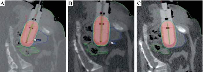
Three consecutive CT images were obtained for each patient by setting the cylinder in neutral (N), parallel (P), and angled (A) positions according to the cranial-caudal axis of the patient (Figure 1). In the N position, the cylin-der was inserted into the vagina in a manner consistent with the patient’s anatomy, and no effort was made to correct the natural insertion angle of the cylinder. In the P position, the cylinder was tilted downwards, so that it was as parallel as possible to the cranial-caudal axis of the patient. In the A position, the tip of the cylinder was moved upwards from the patient’s axial plane, keeping the caudal end of the cylinder stationary. After each
ma-neuver, the cylinder was immobilized with a universal applicator clamping device (Varian Medical Systems, Inc., Palo Alto, CA, USA), which was located under-neath the patient. A CT scan with 2.5 mm slice thickness through the pelvis was used for treatment planning. Ev-ery patient was prompted to empty the bladder before the initial CT process.
All CT slices were transferred to a 3D treatment plan-ning system (BrachyVision™ Eclipse; Varian Medical Systems, Palo Alto, CA, USA). A dose of 12 Gy in three fractions was applied to a depth of 5 mm from the cylin-der surface to treat the entire vaginal cuff. The vaginal length was measured on CT images, and the apical two-thirds of the vaginal cylinder were activated, except in patients with a short vaginal length, in accordance with our institutional practice. As a result, a total of 45 CT scans, including three cylinder positions for each patient, were analyzed.
Organs at risk
In order to minimize inter-observer variability, a single physician contoured the clinical target volume (CTV), blad-der, and rectum of each patient. The contour of the CTV was determined by expanding the upper two segments of the cylinder by 5 mm in all directions from the cylinder surface. The outer wall of the rectum and the entire blad-der were delineated as critical organs. To accommodate the volumetric rectal variations relative to the position of the cylinder, the rectum was contoured in a manner sug-gested in a previous study [16]. Thus, the rectum was de-lineated from 1 cm above the cylinder apex to 1.5 cm below the last activated cylinder segment. We also recorded the volumes of the rectum and bladder to assess the volumet-ric variations due to the different cylinder positions. For the critical organs, we calculated the minimum dose to the highest irradiated 2 cc (D2cc), 1 cc (D1cc), and 0.1 cc (D0.1cc) of
the rectum, and to the D2cc and D0.1cc of the bladder in each application. The dose statistics for the rectum are shown in Table 1. We converted the physical dose distributions in Table 1, in terms of the VCB (for one fraction) and the previous EBRT (for 25 fractions), to biologically equivalent
Fig. 1. The 4 Gy isodose area (red) according to the vaginal cylinder in the (A) neutral, (B) parallel, and (C) angled positions in
a representative patient
doses {BED; BED = n.d. [1 + d/(a/b)]} to account for the dose per fraction as given in fractions of 2 Gy {EQD2; EQD2
= BED/[1 + 2/(a/b)]} using a linear quadratic model with a/b = 3 Gy [17]. Additionally, we estimated an approxi-mate EQD2 VCB value for the three fractions of VCB, based
on the doses shown in Table 1.
Statistical analysis
We analyzed the data using SPSS version 20 (SPSS, Chicago, IL, USA). We determined the volumes of all specified organs at risk and compared the dose-volume histograms for each of the cylinder positions. We used the Wilcoxon matched-pairs test to determine significant differences in volumes and doses between the cylinder positions. We considered differences statistically signif-icant at p < 0.05.
Results
We analyzed a total of 45 CT scans from 15 patients with cylinders in the N, P, and A positions. The cylin-der diameter was 3.5 cm for all of the patients. The mean rectal volume for the P, A, and N positions was 79.3 ± 33.7 cc, 74.2 ± 29.6 cc, and 68.2 ± 22.7 cc, respectively. The mean rectum volume for the P position was significantly larger than that for the N position (p = 0.03). There was no significant difference in bladder volume among the three cylinder positions.
The D2cc, D0.1cc, and D1cc of the rectum in the three
po-sitions are shown in Table 2. The BEDEBRT and EQD2EBRT were 72 Gy and 43.2 Gy, respectively. The sum of the two modalities (EQD2 EBRT and EQD2 VCB) in the rectal D2cc, D0.1cc, and D1cc is listed for three fractions of VCB in
Table 3. The total EQD2 in the D2cc (p = 0.001), D0.1cc Table 1. Dose levels to the rectum according to the cylinder positions in vaginal cuff brachytherapy (VCB) and equivalent dose calculated in 2 Gy (EQD2) of external beam radiotherapy
Cases EQD2 EBRT (1.8 Gy × 25 fx)
VCB (fx)
D2cc (fx/Gy) D0.1cc (fx/Gy) D1cc (fx/Gy)
N P A N P A N P A 1 43.2 3 4.9 4.5 4.5 6.4 6.3 5.6 5.2 4.8 5.1 2 43.2 3 5.2 4.5 4.5 6.5 6.2 5.4 5.5 4.8 4.9 3 43.2 3 5.3 4.8 3.7 6.2 5.9 4.6 5.8 5.1 4.0 4 43.2 3 5.6 3.2 4.4 7.2 3.9 5.5 6.2 3.5 4.7 5 43.2 3 4.9 4.8 5.6 6.3 5.7 8.3 5.2 5.1 6.0 6 43.2 3 5.4 4.9 4.2 6.4 6.3 5.1 5.4 5.5 4.4 7 43.2 3 5.2 5.2 4.7 5.3 5.6 5.6 5.4 5.4 4.9 8 43.2 3 5.1 4.7 4.1 6.6 5.4 4.9 5.4 4.9 4.4 9 43.2 3 4.0 3.6 3.8 4.9 4.1 4.5 4.3 3.8 4.0 10 43.2 3 6.0 5.1 5.0 8.4 6.1 5.5 7.2 5.5 5.3 11 43.2 3 5.3 4.7 4.9 6.3 5.9 5.9 5.6 4.9 5.2 12 43.2 3 5.0 4.4 4.2 6.3 5.1 5.1 5.2 4.6 4.5 13 43.2 3 3.8 5.2 3.8 5.1 8.1 5.1 4.2 5.8 4.2 14 43.2 3 6.4 4.7 4.6 9.2 8.8 5.6 7.5 5.7 5.1 15 43.2 3 4.1 4.1 4.1 4.9 4.6 5.1 4.3 3.6 4.4
EQD2 – equivalent dose calculated in 2 Gy, Gy – gray, fx –fractions, D 2cc, 0.1cc, 1cc – the minimum dose to the most irradiated of 2 cc, 0.1 cc, and 1 cc of organ,
VCB – vaginal cuff brachytherapy, N, P, A – neutral, parallel, and angled cylinder positions, respectively
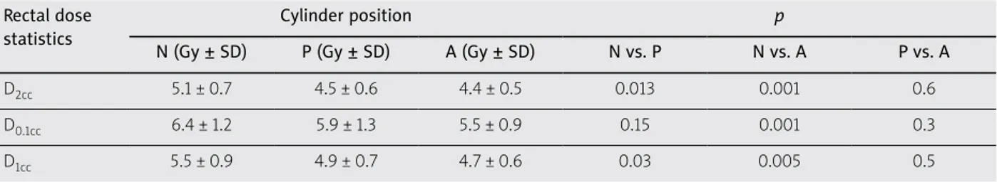
Table 2. Mean rectum dose statistics according to the vaginal cylinder positions for one fraction of brachy-therapy
Rectal dose statistics
Cylinder position p
N (Gy ± SD) P (Gy ± SD) A (Gy ± SD) N vs. P N vs. A P vs. A
D2cc 5.1 ± 0.7 4.5 ± 0.6 4.4 ± 0.5 0.013 0.001 0.6
D0.1cc 6.4 ± 1.2 5.9 ± 1.3 5.5 ± 0.9 0.15 0.001 0.3
D1cc 5.5 ± 0.9 4.9 ± 0.7 4.7 ± 0.6 0.03 0.005 0.5
(p = 0.047), and D1cc (p = 0.004) of the rectum for the A po-sition was significantly lower than the corresponding values for the N position. Similarly, the EQD2 in the D2cc
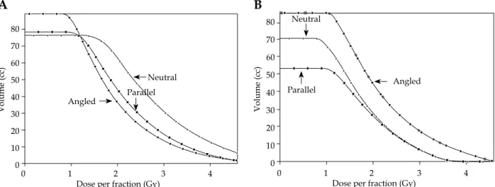
(p = 0.018) and D1cc (p = 0.024) of the rectum for the P position was significantly lower than the corresponding values for the N position. The differences in the dose sta-tistics maintained their significance when we estimated the EQD2 for three fractions of VCB (Table 3). Despite the significant differences between the N and P positions and between the N and A positions, there was no difference in terms of rectal doses between the P and A positions (Figure 2).
Regarding the bladder, the EQD2 in the D2cc for the P position was significantly higher than that for the N po-sition (2.2 ± 0.5 vs. 2.0 ± 0.4; p = 0.02).
Discussion
Although some brachytherapy studies have focused on the association between the angles or positions of the vaginal cylinder and rectal doses in patients with EC, there is little data showing the impact of the vaginal cyl-inder position on the critical organ doses in the context of EQD2. Our analysis demonstrated that the N position was associated with unfavorable EQD2 values compared with the P and A positions in terms of the rectal D2cc,
D0.1cc, and D1cc. Additionally, manipulating the cylinder
in a more angular manner, moving the cylinder from the P position to the A position, did not make any difference in the rectal doses. Conversely, despite the disadvantages of the N position in terms of the rectal doses, the bladder doses for the N position were lower than those for the P and A positions. However, the bladder was not the main topic in our study.
The relationship between the dose and the effect in radiotherapy is linear-quadratic rather than linear. There-fore, the cumulative effect of different treatment methods cannot be identified by adding the doses linearly [18]. The various fractions of VCB and EBRT used in clinical practice can be considered together by converting the dif-ferent units into a single unit, a concept known as EQD2, which allows clinicians to compare different dose rates and dose fractions.
Regarding the rectal dose statistics, the D2cc (EQD2) is the most established parameter as a predictor of rectal toxicity in studies of intracavitary brachytherapy (ICB) and interstitial brachytherapy (IB) [19,20,21,22]. In a pre-vious study, Georg et al. assessed the D2cc in patients with cervical carcinoma who were treated with EBRT and ICB in terms of Grade 1-4 rectal toxicities on the Late Effects on Normal Tissues-Subjective, Objective, Management, and Analytic (LENT-SOMA) scale and found that 72 Gy was more relevant than 63 Gy [21]. Chopra et al. [20] sug-gested that limiting the D2cc exposure to < 55 Gy was
as-Table 3. Total* rectal doses according to cylinder position Dose-volume
parameters
Cylinder position p
N (Gy ± SD) P (Gy ± SD) A (Gy ± SD) N vs. P N vs. A P vs. A
D2cc 67.3 ± 5.5 63.4 ± 4.8 62.1 ± 3.8 0.028 0.002 0.34
D0.1cc 79.3 ± 12 73.1 ± 12.3 70.5 ± 8.4 0.08 0.028 0.49
D1cc 70.9 ± 8.4 66.1 ± 5.6 64.5 ± 4.3 0.043 0.007 0.35
*Total dose is provided in equivalent dose calculated in 2 Gy (EQD2) and EQD2 is calculated as EQD2 EBRT + EQD2 VCB. The EQD2 VCB was calculated based on the
D2cc, 0.1cc, 1cc values presented in Table 1. The EQD2 EBRT was calculated for the total EBRT fractions
EBRT – external beam radiotherapy, VCB – vaginal cuff brachytherapy, Gy – gray, N – neutral, P – parallel, A – angled, D2cc, 0.1cc, 1cc – the minimum dose to the most
irradiated of 2 cc, 0.1 cc, and 1 cc of organ
Volume (cc) Volume (cc) 80 70 60 50 40 30 20 10 0 80 70 60 50 40 30 20 10 0 0 1 2 3 4
Dose per fraction (Gy) 0 1 2 3 4Dose per fraction (Gy)
Neutral
Neutral
Parallel Parallel
Angled
Angled
Fig. 2. Comparative dose-volume histogram of the (A) rectum and (B) bladder according to the vaginal cylinder in the neutral
(triangle), parallel (square), and angled (sphere) positions
sociated with less Grade 2 rectal toxicity in patients with recurrent cervical carcinoma. In addition, Sakata et al. [23] found that patients with maximal rectal EQD2 > 60 Gy
experienced more rectal toxicity. Supporting the results of Sakata et al., another report showed that a cumulative dose of > 65 Gy (EQD2) was associated with more Grade 2 LENT-SOMA rectal morbidities [24].
Our study demonstrates only a dosimetric pattern and lacks toxicity data; however, the rectal dose sta-tistics, shown in Table 3, may offer some foresight into possible rectal toxicities in the period following treat-ment. For the total EQD2 VCB, we used the initial rectal dose statistics in the first fraction of VCB for two subse-quent imaginary fractions. Significant variation in rectal dose should not be expected among the three fractions of VCB when differences in cylinder position or angle are excluded, considering the common clinical practice of performing the fractions of VCB based on the initial planning CT [25].
According to our results, the mean values of the dose statistics (D2cc, D0.1cc, and D1cc) in terms of EQD2 were all
significantly higher for the N position than for the P and A positions (Table 3). Despite the lack of a symptomatic assessment of rectal toxicity in our study, the mean EQD2 for the D2cc (67.3 ± 5.5), D0.1cc (79.3 ± 12), and D1cc (70.9 ±
8.4) of the rectum in our study appeared to be safer than those observed by Georg et al. (72 ± 6, 88 ± 10, and 76 ± 7, respectively) [21].
There have been some reports of the advantages of 3D VCB compared with two-dimensional (2D) VCB [26,27]. In contrast to the dose specification process of 2D plan-ning, which is based on the pelvic bone, 3D VCB uses vol-umetric treatment planning, and hence has more precise target coverage, which is critical for certain patients. For instance, while patients who undergo VCB without EBRT are expected to experience limited rectum and bladder toxicities, the VCB-related dose may become important in patients who receive an EBRT dose of 45-50.4 Gy prior to VCB. Additionally, further techniques have been uti-lized to minimize the critical organ doses, including mod-ifications of the bladder [13,28] or rectal filling [29], the position of the patient on the couch [30], or the geometry of the vaginal cylinder [31].
In a previous study, Hoskin et al. moved the vaginal cylinder from the N position to the P position in 30 pa-tients with EC who were treated with VCB and report-ed an average rectal dose rreport-eduction of 1.3 Gy for a pre-scribed dose of 5.5 Gy [31]. Our results differ from those of Hoskin et al. in some instances. First, we performed a dosimetric analysis of three different positions of the vaginal cylinder according to the cranial-caudal plane of the patient. Second, we used CT images, which are more reliable to assess the variability of doses and critical-or-gan delineations, among different set-up positions com-pared with the radiographs used by Hoskin et al. Third, our report is more comparable to other reports in terms of cumulative doses and possible side effects because of our use of EQD2 in our analyses. However, our results agree
with those of Hoskin et al. in that the rectal doses in our patients diminished as the tip of the cylinder was moved upward from the axial plane of the patient (p < 0.05 for
D1cc and D2cc in the A position) at the cost of increasing
the mean bladder dose.
Another previous dosimetric study lends indirect support to our results. Lati et al. [30] investigated the in-fluence of two different patient positions without correct-ing the cylinder angle in the vagina. The rectal dose in pa-tients positioned with a leg extended was lower than that in patients in the lithotomy position (D2cc, 4.24 Gy vs. 5.14 Gy; p = 0.003). The lithotomy position may be considered as the opposite of our A position, because the vaginal cuff in the lithotomy position is moved posteriorly, approach-ing the rectum, and consequently, the rectum is exposed to higher radiation doses because of its proximity to the vaginal cylinder in the cuff.
Our volumetric analyses showed that the rectal vol-ume for the P position was significantly larger than that for the N position (79.3 ± 33.7 cc vs. 68.2 ± 22.7 cc; p = 0.03). The movement of rectal contents and gas through the hollow rectum during the application in different cylinder positions may be the major reason for the vol-umetric variation. Additionally, CT may have limited sensitivity to delineate the rectal wall, especially for the distal part of the organ.
Our study has several limitations. First, our patient cohort was small, and all of the patients were treated at a single institution. Second, the angled application of the cylinder was performed arbitrarily without a fixed an-gle, which might cause some angular variability. Third, although the P and A positions had advantages relative to the N position in terms of the rectal radiation dose, we did not attempt to determine the optimal cylinder angles.
Conclusions
We examined the dosimetric influence of three differ-ent cylinder positions for the application of VCB in terms of EQD2 in patients with EC. Our results demonstrate that VCB in the P and A positions results in significant-ly lower rectal radiation doses compared with that in the N position, at the expense of higher bladder doses. De-spite the similar rectal dosimetry of the P and A posi-tions, the bladder dose may increase when the cylinder is moved upward from the patient’s axial plane. Further studies, including toxicity analysis in larger cohorts, may be more demonstrative of the optimal cylinder position for the treatment of the vaginal cuff.
Acknowledgement
Supported by the Baskent University Research Fund.
Disclosure
Authors report no conflict of interest.
References
1. Jemal A, Bray F, Center MM et al. Global cancer statistics. CA
Cancer J Clin 2011; 61: 69-90.
2. Benedet JL, Bender H, Jones H 3rd et al. FIGO staging classifi-cations and clinical practice guidelines in the management of gynecologic cancers. FIGO Committee on Gynecologic On-cology. Int J Gynaecol Obstet 2000; 70: 209-262.
3. Nout RA, Smit VT, Putter H et al. Vaginal brachytherapy versus pelvic external beam radiotherapy for patients with endometrial cancer of high-intermediate risk (PORTEC-2): An open-label, non-inferiority, randomised trial. Lancet 2010; 375: 816-823.
4. Meyer LA, Bohlke K, Powell MA et al. Postoperative radi-ation therapy for endometrial cancer: American Society of Clinical Oncology clinical practice guideline endorsement of the American Society for Radiation Oncology evidence-based guideline. J Clin Oncol 2015; 33: 2908-2913.
5. Keys HM, Roberts JA, Brunetto VL et al. A phase III trial of surgery with or without adjunctive external pelvic radiation therapy in intermediate risk endometrial adenocarcinoma: A Gynecologic Oncology Group Study. Gynecol Oncol 2004; 92: 744-751.
6. Nout RA, van de Poll-Franse LV, Lybeert ML et al. Long-term outcome and quality of life of patients with endometrial carcinoma treated with or without pelvic radiotherapy in the post operative radiation therapy in endometrial carcinoma 1 (PORTEC-1) trial. J Clin Oncol 2011; 29: 1692-1700.
7. Anderson JM, Stea B, Hallum AV et al. High-dose-rate post-operative vaginal cuff irradiation alone for stage Ib and Ic endometrial cancer. Int J Radiat Oncol Biol Phys 2000; 46: 417-425.
8. Nag S, Erickson B, Parikh S et al. The American Brachythera-py Society recommendations for high-dose-rate brachythera-py for carcinoma of the endometrium. Int J Radiat Oncol Biol
Phys 2000; 48: 779-790.
9. Mitra D, Klopp AH, Viswanathan AN. Pros and cons of vag-inal brachytherapy after external beam radiation therapy in endometrial cancer. Gynecol Oncol 2016; 140: 167-175. 10. Rovirosa A, Ascaso C, Arenas M et al. Can we shorten the
overall treatment time in postoperative brachytherapy of endometrial carcinoma? Comparison of two brachytherapy schedules. Radiother Oncol 2015; 116: 143-148.
11. Rakhra SS, Weaver C, Donnelly ED et al. The effect of pelvic radiotherapy on vaginal brachytherapy cylinder diameter: Implications for optimal treatment order. Brachytherapy 2016; 15: 549-553.
12. Bahadur YA, Constantinescu C, Hassouna AH et al. Single versus multichannel applicator in high-dose-rate vaginal brachytherapy optimized by inverse treatment planning.
J Contemp Brachytherapy 2015; 6: 362-370.
13. Guler OC, Onal C, Acibuci I. Effects of bladder distension on dose distribution of vaginal vault brachytherapy in pa-tients with endometrial cancer. J Contemp Brachytherapy 2015; 6: 371-376.
14. Sapienza LG, Aiza A, Gomes MJ et al. Bladder (ICRU) dose point does not predict urinary acute toxicity in adjuvant isolated vaginal vault high-dose-rate brachytherapy for in-termediate-risk endometrial cancer. J Contemp Brachytherapy 2015; 7: 357-362.
15. Damast S, Higgins SA, Ratner E et al. High-dose-rate vagi-nal brachytherapy with chemotherapy for surgically staged localized uterine serous carcinoma. J Contemp Brachytherapy 2015; 7: 35-40.
16. Sabater S, Andres I, Jimenez-Jimenez E et al. Rectal contrast increases rectal dose during vaginal cuff brachytherapy.
Brachytherapy 2016; 15: 35-39.
17. Dale RG. The application of the linear-quadratic dose-effect equation to fractionated and protracted radiotherapy. Br J
Radiol 1985; 58: 515-528.
18. Nag S, Gupta N. A simple method of obtaining equivalent doses for use in HDR brachytherapy. Int J Radiat Oncol Biol
Phys 2000; 46: 507-513.
19. Pötter R, Haie-Meder C, Van Limbergen E et al. Recom-mendations from Gynaecological (GYN) Gec Estro Working
Group (II): Concepts and terms in 3D image-based treatment planning in cervix cancer brachytherapy-3D dose volume pa-rameters and aspects of 3D image-based anatomy, radiation physics, radiobiology. Radiother Oncol 2006; 78: 67-77. 20. Chopra S, Dora T, Engineer R et al. Late rectal toxicity after
image-based high-dose-rate interstitial brachytherapy for postoperative recurrent and/or residual cervical cancers: EQD2 predictors for grade ? II toxicity. Brachytherapy 2015; 14: 881-888.
21. Georg P, Kirisits C, Goldner G et al. Correlation of dose-vol-ume parameters, endoscopic and clinical rectal side effects in cervix cancer patients treated with definitive radiotherapy including MRI-based brachytherapy. Radiother Oncol 2009; 91: 173-180.
22. Koom WS, Sohn DK, Kim JY et al. Computed tomogra-phy-based high-dose-rate intracavitary brachytherapy for uterine cervical cancer: Preliminary demonstration of cor-relation between dose-volume parameters and rectal muco-sal changes observed by flexible sigmoidoscopy. Int J Radiat
Oncol Biol Phys 2007; 68: 1446-1454.
23. Sakata K, Nagakura H, Oouchi A et al. High-dose-rate in-tracavitary brachytherapy: Results of analyses of late rectal complications. Int J Radiat Oncol Biol Phys 2002; 54: 1369-1376. 24. Tharavichtikul E, Meungwong P, Chitapanarux T et al. The association of rectal equivalent dose in 2 Gy fractions (EQD2) to late rectal toxicity in locally advanced cervical cancer pa-tients who were evaluated by rectosigmoidoscopy in faculty of medicine, chiang mai university. Radiat Oncol J 2014; 32: 57-62.
25. Patel S, Mehta KJ, Kuo HC et al. Do changes in interfraction organ at risk volume and cylinder insertion geometry impact delivered dose in high-dose-rate vaginal cuff brachytherapy?
Brachytherapy 2016; 15: 185-190.
26. Haie-Meder C, Pötter R, Van Limbergen E et al. Recom-mendations from gynaecological (GYN) Gec-Estro Working Group (i): Concepts and terms in 3D image based 3D treat-ment planning in cervix cancer brachytherapy with emphasis on MRI assessment of GTV and CTV. Radiother Oncol 2005; 74: 235-245.
27. Viswanathan AN, Erickson BA. Seeing is saving: The benefit of 3D imaging in gynecologic brachytherapy. Gynecol Oncol 2015; 138: 207-215.
28. Stewart AJ, Cormack RA, Lee H et al. Prospective clinical trial of bladder filling and three-dimensional dosimetry in high-dose-rate vaginal cuff brachytherapy. Int J Radiat Oncol
Biol Phys 2008; 72: 843-848.
29. Sabater S, Arenas M, Berenguer R et al. Dosimetric analysis of rectal filling on rectal doses during vaginal cuff brachythera-py. Brachytherapy 2015; 14: 458-463.
30. Iati G, Pontoriero A, Mondello S et al. Three-dimensional treatment planning for vaginal cuff brachytherapy: Dosim-etric effects on organs at risk according to patients position.
Brachytherapy 2014; 13: 568-571.
31. Hoskin PJ, Bownes P, Summers A. The influence of applica-tor angle on dosimetry in vaginal vault brachytherapy. Br J