* Corresponding Author
Myelosuppression and Oxidative Stress Induced by Cyclophosphamide in Rats: The Protective Role of Selenium
Adnan AYHANCI1, Nilhan HEYBELİ1, İlknur KULCANAY ŞAHİN2, Mustafa CENGİZ3, *
1Eskisehir Osmangazi University, Faculty of Art and Science, Department of Biology, Eskisehir, Turkey [email protected], ORCID: 0000-0003-4866-9814
1Eskisehir Osmangazi University, Faculty of Art and Science, Department of Biology, Eskisehir, Turkey [email protected], ORCID: 0000-0001-8425-9965
2 Kırıkkale University, Vocational School of Health Services, Kırıkkale, Turkey ilknurkulcanay@kırıkkale.edu.tr, ORCID: 0000-0003-1948-6912
3 Siirt University, Faculty of Education, Department of Elementary Education, Siirt, Turkey [email protected], ORCID: 0000-0002-6925-8371
Abstract
Aim of this study is to detect the protective role of selenium (Se) against bone marrow and blood toxicity in CP-induced hematoxicity model. We categorized the rats into 6 groups of 7 animals in each group (control; 150 mg/kg CP; 0.5 mg/kg Se; 1 mg/kg Se; 150+0.5 mg/kg CP+Se; 150+1 mg/kg CP+Se). Se injections started 5 days before the CP injections and carried on until the end of the experiment (6th day) for the groups to which CP was injected together with Se. CP was applied as a single dose before anesthesia. For that reason, on the 7th day, blood was taken with cardiac puncture and bone marrow was taken by flushing the femur. Peripheral blood cells and bone marrow nucleated cells were counted on a cell counter. Intraperitoneal CP injection was found to reduce the number of leukocytes by 317%, thrombocyte by 36% and bone marrow nucleated cells by 481% compared to the control group. In the groups where CP was given after 0.5 and 1 mg/kg Se, numbers of leukocyte, thrombocyte and bone marrow nucleated cells were considerably improved compared to the group to which CP was
Adıyaman University Journal of Science
https://dergipark.org.tr/en/pub/adyujsci DOI: 10.37094/adyujsci.617016
ADYUJSCI
9 (2) (2019) 252-265
given only (p<0.001). Results show that 1 mg/kg Se has a better protection than 0.5 mg/kg against CP associated hematoxicity and myelosuppression. Our results also imply that the doses of Se could be adjusted according to enhance in CP dose so as to gain a stronger protective effect. We believe there is a need of further studies in which different doses of Se will be used against CP induced hematoxicity. Se can provide protection against CP-induced myelosupression and lipid peroxidation.
Keywords: Cyclophosphamide, Hematoxicity, Selenium, Myelosupression, Lipid
peroxidation
Sıçanlarda Siklofosfamid'in Neden Olduğu Miyelosupresyon ve Oksidatif Stres: Selenyumun Koruyucu Rolü
Öz
Bu çalışmanın amacı, selenyumun (Se), CP kaynaklı hematoksisite modelinde kemik iliği ve kan toksisitesine karşı koruyucu rolünü tespit etmekti. Her grupta sıçanları 7 hayvandan oluşan 6 gruba ayırdık (kontrol; 150 mg/kg CP; 0,5 mg/kg Se; 1 mg/kg Se; 150+0,5 mg/kg CP+Se; 150+1 mg/kg). CP+Se). Se enjeksiyonları, CP enjeksiyonlarından 5 gün önce başlamış ve Se ile birlikte CP enjekte edilen gruplar için deneyin sonuna (6. gün) kadar devam etmiştir. CP anestezi öncesi tek doz olarak uygulandı. Bu nedenle 7. günde kardiyak delinme ile kan alındı ve femur yıkanarak kemik iliği alındı. Periferik kan hücreleri ve kemik iliği çekirdekli hücreler, bir hücre sayacında sayıldı. İntraperitoneal CP enjeksiyonunun lökosit sayısını %317, trombositi %36 ve kemik iliği çekirdekli hücreleri %481 azalttığı bulundu. CP' nin 0,5 ve 1 mg/kg Se'den sonra verildiği gruplarda, sadece CP verilen gruba kıyasla lökosit, trombosit ve kemik iliği çekirdekli hücrelerin sayısı önemli ölçüde artmıştır (p<0.001). Sonuçlar, 1 mg/kg Se'nin, CP ile ilişkili hematoksisite ve miyelosupresyona karşı 0,5 mg / kg'dan daha iyi bir korumaya sahip olduğunu göstermektedir. Sonuçlarımız ayrıca, Se dozlarının daha güçlü bir koruyucu etki elde etmek için CP dozundaki artışa göre ayarlanabileceği anlamına gelir. CP'nin neden olduğu hematoksisiteye karşı farklı Se dozlarının kullanılacağı konusunda daha fazla çalışmaya ihtiyaç olduğuna inanıyoruz. Se, CP kaynaklı miyelosupresyon ve lipit peroksidasyonuna karşı koruma sağlayabilir.
Anahtar Kelimeler: Siklofosfamid, Hematoksisite, Selenyum, Miyelosupresyon,
Lipit peroksidasyonu
1. Introduction
Cyclophosphamide (CP) is an antineoplastic chemotherapeutic agent, which is the most commonly used immunosuppressive and anticancer drug [1, 2]. CP is a strong oxazophosphine, an alkylating agent commonly used alone or in combination with diverse drugs in the treatment of various solid tumours. It is also used for the treatment of non-neoplastic diseases including rheumatoid arthritis, nephrotic syndrome, systemic lupus erythromatosis, and conditions before bone marrow transplantation. CP is a prodrug with its two active metabolites, phosphoramide mustard (PM) and acrolein. PM is an anticancer metabolite whereas acrolein is a toxic metabolite, responsible for hematological and myelotoxicity [3]. CP or its metabolite reacts with GSH, restrict its antioxidant activity, increases the production of reactive oxygen species and causes lipid peroxidation that leads to oxidative stress [4].
Free radicals and lipid peroxides caused by these radicals are known to be lined to the pathogenesis of several illnesses. In studies, the effects of radiotherapy on antioxidants and oxidant activities were investigated, which found that radiotherapy had raised lipid peroxidation and suppressed the activity of antioxidant enzymes [5]. On the other hand, antineoplastic agents are biological sources of free radicals. These agents increase lipid peroxidation due to their cytotoxic effects [6]. Lipid peroxidation is considered among the causes of cancer; studies have investigated the effects of various nutritional antioxidants on cancer formation [7]. Antioxidants have been found to inhibit the onset and development of carcinogenesis, as well as preventing cell death and change [8].
Selenium (Se) is the cofactor of many enzymes in our body, and it is basically an essential trace element known for its anti-oxidative function. It also plays a role in many metabolisms, including glutathione peroxidases (GPx), and selenoprotein P which protect the organism from oxidative damage [9]. GPx is an antioxidant enzyme in the cell cytoplasm and serves as the Se depot [10]. It also interacts with vitamin E to protect the cell membrane from the oxidative effect of peroxides, a product of lipid metabolism [11].
In addition to the protective function of the cell membrane by suppressing lipid peroxidation [12], it has also been shown that it has a synergistic effect with chemotherapeutic agents due to its interaction with antioxidants [13]. In this study, we planned to examine the potential cytoprotective effects of the well-known anti-oxidant properties in peripheral blood and bone marrow nuclei cells against hematoxicity due to experimentally-given CP.
2. Material and Method
In our experimental study, healthy, male, 220 ± 20-gram weight, 3-month-old Spraque-Dawley albino rats were used. Experimental animals were obtained from Eskisehir Osmangazi University (ESOGU) Medical and Surgical Research Center. All experimental animals were placed in a weekly quarantine prior to injection and were housed in polycarbonate transparent cages, in which the heat (22 °C) and humidity (45-50%) were automatically adjusted in rooms with light/dark illumination during the experiment, and pellet feed and fountain as required water was given. Our study gained approval from the Animal Ethics Committee of ESOGU.
2.1. Experimental Groups
In current study, CP treatment protocol used to develop bone marrow and blood toxicity [14] and the defensive doses of Se [14] have been previously reported. Forty-two male Sprague-Dawley rats were used for the intraperitoneal (i.p.) injection of Se (sodium selenite 98% powder; Sigma S5261, Sigma-Aldrich), CP (Sigma-Aldrich, Darmstadt, Germany), and saline (Sigma-Aldrich). The animals were given standard pellet and water ad libitum. Rats were randomly divided into the following experimental groups, each including 7 animals:
1. Group 1 served as the normal saline treated controls.
2. Group 2 animals received CP i.p. dissolved in saline, in a single dose of 150 mg/kg b.w.
4. Group 5 and 6 received respective Se for 6 days and then a single dose of CP administered on the sixth day.
2.2. Sample Collection
All animals were anesthetized with an i.p. injection of 50 mg/kg ketamine and 10 mg/kg xylazine, respectively (Sigma Aldrich), the blood samples were collected by cardiac puncture, and then the rats were killed by cervical dislocation after 24 h of the final saline, CP or Se injections. After blood collection, a femur of the killed animals was thoroughly removed from the muscles.
2.3. Chemical Substances and Injections
CP (Sigma C0768-10G, America) and Se (Sigma 209643-50G, America) were commercially obtained and applied to the related groups (Table 1). In our experiment, two different doses (0.5 and 1 mg/kg) of Se were used. In rats, the lethal dose (LD50) was 4.25 mg/kg. 500 mg of CP was dissolved in 25 mL bidistile water and 25 mL/500 mg CP-containing solution was prepared, which was prepared by dissolving 0.5 and 1 mg/kgSe in 0.5 mL saline. These chemicals and the control group were administered the necessary doses of saline i.p. All the animals were weighed prior to the first injection and sacrifice. The animals in group 1 only CP was anesthetized 24 hours after CP injection (day 7). In the groups given CP plus Se, Se was started five days before the CP application and continued until the end of the experiment. On the sixth day, the rats were reweighed to determine the dose of CP to be applied and thus CP+Se was given. Apart from these groups, we formed another two groups to which only Se was given (0.5 and 1 mg/kg, respectively). On the seventh day, the animals were anesthetized so that their blood and bone marrow could be taken [15].
2.4. Analysis of Biochemical Data
2.4.1. Determining Malondialdehyde (MDA) Level
Plasma MDA levels were calculated by colorimetric method, the basis of which consists of reactions of one molecule of MDA and two molecules of thiobarbituric acid (TBA) in forming a red MDA-TBA compound. The pH of the TBA (Sigma-Aldrich) solution prepared as a 0.67% solution was modified to 7.2 using 0.1 mol/L NaOH. To a
10 mL centrifuge tube, 2.5 mL of 10% trichloroacetic acid (TCA) (Sigma-Aldrich) was added plasma (0.5 mL) and then the mixture was keep warm for 15 minutes in a boiling water bath, then exposed to under tap water to cool off before being centrifuged at rpm for 10 minutes (protein precipitated). After adding 2 mL of supernatant to 1 mL of TBA solution, it was kept in a boiling water bath for as long as fifteen minutes prior to being cooled off under tap water. The MDA concentration was then measured by spectrophotometer at 535 nm absorption. Results have been shown as nmol/mL plasma [16].
2.4.2. Determining Serum Glutathione (GSH) Level
We determined the GSH level changes using the colorimetric assay kit (Cayman Chemical Company, Michigan, USA), following the manufacturer's protocols. We measured the absorption of the samples through an adjustable VERSA max microplate reader (Molecular Devices in California, USA) at 405 nm, the results of which have been shown as µmol/L GSH.
2.5. Statistical Evaluations
The donnee obtained from the animals was shown as standard error of the mean (±SEM). The independent measurements, as well as continuous data showing a normal distribution, were analyzed via One Way Anova program. The differences watched in the groups were acknowledged to be important if the p value was <0.001 and <0.05.
3. Results
3.1. Se Protects the Cell Against Lipid Peroxidation
Table 1, as well as Fig. 1 and Fig. 2, exhibit show Se, CP, and their co-administration, affects oxidative stress biomarkers like GSH and MDA, respectively. Although we observed a change in the levels of MDA and GSH in saline and Se (0.5 and 1 mg/kg) groups, this change achieved no statistical significance. Application of CP drastically increased MDA levels in plasma by comparison with the control group (Fig. 1). Concurrently, CP caused a remarkable decrease (p<0.001) in GSH levels by comparison with the control group (Fig. 2). As to treating CP, Se (0.5 or 1 mg/kg) remarkably lowered plasma MDA levels (p<0.001) and significantly increased the levels
of serum GSH by comparison with the CP group. That is to say, 1 mg/kg of Se yielded an added a value greater than 0.5 mg/kg by increasing its antioxidant capacity and eliminating the ill effects of lipid peroxidation.
Table 1. Plasma MDA and serum GSH levels of blood tissue taken from rats
Groups (n=7) MDA (nmol/mL) GSH (µM GSSG)
1 0.485±0.0795 5.809±1.107 2 0.798±0.0890*** 2.820±0.526*** 3 0.412±0.0948ns 5.580±1.105 ns 4 0.445±0.0855 ns 6.336±1.451 ns 5 0.476±0.0895 ns 4.414±0.688*** 6 0.490±0.102 ns 7.659±1.146***
ns: not significant, ***p; Significant difference compared to control
Figure 1. Malondialdehyde (MDA) values of blood samples. All of the values have been represented as mean ± standard deviation (n=7). It was thanks to ANOVA that the statistical significance concerning the means was performed, which was followed by Tukey-Kramer and ANOVA (p<0.001). *p<0.001 refers to a significant difference by comparison with control and CP groups
Figure 2. Glutathione (GSH) levels of the blood samples. All of the values were expressed as mean ± standard deviation (n=7). It was thanks to ANOVA that the statistical significance concerning the means was performed, which was followed by Tukey-Kramer and ANOVA (p<0.001). *p<0.001 refers to a
significant difference by comparison with control and CP groups
3.2. Se Improves Myelosuppresion
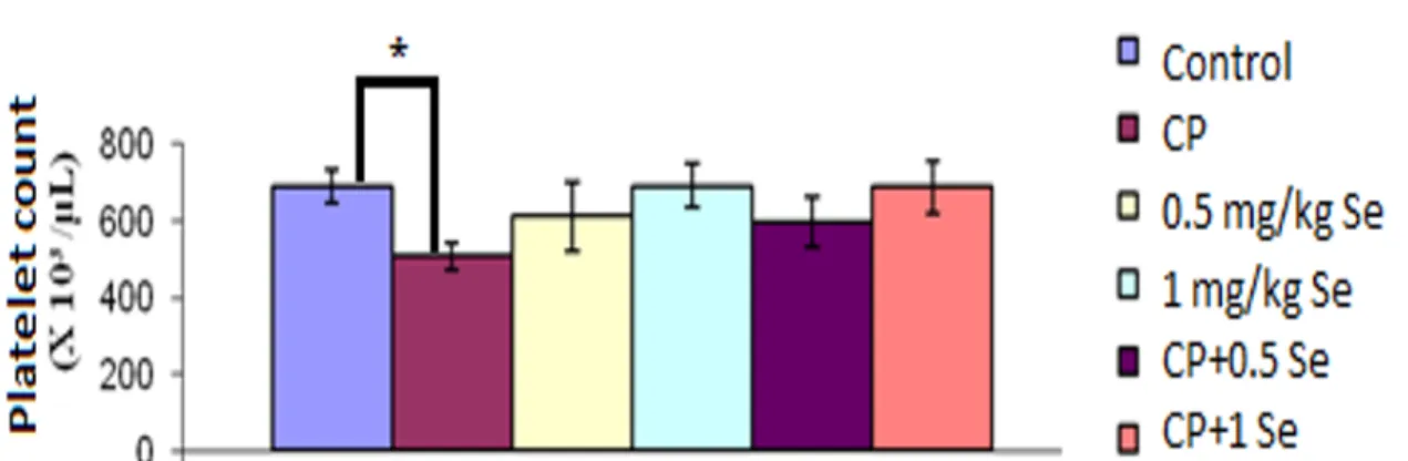
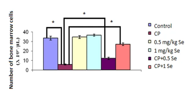
Figs. 3-5 present the statistical comparison of mean values as regards leukocyte, platelet and bone marrow nucleated cells of the experimental groups. As these two figures
show, CP resulted in a decrease in leukocyte, platelet and bone marrow nucleus cell counts by comparison with the control group, respectively (317, 36, 481%) (p<0.001). Conversely, when CP+Se (0.5 and 1 mg/kg) was co-given, a noteworthy rise occurred in leukocyte, platelet and bone marrow-nucleated cell counts by comparison with the CP alone group. In other words, the experimental group of 150+0.5 mg/kg CP+Se exhibited an increase of statistical significance in leukocyte, platelet and bone marrow cell counts by comparison with the CP group (203, 118, and 212%) (p<0.001). The group given 150+1 mg/kg CP+Se, a noticeable rise was observed in leukocyte, platelet and bone marrow cell counts by comparison with 150 mg/kg CP group (320, 38, and 420%)
(p<0.001). We can, thus, conclude that 1 mg/kg of Se is much more effective by
comparison with 0.5 mg/kg Se as far as protection against CP-related myelosuppression concerned.
Figure 3. Peripheral leukocyte levels in the existence of saline, CP or CP+Se groups. *p<0.001 represents a significant difference by comparison with the control and CP groups
Figure 4. Peripheral Platelet count in the existence of saline, CP or CP+Se groups. *p<0.001 refers to asignificant difference by comparison with the control group
Figure 5. Bone marrow cells number in the existence of saline, CP or CP+Se groups. *p<0.001 refers to a significant difference compared to control and CP groups
4. Discussion
Chemotherapeutic practicality of alkylating agents is linked to their capability to form a diversity of DNA adducts which adequately change the structure of DNA or its function, or sometimes both, as far as having a cytotoxic effect on the cells is concerned. A large number of them go through a rather complex activation process prior to triggering reactive intermediates. The initial activation reaction of CP executed by the microsomal oxidation system in the liver results in 4-hydroxy CP, a cytotoxic metabolite, diffusing from hepatocytes into plasma and then travelling across the whole the body. Then and there, 4-hydroxy CP gets converted into other cytotoxic metabolites, such as ACR and PAM, which is renowned to be the cause of myelosuppression [1, 17]. Myelosuppression is known to be a chief potential toxic, as well as a dose-limiting, adverse effect of CP. CP leads to cross-linking of DNA and inhibition of DNA synthesis in that it acts upon not only cyclic but also upon inter-mitotic cells as well, thus causing a general depletion of immune component cells [18]. One study report that 150 mg/kg CP resulted in a decrease of 92% in the leukocyte count, 54% in the platelet count and 94% in the bone marrow [15]. Correspondingly, following a single dose of CP injection, there occurred a fall in the leukocyte counts [19]. Furthermore, when a single dose of 40 mg/kg CP was given to baboons, it caused a temporary fall in WBC counts [20]. Another experimental study has emphasized that CP (20 and 40 mg/kg) has a mutagenic effect on spleen and bone marrow
[21]. Trasler et al. (1987) emphasized that the effects of erythrocyte and leukocyte counts on bone marrow cell count and bone marrow in CP mice treated with erythrocyte and leukocyte counts were very dramatic if a high dose CP was given [22]. In another study using 200 mg/kg CP, the number of erythrocytes and hemoglobin decreased by 20% and the number of hematocrits decreased by 21% compared to the control group. When used alone, 200 mg/kg of CP caused 96% reduction in leukocytes, (p<0.001) compared to control group, which means that bone marrow was being seriously repress [23]. Still another study reported that injection of 50, 100, or 150 mg/kg CP intraperitoneally for 3 days resulted in a dose-dependent manner, apart from causing reduction in thrombocytes, leukocytes and bone marrow nucleated cells [24]. Incidentally, another study reported a similar result, indicating that reduction in platelets only occur in the cases where high
doses of CP are given [25]. The results of our study are also very similar to those of the
abovementioned studies (Figs. 3-5).
Se, a basic trace element, is very important for many biological functions like body's antioxidant defense systems, thyroid hormone metabolism, adaptive and acquired immune system, apart from being important for prevention of the bone marrow suppression. It also has hemaprotective features, besides helping in preventing certain cancers [26]. However, a study has been found in the literature on myelosuppression. Our study is unique in this aspect. In experimental study on the elimination of myesupression, the doses of Se (0.4 and 0.8 mg/kg etc.) were found to be successful [24]. However, in order to increase this level of success a little higher, we aimed to provide more protection by applying these doses (0.5 and 1 mg/kg) a little higher in our study. The least toxic effect of CP has been observed on the circulating thrombocytes and that 150 mg/kg CP caused only about 52% reduction in blood platelet counts. There occurred complete protection when we applied 0.4 mg/kg of Se together with 50 mg/kg of CP. Platelet numbers in the rats treated with 50, 100, or 150 mg/kg of CP had decreased by 22, 33, and 52%, respectively. Injection of respective doses of CP, 0.4mg/kg of Se lowered thrombocyte numbers by 5, 22, and 49%, respectively. A recovery of about 83% was achieved solely in the 50+0.4 mg/kg CP+Se group. By comparison with the 100 mg/kg CP group, the platelet numbers in the 100+0.4 mg/kg CP+Se group had increased by 33%, whilst the rise in platelet numbers in the 150+0.4 mg/kg CP+Se group was estimated to be 6%. Injected along with respective doses of CP, by comparison with the control, 0.8
mg/kg of Se lowered thrombocyte numbers by 22, 25, and 25%, respectively, whilst 0.8 mg/kg of Se was found ineffective in preventing thrombocytopenia attributable to 50 mg/kg of CP. On the other hand, it was determined to be highly effective in preventing thrombocytopenia attributable to 100 and 150 mg/kgof CP (25 and 52%, respectively). We are of the opinion that further studies should be conducted to verify the effects of Se upon CP toxicity of the platelets [24]. Olas and Wachowicz (1997) reported that sodium selenite reduces cisplatin toxicity without constraining the antitumor activity of cisplatin [27]. In later times, Weijl et al. (2004) determined that a higher dose of Se with further antioxidants (eg. vitamins C and E) may be linked to cisplatin-induced ototoxicity and nephrotoxicity [28]. In addition, the 3-month management of WBC has increased significantly [29]. Appearing to have acquired a selective survival advantage, deprivation of Se can decrease protection against oxidative stress and weaken immune deficiency, which is evident under Se deficiency, as well as under oxidative stress conditions in some cancer cells [30]. Our study results for Se are consistent with those of other studies in the literature.
As a result of the changes in antioxidant parameters, CP has been shown to increase lipid peroxidation in rat blood serum. Injection of CP (150 mg/kg) caused a significant increase in the level of final lipid peroxidation product –MDA on the 1st, 5th, and 14th days (by about 38, 26, and 35%, respectively). In Stankiewicz and Skrzydlewska, 2005, injection of CP reported a significant increase in the level of MDA on the 1st, 5th, and 14th days (about 38%, 26% and 35%, respectively) [31]. In this study, CP caused a 64% decrease in MDA and a 105% decrease in GSH when compared to the control group.GSH level in the experimental group with 150+0.5 mg/kg CP+Se increased by 31% compared to the control and MDA level was decreased by 1%. Moreover, when the experimental group given 150+0.5 mg/kg CP+Se was compared with the group given 150 mg/kg CP, the GSH level increased by 56% and the MDA level decreased by 67%. The change in GSH and MDA levels can probably be explained by the significant elimination of free radicals by increasing GPx enzyme activity and reduced glutathione levels in the presence of Se. On the other hand, in the CP+Se group (1 mg/kg), the GSH level increased by 31% and the MDA level by 1% compared to the control group. When compared with the CP group, in the experimental group of 150+1 mg/kg CP+Se, the GSH level increased by 171% and the MDA level decreased by 38%, which means the antioxidant activity of Se
increases in the presence of oxidative stress and seems to be more effective then. According to our experimental results, when a low dose (0.5 mg/kg) Se was applied, an improvement was observed in the hematopoietic system but it was not as effective as 1 mg/kg Se, which could mean that relatively high doses of Se (1 mg/kg) are more protective with a high-dose CP. According to our experimental data, Se is an effective agent in preventing CP-induced toxicities and may be included in chemotherapeutic drug protocols, but different doses of the test may reveal a more useful dose.
As our results indicate, CP administration can cause oxidative stress in peripheral blood and bone marrow due to toxic metabolites. Pre-treatment was highly effective in preventing myelosuppression due to CP. This can be interpreted as increasing the antioxidant capacity and significantly reducing the effects of CP's toxic metabolites. We, nevertheless, believe that further comprehensive studies are needed to elucidate this issue.
References
[1] Kumar, K.B., Kuttan, R., Chemoprotective activity of an extract of Phyllanthus
amarus against cyclophosphamide-induced toxicity in mice, Phytomedicine, 12, 494-500,
2004.
[2]Senthilkumar, S., Devaki, T., Manohar, B.M., Babu, M.S., Effect of squalene on
cyclophosphamide-induced toxicity, Clinica Chimitica Acta, 364(1-2), 335-42, 2006.
[3]Kawabata, T.T., Chapman, M.Y., Kim, D.H., Stevens, W.D., Holsapple, M.P.,
Mechanism of in vitro immunu suppression by hepatocyte generated cyclophosphamide metabolites and 4-Hydroxi cyclophosphamide, Biochemical Pharmacology, 40(5), 927-
935, 1990.
[4] Kehrer, J.P., Biswal, S.S., The molecular effects of acrolein, Toxicological Sciences, 57(1), 6-15, 2000.
[5]Sabitha, K.E., Shyamaladevi, C.S., Oxidant and antioxidant activity changes in
patients with oral cancer and treated with radiotherapy, Oral Oncology, 35(3), 273-7,
1999.
[6] Akkuş, İ., Serbest radikaller ve fizyopatolojik etkileri. Mimoza Yayınları, Konya, 1995.
[7]Byers, T., Perry, G., Dietary carotenes, vitamin C, and vitamin E as protective
[8] İşcan, M., Çoban, T., Normal veneoplastik meme dokusunda antioksidan
enzimler. Klinik Gelişim, 11, 392-395, 1998.
[9]Kohrle, J., Jakob, F., Contempre, B., Dumont, J.E., Selenium, the thyroid, and
the endocrine system, Endocrine Reviews, 26:944-84, 2005.
[10] Brown, K.M.., Arthur, J.R., Selenium, selenoproteins and human health: a
review, Public Health Nutrition, 4, 593-599, 2001.
[11] Rayman, M.P., The importance of selenium to human health, Lancet, 356(9225), 233-41, 2000.
[12] Ilio, C.D., Boccio, G.D., Casaccia, R., Aceto, A. Giacomo, F., Federici, G.,
Selenium level and glutathione-depent enzyme activities in normal and neoplastic human lung tissues, Carcinogenesis, 8(2), 281-284, 1987.
[13] Dai, J., Weinberg, R.S., Waxman, S., Jing, Y., Malignant cells can be
sensitized to undergo growth inhibition and apoptosis by arsenic trioxide through modulation of the glutathione redox system, American Society of Hematology,
93(1),268-277, 1999.
[14] Ayhanci, A., Gunes, S., Sahinturk, V., Appak, S., Uyar, R., Cengiz, M.,
Altuner, Y., Yaman, S., Seleno L-methionine acts on cyclophosphamide-induced kidney toxicity, Biological Trace Element Research, 136, 171–179, 2010.
[15] Ayhanci, A., Uyar, R., Aral, E., Kabader, S., Appak, S., Protective effect of
zinc on cyclophosphamide-induced hematoxicity and urotoxicity, Biological Trace
Element Research, 126, 186–193, 2008.
[16] Esterbauer, H., Cheeseman, K. H., Determination of aldehydic lipid
peroxidation products: malonaldehyde and 4-hydroxynonenal, Methods in Enzymology,
186, 407–42, 1990.
[17] Liang, J., Huang, M., Duan, W., Yu, X. Q., Zhou, S., Design of new
oxazaphosphorine anticancer drugs, Current Pharmaceutical Design, 13, 963–978, 2007.
[18]George, K.S., Rajesh, R., Sunil, K.S., Sulekha, B., Balaram, P., A polyherbal
ayurvedic drug—Indukantha Ghritham as an adjuvant to cancer chemotherapy via immunomodulation, Immunobiology, 213,641–649, 2008.
[19] Fraiser, L.H., Kanekal, S., Kehrer, J.P., Cyclophosphamide toxicity:
characterizing and avoiding the problem, Drugs, 42,781–795, 1991.
[20] Schuurman, H.J., Smith, H.T., Cozzi, E., Tolerability of cyclophosphamide
and methotrexate induction immunosuppression in nonhuman primates, Toxicology, 213,
[21]Moore, F.R., Urda, G. A., Krishna, G., Theiss, J. C., An invivo/invitro method
for assessing micronucleus and chromosome aberration induction in rat bone morrow and spleen. 1. Studies with cyclophosphamide, Mutataion Research/Environmental
Mutagenesis and Related Subjects, 335(2), 191-199, 1995.
[22]Trasler, J.M., Hales, B.F., Robaire, B., A time-course study of chronic paternal
cyclophosphamide treatment in rats: effects on pregnancy outcome and themalere productive and hematologic systems, Biology of Reproduction, 37(2),317-26, 1987.
[23]Cengiz, M., Hematoprotective effect of boron on cyclophosphamide toxicity in
rats, Cellular and Molecular Biology (Noisy le Grand), 64(5),62-65, 2018.
[24] Ayhanci, A., Yaman, S., Appak, S., Gunes, S., Hematoprotective effect of
seleno-L-methionine on cyclophosphamide toxicity in rats, Drug and Chemical
Toxicology, 32(4), 424-428, 2009.
[25] Korkmaz, A., Topal, T., Oter, S., Pathophysiological aspects of
cyclophosphamide and ifosfamide induced hemorrhagic cystitis; implication of reactive oxygen and nitrogen species as well as PARP activation, Cell Biology and Toxicology,
23, 303–312, 2007.
[26] Spallholz, J.E., Use of selenium to prevent and treat cancer in the new
millennium, Journal of Korean Association of Cancer Prevention, 8:9–23, 2003.
[27] Olas, B., Wachowicz, B., Selenium in the cytotoxicity of cisplatin, Postępy Higieny i Medycyny Doświadczalnej, 51, 95–108, 1997.
[28]Weijl, N.I., Elsendoorn, T.J., Lentjes, E.G., Hopman, G.D., Wipkink-Bakker, A., Zwinderman, A.H., Supplementation with antioxidant micronutrients and
chemotherapy-induced toxicity in cancer patients treated with cisplatin-based chemotherapy: a randomised, double- blind, placebo-controlled study, European Journal
of Cancer, 40, 1713–1723, 2004.
[29] Sieja, K., Talerczyk, M., Selenium as an element in the treatment of ovarian
cancer in women receiving chemotherapy, Gynecologic Oncology, 93, 320–327, 2004.
[30] Zeng, H., Combs, J.G.F., Selenium as an anticancer nutrient: roles in cell
proliferation and tumor cell invasion, Journal of Nutritional Biochemistry, 19, 1–7, 2008.
[31]Stankiewicz, A., Skrzydlewska, E., Amifostine Antioxidant Effect on Serum of
Rats Treated with Cyclophosphodamide, Polish Journal of Environmental Studies, 14(3),