164
Original Article / Özgün Araştırma
DO I:10.4274/tnd.2019.00086Turk J Neurol 2019;25:164-167
Objective: Thirty percent of all acute ischemic strokes cause is the internal carotid artery stenosis or occlusion. The carotid artery stenting (CAS) was rarely performed in early time, was used more frequently with the development of angiographic method and materials. In this study, we aimed to evaluate the success rates, clinical outcomes and complications of carotid stenting due to carotid stenosis in the Neurology Department of Selçuk University.
Materials and Methods: The patients who underwent conventional angiography between November 2017 and August 2018 were evaluated retrospectively in the Neurology Department of Selçuk University. Ninety four patients who underwent conventional angiography due to carotid artery stenosis were included in the study. Fifty four of 94 patients underwent CAS. Control carotid doppler and/or carotid CT angiography was performed at 1 month after discharge. Neurological examinations of the patients were also repeated.
Results: The operation success rate was 96.3% (two patients developed minor stroke at the 2 hours after the procedure). No deaths or major stroke occurred in any of these patients with CAS. No ischemic cerebrovascular event or transient ischemic attack occurred in any of the patients at 1 months follow-up. No restenosis was observed in any of the carotid doppler follow-ups or CT anjiyography.
Conclusion: In conclusion, we have shown that CAS performed by neurology clinics is effective and reliable. We have also shown that a protective device is not necessary for the risk of distal embolism in these patients.
Keywords: Carotid stenosis, carotid artery stenting (CAS), carotid end arterectomy (CEA), conventional angiography
Amaç: Tüm akut iskemik inmelerin yaklaşık %30’unda neden internal karotis arter darlık veya tıkanıklarıdır. Başta nadir olarak yapılan karotis arter stentlemesi (KAS) zamanla anjiyografik yöntem ve kullanılan malzemelerin gelişmesiyle daha sık kullanılır hale geldi. Bu çalışmamızda Selçuk Üniversitesi Nöroloji Kliniğince karotis darlığı nedeniyle karotis stentlemesi yapılan hastaların başarı oranlarını, klinik sonlanımlarını ve komplikasyon sonuçlarını göstermeyi amaçladık. Gereç ve Yöntem: Selçuk Üniversitesi Tıp Fakültesi Hastanesi Nöroloji Kliniğince Kasım 2017-Ağustos 2018 tarihleri arasında konvansiyonel anjiyografisi yapılan hastalar retrospektif olarak değerlendirildi. Bunlardan, karotis arter darlığı nedeniyle konvansiyonel anjiyografisi yapılan 94 hasta çalışmaya alındı. Doksan dört hastanın da 54 tanesinde KAS uygulandı. Karotis stenti yerleştirilen hastaların, taburculuklarından sonraki 1. ayda kontrol karotis doppleri ve/veya karotis bilgisayarlı tomografi (CT) anjiyografi yapıldı. Bu kontrolde ayrıca hastaların nörolojik muayeneleri tekrar edildi.
Bulgular: Müdahale başarı oranı %96,3’tür (iki olguda müdahaleden 2 saat sonra minör inme gelişti). Müdahale ettiğimiz bu olguaların hiçbirinde KAS işlemine bağlı olarak ölüm veya majör inme görülmedi. Bu hastaların 1 ay sonraki kontrollerinde; hiçbir hastada geçici veya kalıcı serebrovasküler hastalık izlenmedi. Ayrıca kontrol karotis doppler veya CT anjiyografilerde hiçbir hastada restenoz izlenmedi.
Sonuç: Sonuç olarak, nöroloji klinikleri tarafından yapılan KAS işlemlerinin etkili ve güvenilir olduğunu gösterdik. Ayrıca bu hastalarda distal emboli riski için koruma cihazının şart olmadığını göstermiş olduk.
Anahtar Kelimeler: Karotis darlığı, karotis arter stentlemesi (KAS), karotis end arterektomisi (KEA), konvansiyonel anjiyografi
Retrospective Evaluation of Carotid Artery Stenting Experience of
Neurology Clinic of Selçuk University
Selçuk Üniversitesi Nöroloji Kliniği’nin Karotis Arter Stentlemesi Deneyiminin
Retrospektif Değerlendirilmesi
Gökhan Özdemir, Şerefnur Öztürk Selcuk University Faculty of Medicine, Department of Neurology, Konya, Turkey
Abstract
Öz
Ad dress for Cor res pon den ce/Ya z›fl ma Ad re si: Gökhan Özdemir MD, Selcuk University Faculty of Medicine, Department Of Neurology, Konya, Turkey Phone: +90 530 118 77 86 E-mail: [email protected] ORCID: orcid.org/0000-0001-8140-6333
Re cei ved/Ge lifl Ta ri hi: 30.10.2018 Ac cep ted/Ka bul Ta ri hi: 27.04.2019
©Copyright 2019 by Turkish Neurological Society
165 Turk J Neurol 2019;25:164-167 Özdemir and Öztürk; Carotid Artery Stenting of Neurology
Introduction
Ischemic stroke is one of the leading causes of death in our country and around the world. In addition, it is the leading cause of long-term disability (1). Mortality and permanent disability are more common in supraaortic large artery occlusions such as in the internal carotid artery (ICA). ICA obstructions are the cause of approximately 30% of all acute ischemic strokes (2). In particular, the origin and proximal segments of the ICA are occluded because these segments are subjected to turbulence flow. As a result, the development of atherothrombosis is facilitated. Therefore, it is necessary to detect and treat the stenosis before a permanent stroke develops. In patients suspected of having carotid stenosis, computed tomography (CT) brain angiography, magnetic resonance (MR) brain angiography and carotid artery Doppler ultrasonography (USG) can be used for the detection, degree determination, and follow-up of stenosis. Although the main advantage of these diagnostic methods is their being non-invasive, conventional angiography is still the gold standard for demonstrating true stenosis (3).
Carotid artery endarterectomy (CAE) was previously performed for carotid artery stenosis. However, some patients could not undergo surgery. Carotid artery stenting (CAS), which was rarely performed, has become more common with the development of angiographic methods and materials. Nowadays, it has been shown that there is no significant difference between the complications and success rates of the two methods (4,5). In addition, restenosis rates were shown to be similar in long-term follow-up (6). The lack of general anesthesia, the short duration of the procedure and hospitalization, and the lack of surgical procedures in stenting have led to the increasing application of this method. In Turkey, CAS is also applied successfully in many neurology clinics (5). In this study, we aimed to show the success rates, clinical outcomes, and complications of patients who underwent CAS due to carotid stenosis at Selçuk University Neurology Clinic.
Materials and Methods
Patients who underwent conventional angiography between November 2017 and August 2018 by the Neurology Clinic of Selçuk University Medical Faculty Hospital were evaluated retrospectively. Ninety-four patients who underwent conventional angiography for carotid artery stenosis were included in the study. CAS was performed in 54 of 94 patients. These patients had one or more non-invasive carotid angiographic imaging (CT angiography, MR angiography, carotid Doppler USG) before conventional angiography. The patients were informed about the procedure before the intervention and detailed written consent was obtained from the patient or next of kin.
Carotid artery stenosis leading to transient ischemic attack (TIA) or hemispheric infarction in the last 6 months was evaluated as symptomatic carotid artery stenosis. TIA was accepted if symptoms resolved without sequelae within 24 hours and a neuroradiologic examination was evaluated as completely normal. Complete blood count, routine biochemistry values, and electrocardiograms of all patients were evaluated prior to intervention. In addition, all patients underwent brain MR imaging (MRI) and/or cranial CT before the procedure. Neurologic examinations were performed
before and after conventional angiography. After the stenting, two patients developed symptoms and diffusion MRI was performed urgently. All patients received 75 mg clopidogrel and 150 mg aspirin before the procedure. Both antiaggregant drugs were continued for at least three months after stenting. Control carotid Doppler and/or carotid CT angiography was performed in the first month after discharge of the patients. Neurologic examinations of the patients were also repeated during this follow-up examination.
The femoral artery was used for the intervention. A sheath was placed in the femoral artery, varying in size from 6F to 8F. In one patient, because both femoral arteries were occluded, access was performed through the brachial artery. During the procedure, cardiac and direct arterial blood pressure monitoring was performed. Aortography was performed with a 6-F pigtail from the aortic arch before the stenting procedure. Thus, the origins and proximal segments of both the aorta and supraaortic arteries were evaluated. Before stenting, unfractionated heparin was administered in doses varying between 3000-7000 U according to the weight of the patients. Selective access was performed into each common carotid artery. For diagnostics, a 5-F vertebral catheter or Judkins right coronary catheter, which is commonly used in coronary arteries, was used. A stiff wire was directed to the external carotid artery in patients with change after road-map. The diagnostic catheter was then withdrawn, but the stiff wire was left in place. A 7-F guiding catheter or 6-F long-sheath was placed over the stiff wire distal to the common carotid artery. In some patients, a 7-F or 8-F guiding catheter was inserted directly into the common carotid artery without change. Conventional angiographic imaging of the intracranial anterior system was performed in multiple poses before and after stenting.
Ethical consent was obtained from the Ethics Committee of Selçuk University Faculty of Medicine (Decision No. 2018/354). All participants were filled out consent form.
Statistical Analysis
The SPSS 18 software was used to analyze the data. There was no control group. Therefore, no comparative analysis was performed. The normality of the variables was determined using the Kolmogorov-Smirnov test. Mean ± standard deviation was evaluated for normally distributed continuous variables. Data without normal distribution are expressed as median. The evaluation of categorical data is shown as percentage.
Results
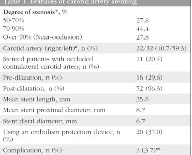
In 6 patients who underwent conventional angiography, the anatomic structure of the arch was not suitable for stenting, so these patients were referred for CAE. The reason for stenosis was fibromuscular dysplasia in two patients and the stenosis was not severe enough to require intervention in 32 patients. Thus, stents could be placed in 54 of 94 patients who underwent conventional angiography for CAS. The degree of stenosis in patients undergoing stenting was calculated according to the North American Symptomatic Carotid Endarterectomy Trial method. Distal protection filter was used in 20 patients. The remaining 34 patients were treated without a protection filter. Pre-dilatation was performed in 16 patients before stenting, and post-dilatation was performed with appropriate non-compatible balloon in 52
166
Turk J Neurol 2019;25:164-167 Özdemir and Öztürk; Carotid Artery Stenting of Neurology
patients who had residual stenosis after stenting. All patients who underwent pre- or post-dilatation and had bifurcation stenosis were also given 0.5-2 mg atropine intravenously during the intervention. Forty-one (75.9%) of the stented patients were male. Fifty-one patients (94.4%) had a history of TIA or previous minor ischemic stroke in the last 6 months. A self-expandable stent was placed in all patients. Another feature of these stents was that their proximal and distal diameters were different from each other. A stent was placed into the right carotid artery in 22 patients and the right carotid artery in 32 patients. In one case, an additional stent was used because the lesion was too long and one stent was not sufficient. The contralateral carotid artery was occluded in 11 (20.4%) stented patients. In 15 (27.8%) patients, the degree of stenosis was near- or pre-occlusion. The data related to the intervention can be seen in Table 1.
Discussion
The success rate was high in CAS performed by the Neurology Department of Selçuk University Medical Faculty Hospital. There were few complications and clinical outcomes were good. In addition, we proved that CAS procedures performed by the neurology clinic were effective and reliable.
Carotid artery disease is the main cause in the majority of anterior system ischemic strokes. Critical stenosis or occlusion of the carotid artery may result in permanent sequelae or death (7). The most common cause of carotid artery stenosis or occlusion is atherosclerosis. The risk is even greater for near or pre-occlusion and ulcerative stenosis (8). Atherothrombosis itself may lead to a direct symptom, as well as a fragment of a clot that breaks out of this plaque, causing distal occlusion of another vessel (artery-to-artery embolism). Therefore, when carotid artery stenosis is detected, appropriate treatment should be initiated rapidly. If not, we may encounter uncompensated results in a short time. Patients with symptomatic or severe stenosis in particular are among the
neurologic emergencies. In addition to treatment strategies for atherosclerotic risk factors, antiaggregant initiation and stenting or CAE should be performed without delay. Our cases were intervened in no more than 2 weeks.
Carotid artery stenosis is usually seen in the elderly population. CAS was developed as an alternative to carotid surgery because most of these patients could not handle surgery and/ or anesthesia. However, complication rates were higher in the initial CAS compared with CEA (9,10). Over time, there have been improvements in CAS in parallel with the improvements in materials used in endovascular interventions and the increase in experience. Recent studies have shown similar results and complication rates to surgical treatment (11). Nowadays, stenting applications have outnumbered CEA procedures (12). The stent used in carotid artery stenosis gradually expands in the first year after its insertion into the vein and fits well to the wall of the vessel. Since 2017, these self-expandable stents have been successfully installed by our neurology clinic.
During or after the procedure, embolism in the aorta or the intervention site is an important problem. During the procedure, protective devices may be used to prevent embolism from spreading distally. These devices are used by placing them proximally or distally. However, there are studies suggesting that they should not be used (5,13). Embolism did not develop in any patients with stent placements without a protective device in our study. In addition, the procedure was shorter and the cost has also decreased in these patients. In two cases, minor stroke developed a few hours after stenting. Diffusion MRIs of these patients were performed immediately and small infarct areas that did not require intervention were observed.
Our clinical results in patients with stenting are good, even when compared with studies in the world and in our country. Studies have shown a correlation between the degree of stenosis and poor clinical outcomes and complication rates. When we evaluate this aspect, the ratio of patients who were stented due to more than 70% stenosis was approximately 75%. In other words, although the procedure was more difficult and there was a high risk of complications, we successfully performed stenting. There were also few complications. The diagnosis, treatment, and follow-up of the patients were performed by our own neurology clinic. Thus, patients were followed up better, and it was also comfortable for patients.
Study Limitations
The most important problem related to the study is the absence of a CAE patient group for comparison, and in addition, the study was conducted in a single center.
Conclusion
In conclusion, we demonstrated that CAS performed by neurology clinics is effective and reliable. We have also demonstrated that a protective device is not necessary for the risk of distal embolism in these patients.
Acknowledgements
We would like to thank the technicians and nurses of the Angio Laboratory of Selçuk University Hospital.
Table 1. Features of carotid artery stenting
Degree of stenosis*, % 50-70% 70-90% Over 90% (Near-occlusion) 27.8 44.4 27.8
Carotid artery (right/left)*, n (%) 22/32 (40.7/59.3) Stented patients with occluded
contralateral carotid artery, n (%) 11 (20.4)
Pre-dilatation, n (%) 16 (29.6)
Post-dilatation, n (%) 52 (96.3)
Mean stent length, mm 35.6
Mean stent proximal diameter, mm 8.7
Stent distal diameter, mm 6.7
Using an embolism protection device, n
(%) 20 (37.0)
Complication, n (%) 2 (3.7)**
*The stenosis was not severe enough to require intervention in 32 patients. **None of the 32 patients who underwent only diagnostic angiography without stenting did not develop complications.
167 Turk J Neurol 2019;25:164-167 Özdemir and Öztürk; Carotid Artery Stenting of Neurology
Ethics
Ethics Committee Approval: Ethical consent was obtained
from the Ethics Committee of Selçuk University Faculty of Medicine (Decision No. 2018/354).
Informed Consent: All participants were filled out consent
form.
Peer-review: Externally peer-reviewed. Authorship Contributions
Cerrahi ve Medikal Uygulama: G.Ö., Ş.Ö., Konsept: G.Ö., Ş.Ö., Dizayn: G.Ö., Ş.Ö., Veri Toplama veya İşleme: G.Ö., Ş.Ö., Analiz veya Yorumlama: G.Ö., Ş.Ö., Literatür Arama:G.Ö., Ş.Ö., Yazan: G.Ö., Ş.Ö.
Conflict of Interest: No conflict of interest was declared by
the authors.
Financial Disclosure: The authors declared that this study
received no financial support.
References
1. Ovbiagele B, Nguyen-Huynh MN. Stroke Epidemiology: Advancing Our Understanding of Disease Mechanism and Therapy. Neurotherapeutics 2011;8:319-329.
2. Park JH, Lee JH. Carotid Artery Stenting. Korean Circ J 2018;48:97-113. 3. Wardlaw JM, Chappell FM, Stevenson M, et al. Accurate, practical and
cost-effective assessment of carotid stenosis in the UK. Health Technol Assess 2006:10.
4. Dumont TM, Rughani AI. National trends in carotid artery revascularization surgery. J Neurosurg 2012;116:1251-1257.
5. Özdemir G, Kızıldağ N, Ekmekçi AH, Gümüş H, Aygül R, Öztürk Ş. Üçüncü basamak bir nöroloji kliniğinin karotis arter stentlemesi deneyiminin retrospektif değerlendirilmesi. Türk Beyin Damar Hastalıkları Dergisi 2018;24:14-18.
6. Shao A, Dong X, Zhou J, Liu F, Hong Y, Zhang J. Comparison of carotid artery end-arterectomy and carotid artery stenting in patients with atherosclerotic carotid stenosis. J Cranio facial Surg 2014;25:1441-1447.
7. Regenhardt RW, Das AS, Stapleton CJ. Blood Pressure and Penumbral Sustenance in Stroke from Large Vessel Occlusion. Front Neurol 2017;8:317. 8. No authors listed. Endarterectomy for asymptomatic carotid artery stenosis. Executive Committee for the Asymptomatic Carotid Atherosclerosis Study. JAMA 1995;273:1421-1428.
9. Naylor AR, Bolia A, Abbott RJ. Randomized study of carotid angioplasty and stenting versus carotid endarterectomy: a stoppedtrial. J Vascular Surg 1998;28:326-334.
10. Jordan WD, Schroeder PT, Fisher WS, McDowell HA. A comparison of angioplasty with stenting versus endarterectomy for the treatment of carotid artery stenosis. Ann Vascr Surg 1997;11:2-8.
11. Gurm HS, Yadav JS, Fayad P, et al. Long-term results of carotid stenting versus endarterectomy in high-risk patients. N Engl J Med 2008;358:1572-1579.
12. Kim LK, Yang DC, Swaminathan RV, et al. Comparison of trendsand outcomes of carotid artery stenting and endarterectomy in the United States, 2001 to 2010. Circ Cardiovasc Interv 2014;7:692-700.
13. Saw J. Carotid artery stenting for stroke prevention. Can J Cardiol 2014;30:22-34.