Acute phase response and clinical changes in calves with lipopolysaccharide induced
endotoxemia
Alparslan Coskun1*, lsmail Sen2 Özet
Coskun A, Sen I. Lipopolisakkarit ile endotoksemi oluştu-rulan buzağılarda akut faz cevap ve klinik değişimler. Eura-sian J Vet Sci, 2012, 28, 1, 21-26
Amaç: Lipopolisakkarit ile deneysel endotoksemi oluşturu-lan buzağılarda akut faz proteinlerin ve klinik değişimlerin belirlenmesi amaçlanmıştır.
Gereç ve Yöntem: Çalışmada yaşları 25-42 gün arasında değişen sekiz adet Holstein ırkı buzağı kullanıldı. Lipopoli-sakkarit damar içi 0.1 µg/kg dozunda 50 mL % 0.9 NaCl’de seyreltilip 30 dakika süresince verilerek endotoksemi oluş-turuldu. Çalışma boyunca endotoksemi gelişen buzağılarda klinik olarak gözlenen değişiklikler kayıt edilirken ve akut faz proeinlerinin konsantrasyonları belirlendi.
Bulgular: Lipopolisakkarit uygulanması sonrasında buzağı-ların tamamında solunum sayısında ve kalp atım sayısında artış, yerde yatma, depresyon, hipotermi/hipertermi muko-zada hiperemi/siyanoz ve emme refleksinde azalma gözlen-di. Klinik olarak gelişen değişimler 1. ve 3. saatler arasında daha şiddetli olduğu belirlendi. Damar içi lipopolisakkarit verilmesinden sonra haptoglobin 36. saatte (500±93.2 µg/ mL) ve serum amyloid-A 24. saatte (185± 46.6 µg/mL) en üst seviyeye ulaştı.
Öneri: Akut faz proteinlerde ve klinik bulgulardaki gözle-nen değişim, buzağılarda gelişen endotoksemi ile ilişkili-dir. Buzağılarda serum haptoglobin ve serum amyloid-A konsantrasyonlardaki artışlar endotokseminin bir belirteçi olarak değerlendirilebilir.
Abstract
Coskun A, Sen I. Acute phase response and clinical changes in calves with lipopolysaccharide induced endotoxemia. Eurasian J Vet Sci, 2012, 28, 1, 21-26
Aim: The purpose of the study was to determine the levels of acute phase proteins and clinical changes in calves with lipopolysaccharide induced experimental endotoxemia. Materials and Methods: Eight Holstein breed calves were used in the study. Endotoxemia was induced via intravenous administration of 0.1 µg/kg dose of lipopolysaccharide in 50 mL of physiological 0.9% NaCl over 30 min. The calves were continuously observed for clinical changes during the experiment, and serum acute phase protein levels were measured.
Results: Increase in respiratory frequency, tachycardi, mucosal hyperemia/cyanosis, recumbency, depression, hyperthermia/hypothermia, and poor suckle reflexes were observed in all calves after lipopolysaccharide administra-tion. After lipopolysaccharide infusion, serum haptoglobin (500±93.2 µg/mL) and serum amyloid A (185±46.6) con-centrations reached peak levels of at 36 h and 24 h, respec-tively.
Conclusion: Changes in acute phase proteins and clinical findings were related to endotoxemia. A moderate to high increase in haptoglobin and serum amyloid A concentra-tions may indicate the presence of endotoxemia in calves.
1Department of Internal Medicine, Faculty of Veterinary Medicine, Cumhuriyet University, 58140, Sivas, 2Department of Internal Medicine, Faculty of Veterinary Medicine, Selcuk University, 42075, Konya, Turkey
Received: 21.10.2011, Accepted: 11.12.2011 *[email protected]
Anahtar kelimeler: Akut faz protein, endotoksemi, buzağı, klinik skor
Keywords: Acute phase proteins, endotoxemia, calves, clinical score
RESEARCH ARTICLE
Journal of Veterinary Sciences
www.eurasianjvetsci.org - www.ejvs.selcuk.edu.trIntroduction
The presence of endotoxins in the blood is called en-dotoxemia. It is seen in patients with sepsis and sep-tic shock and can also be seen experimentally with li-popolysaccharide (LPS) infusion (Mackay 1996). LPS has been used to induce endotoxemia in animals such as rat (Er and Yazar 2010), rabbit (Yazar et al 2004, Turgut et al 2006), horse (Danek 2006) and cattle (Biniek et al 1998, Jacobsen et al 2005). Endotoxin, a part of the cell wall of gram (-) bacteria, initiates acute inflammation when injected in vivo (Lohuis et al 1988a). LPS causes hemodynamic, respiratory, metabolic and pyrogenic responses, which are similar to those seen in calves with naturally occurring sepsis (Templeton et al 1988, Biniek et al 1998). After LPS administration, it causes physiological acute phase responses such as fever, systemic hypotension, brady-cardia, disseminated intravascular coagulation (DIC), tissue necrosis, and the production of inflammatory mediators including cytokines, interferon and eicosa-noids (Adams et al 1990).
Endotoxins contribute to the development of condi-tions commonly observed with gram (-) infeccondi-tions such as coliform mastitis, neonatal coliform septi-cemia, pasteurellosis and salmonellosis. In addition, endotoxins are associated with non-infectious diseas-es such as ruminal acidosis, laminitis and abomasal displacement (Jacobsen et al 2005). Endotoxemia is most commonly associated with bacteremia or sep-ticemia due to gram (-) organisms, especially E. coli (Constable 2007). The clinical symptoms of severe endotoxemia (Lohuis et al 1988, Gerros et al 1995, Constable 2007); depression, hyperthermia following hypothermia, tachycardia followed by decreased car-diac output, decreased systemic blood pressure, cool skin and extremities, diarrhea, congested mucosa with an increased capillary refill time, and muscular weakness leading to recumbency.
The acute phase of the immunological response is seen following systemic infection, inflammation, tis-sue injury, trauma, burns or neoplastic formations, and the accumulation of acute phase proteins (APPs) occurs (Gruys et al 1994, Niewold et al 2003, Mura-ta et al 2004, Ganheim et al 2007, Nazifi et al 2009). These proteins are synthesized mainly in the liver and have a primarily glycoprotein structure. The secretion of APPs is regulated by proinflamatory cytokines such as IL-6, TNFα, and IL-1β (Yoshioka et al 2002, Murata et al 2004). Increased haptoglobin (Hp) level in acute inflammation is the major APP in cattle (Eckersall and Conner 1988, Murata et al 2004, Eckersall 2007, Orro et al 2008). In healthy animals, serum Hp levels are very low or below the detection limit (Conner et al 1986, Eckersall and Conner, 1988). Serum amyloid A (SAA) is an acute phase apolipoprotein of the high density lipoprotein fraction of plasma (Uriel-Shoval et al 2000, Niewold et al 2003, Murata et al 2004). Although its physiological role in the host defense
during inflammation is not clear, various effects have been reported (Uriel-Shoval et al 2000).
This study have been hypothesized that acute phase proteins have important role in calves with LPS-in-duced experimental endotoxemia. The purpose of this study was to determine the changes in the SAA and Hp concentrations, and clinical changes in calves with LPS-induced experimental endotoxemia.
Materials and Methods
Eight Holstein breed calves (male, 25-42 d, 35-60 kg) were used in the study. Study protocol was approved by Ethical Committee. They were kept unrestrained in stalls that were bedded with wood shavings for 1week before experiment. The routine clinical and hematological findings of all calves were recorded at 1 week before the experiment. The calves were fed whole milk (60 mL/kg) twice a day. The calves had access to fresh water at all times. According to clini-cal and hematologiclini-cal findings, healthy clini-calves were included in the study. A jugular venous catheter was placed aseptically into each calf at 24 h before LPS infusion. Endotoxemia was induced via intravenous administration of LPS (0.1 µg/kg, 0111:B4, Sigma, Germany) in 50 mL of physiological 0.9% NaCl for over 30 min. Time zero (baseline) is meaning start time of LPS infusion. The calves were continuously observed for clinical changes during the experiment. Rectal temperature, heart rate, respiration frequency rate, respiratory type, capillary refill time, mucous membrane examination, mental status and loss of consciousness, ability to stand, appetite, and defeca-tion were recorded during the experiment. A numeri-cal score was given for each clininumeri-cal symptom, which is presented in Table 1. All calves returned to normal health at the end of the study. Venous blood samples were collected anaerobically in 8 mL non-heparinized plastic syringes. Blood samples were collected from the jugular vein of calves at 0 (before LPS infusion, baseline), 0.5, 1, 2, 3, 4, 6, 8, 12, 18, 24, 36, 48, 72, 96, 120, and 144 h after LPS infusion. An aliquot of blood was collected in a glass tube for determination of Hp, SAA and albumine levels and the tubes were centri-fuged after clotting, and the serum was harvested and stored at –20 0C until analysis. Serum Hp (Life Diag-nostics Inc., West Chester, PA, USA) and SAA (Invitro-gen Corporation, Carlsbad, CA, USA) concentrations were measured with a commercially available ELISA kit.
Data are expressed as mean±SD or median. The level of statistical significance was set at p<0.05. Each value compared with ANOVA and Tukey test. Clinical scores at the different time points compared to the baseline at time zero by the Mann-Whitney U test. A statistical software program (SPSS 10.0) was used for statistical analysis.
Results
Clinical Findings
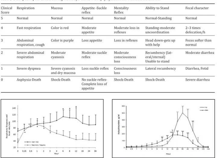
The rectal temperature in all calves showed a biphasic pattern after LPS administration. The rectal tempera-ture was 39 0C within 1 h after LPS administration and then decreased (<37 0C) within 4 h. The rectal tem-perature then increased gradually after 4 h follow-ing LPS administration. The increase or decrease in the rectal temperature was between 1 and 2 0C. Rec-tal temperatures returned to baseline levels within 24–36 h following LPS administration. The changes in rectal temperature was no statistically significant during of the study Abnormal clinical signs were ob-served in all calves after LPS infusion. Clinical changes such as an increase in the respiratory frequency rate, tachycardia, mucosal hyperemia/cyanosis, appetite, lethargy, recumbency, diarrhea, depression, and poor suckle reflexes were observed in all calves (Table 2). In the first 2 h, defecation in all calves increased, and 2 calves had malodorous diarrhea after 4 h. The res-piratory frequency and heart rate increased after LPS infusion (Figure 1). These changes started within ap-proximately 30 min and lasted 24 h after LPS infusion. Capillary refill time increased in all calves within 6 h
and returned to baseline by 24–36. There were some differences with respect to the duration and the se-verity of clinical changes among the calves.
Laboratory findings
The serum Hp baseline concentration was 8.13±6.51 µg/mL. However, serum Hp concentrations increased from 20.8±8.40 µg/mL to 159±90.30 µg/mL within 6 h and 18 h, The Hp concentration reached its peak level (500±93.2 µg/mL) at 36 h, and then it decreased gradually after 48 h and returned to baseline levels within 144 h. There was a statistical difference be-tween the baseline Hp values and Hp concentrations after LPS infusion (Figure 2). The SAA baseline con-centration was 12.4±5.16 mg/mL, which increased to 30.2±24.6 µg/mL and 97.8± 47.4 µg/mL within 3 h and 8 h, respectively. The highest level of SAA was 185±46.6 µg/mL at 24 h, which was maintained until 48 h, and then it decreased gradually and returned to the baseline levels after 96 h in all the calves. There was a statistical difference between the baseline SAA values and SAA concentrations after LPS infusion (Figure 2)
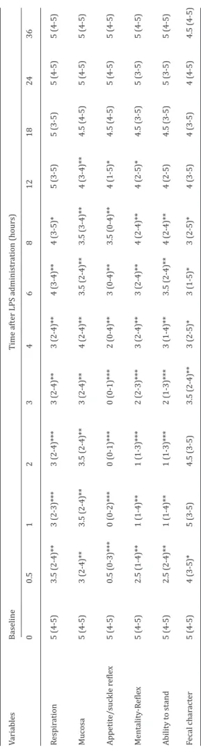
Table 1. Description of the clinical scoring method for calves with lipopolysaccharide induced endotoxemia. Clinical
Score Respiration Mucosa Appetite -Suckle reflex Mentality Reflex Ability to Stand Fecal character
5 Normal Normal Normal Normal Normal-Standing Normal
4 Fast respiration Color is red Moderate
appetite Moderate loss in reflexes Standing-moderate uncoordination 2–3 times defecation/h 3 Abdominal
respiration, cough Color is purple Loss appetite Loss in reflexes Head down-gets up with help Feces softer than normal
2 Severe abdominal
respiration Moderate cyanosis Moderate suckle reflex Moderate consciousness loss
Recumbency (lat-eral/sternal) Unable to stand
Moderate diarrhea 1 Severe dyspnea Severe cyanosis
and dry mucosa Loss suckle reflex Consciousness loss Lateral recumbency Diarrhea, Fetid
0 Asphyxia-Death Shock-Death No suckle
reflex-Complete loss of appetite
Shock-Death Shock-Death Severe diarrhea
Figure 1. Changes in respiratory frequency and heart pulse rate dur-ing the experiment in calves with lipopolysaccharide induced experi-mental endotoxemia (mean±SD, n:8). Asterisked (*) mean values are significantly different (*p<0.05)
Figure 2. Serum Hp and SAA concentrations during the experiment in calves with lipopolysaccharide induced experimental endotoxemia (mean±SD, n:8). Asterisked (*) mean values are significantly different (*p<0.05, **p<0.01, ***p<0.001)
Discussion
Gram (-) sepsis and endotoxemia have a strong as-sociation with mortality in cattle, particularly in neo-natal calves (Gerros et al 1993, Semrad ve Dubeilzig, 1993, Gerros et al 1995). Metabolic, hematological and pathological changes occur in LPS-induced ex-perimental endotoxemia, which show similarities with the results of naturally occurring cases of sepsis (Templeton et al 1988, Gerros et al 1995, Biniek et al 1998).
In the present study, experimental endotoxemia was induced in calves by intravenous administration of LPS at the dose of 0.1 µg/kg. Clinical findings related to endotoxemia were observed in all calves within 30 min following the administration of LPS (Table 2). Lohuis et al (1988) stated that pathophysiological ef-fects of endotoxins were dependent on the dose, and the clinical symptoms induced by endotoxins were generally depression, respiratory distress, vasomo-tor disturbances (possibly terminating in shock), high fever, (sometimes followed by hypothermia) and mo-tility disorders of the gastrointestinal system (retch-ing and diarrhea). Abnormal clinical signs such as in-creased respiratory frequency, tachycardia, mucosal hyperemia/cyanosis, appetite, recumbency, diarrhea, depression and poor suckle reflexes were observed in all calves after LPS infusion (Table 2). Gerros et al (1995) reported that clinical changes became more severe depending on the dose administered within 20–30 min following infusion of LPS. Change in body temperature following the administration of endotox-in occurred dependendotox-ing on the LPS dose and the form of administration (Boosman et al 1989, Jacobsen et al 2005). In the current study, rectal temperatures increased by 1 to 2 0C within 1 h after LPS adminis-tration followed by hypothermia for a certain time (at 4th h) and hyperthermia reoccurred (at 18th h) fol-lowing hypothermia. Monophasic or biphasic fevers have been previously observed depending on the ad-ministered dose of LPS (Lohuis et al 1988, Jacobsen et al 2005). For biphasic fevers, the initial rise of tem-perature is related to the direct effect of the endotoxin on the thermoregulatory center, and the second in-crease in temperature is related to the release of IL-1 (Lohuis et al 1988). Jacobsen et al (2005) stated that an early high fever was caused by endotoxin stimu-lation of the brain to produce PGE2, and the subse-quent high fever was related to IL-1-induced release of PGE2. Various researchers have reported changes in respiration rate, type of respiration and heart rate following endotoxin administration (Nagaraja et al 1979, Gerros et al 1995, Borderas et al 2008). In the current study, heart rate reached the highest level at 6 h, and changes in the respiration frequency were observed at 15 min after LPS administration (Figure 1). Because of its wide microvascular structure, pul-monary symptoms are frequently observed in sepsis and acute respiratory distress syndrome frequently
Table 2. Clinic al sc or e in c alv es wit h lipopolysac charide induc ed e xperiment al endot ox emia (Median-r ang e, n:8). Variables Baseline Time aft er LPS administr ation (hours) 0 0.5 1 2 3 4 6 8 12 18 24 36 Respir ation 5 (4-5) 3.5 (2-4)** 3 (2-3)*** 3 (2-4)*** 3 (2-4)** 3 (2-4)** 4 (3-4)** 4 (3-5)* 5 (3-5) 5 (3-5) 5 (4-5) 5 (4-5) Mucosa 5 (4-5) 3 (2-4)** 3.5 (2-4)** 3.5 (2-4)** 3 (2-4)** 4 (2-4)** 3.5 (2-4)** 3.5 (3-4)** 4 (3-4)** 4.5 (4-5) 5 (4-5) 5 (4-5) Appetit e/suckle r efle x 5 (4-5) 0.5 (0-3)*** 0 (0-2)*** 0 (0-1)*** 0 (0-1)*** 2 (0-4)** 3 (0-4)** 3.5 (0-4)** 4 (1-5)* 4.5 (4-5) 5 (4-5) 5 (4-5) Mentality -R efle x 5 (4-5) 2.5 (1-4)** 1 (1-4)** 1 (1-3)*** 2 (2-3)*** 3 (2-4)** 3 (2-4)** 4 (2-4)** 4 (2-5)* 4.5 (3-5) 5 (3-5) 5 (4-5) Ability t o stand 5 (4-5) 2.5 (2-4)** 1 (1-4)** 1 (1-3)*** 2 (1-3)*** 3 (1-4)** 3.5 (2-4)** 4 (2-4)** 4 (2-5) 4.5 (3-5) 5 (3-5) 5 (4-5) Fecal char act er 5 (4-5) 4 (3-5)* 5 (3-5) 4.5 (3-5) 3.5 (2-4)** 3 (2-5)* 3 (1-5)* 3 (2-5)* 4 (3-5) 4 (3-5) 4 (4-5) 4.5 (4-5) *S ignific ant ly diff er ent fr om base line (*p<0.05, **p<0.01, ***p<0.001).
occurs. Interstitial and alveolar edemas develop as a result of endothelium damage (Baykal et al 2001). The acute phase response is the main systemic reac-tion of an organism following infecreac-tion, inflamma-tion, tissue injury, burns, neoplastic growth and im-munological disorders (Conner et al 1986, Orro et al 2008). Hp, which is a positive APP that increases during acute inflammation, is defined by a number of researchers as the major APP in cattle (Eckersall ve Conner, 1988, Murata et al 2004, Eckersall 2007, Orro et al 2008). Serum Hp levels in healthy animals are very low or undetectable (Conner et al 1986, Eck-ersall and Conner 1988, Godson et al 1996). In this study, Hp concentration was 8.13 µg/mL before LPS infusion, it increased after 6 h and showed statically significant increases by 18 h and reached its maxi-mum level (500±93.2 µg/mL) at 36 h. Murata et al (2004) defines major APPs as those proteins whose levels increase 10 to 100 times over the basal levels as a result of a stimulus such as inflammation or trau-ma. We observed more than a 60-fold increase in the Hp levels over basal levels, which is consistent with the definition of Hp as being a major APP in calves. Furthermore, the sustained increase in Hp concen-trations from 6 h to 144 h is important in evaluating the prognosis of endotoxemia. Adams et al (1990) stated that inflammatory mediator activation (IL-I, IL-6, TNFα etc) occurs rapidly in sepsis, changes in the serum and plasma levels of these inflammatory mediators occur between the first 4 and 18 h. Since the determination of inflammatory mediators can be difficult and time-consuming, measuring the levels of these inflammatory mediators does not have practical significance in evaluating the prognosis of septicemia in cattle. Skinner et al (1991) stated that Hp concen-trations indicate mild inflammation when higher than 200 µg/mL, severe inflammation at 400 µg/mL, and extended pathological lesions at levels of 1–2 mg/dL. Therefore the Hp concentration observed in our study (500±93.2 µg/mL) indicates that severe inflamma-tion developed in calves following LPS infusion. Boosman et al (1989) induced endotoxemia in cattle by administering LPS (0.15 µg/kg, IV) to evaluate the acute phase response and reported that a significant increase in SAA concentration was observed at 5 h after endotoxemia, which reached a maximum level between 17 and 20 h. Some researchers (Werling et al 1996, Heergaard et al 2000, Jacobsen et al 2004) have emphasized that SAA levels increase faster than Hp levels in acute infections in cattle; Hp increased by 36 h and SAA increased by 6 h after intravenous injection of LPS (100 ng/kg). In the present study, SAA concen-trations increased by 3 h; a statistically significant increase was observed by 8 h, which peaked at 24 h. In only 1 calf, SAA concentrations reached their high-est level at 48 h. Therefore, compared to HP levels, SAA levels increased earlier (8 h), but also returned to basal levels sooner (96 h) (Figure 2). Niewold et al
(2003) stated that SAA was a moderate APP in cattle, whereas Murata et al (2004) classified SAA as both a moderate and major APP. In the present study, SAA levels showed a 15-fold increase from basal levels fol-lowing the administration of LPS; therefore, this can be defined as a major APP in calves.
In a study conducted by Semrad and Dubielzig (1993) on neonatal calves, plasma protein values showed a decrease until 96 h following the intravenous admin-istration of LPS at progressively increasing doses from 0.1 µg/kg to 10 µg/kg. Jacobsen et al (2004) reported significant decreases in the level of albumin, which is a negative APP, from 30 min to 5 h following the intra-venous administration of LPS in cattle at doses of 100 ng/kg and 1000 ng/kg. In the current research, the al-bumin concentration decreased within 6 h, which can be explained by the migration of serum proteins into the perivascular tissue as a result of increased vascu-lar permeability induced by inflammation (Werling et al 1996, Boosman et al 1989).
Conclusions
Endotoxemia might be clinically induced by the intra-venous administration of LPS at a dose of 0.1 µg/kg in calves. Changes of acute phase protein levels and clinical signs were related to endotoxemia. Increased Hp and SAA concentrations may indicate the presence of endotoxemia in calves. The result of the study could be of practical use to investigators in the field of host response to infection as baseline data for developing future trials.
Acknowledgements
This study was produced from A. Coskun’s doctoral thesis, and previously presented as a poster at the XXV. WBC Budapest, 2008. Supported by a grant from S. U. Scientific Research Office and The Scientific and Technological Research Council of Turkey.
References
Adams JL, Semrad SD, Czuprynski CJ, 1990. Administitra-tion of bacterial lipopolysaccharide elicits circulating tumor necrosis factor-alpha in neonatal calves. J Clin Microbiol, 28, 998-1001.
Baykal Y, Erikçi S, Azal Ö, Karaayvaz M, Zeybek N, 2001. Sep-tic Shock (in Turkish). GATA, Ankara, Turkey, pp; 30-41. Biniek K, Szuster-Ciesielka A, Kaminska T, Konracki M,
Witek M, Kandefer-Szerszen M, 1998. Tumor necrosis factor and interferon activity in the circulation of calves after reated injection low doses of lipopolisacharide. Vet Immunol Immunopathol, 62, 297-307.
Borderas TF, Pasille AM, Rushen J, 2008. Behavior of dairy calves after a low dose of bacterial endotoxin. J Anim Sci, 86, 2920-2927.
Boosman R, Niewold TA., Mutsaers, CWAAM, Gruys E, 1989. Serum amylod A concentrations in cows given endotox-in as an acute phase stimulant. Am J Vet Res, 50, 1690-1694.
Conner JG, Eckersall, PD, Doherty M, Douglas TA, 1986. Acute phase response and mastitis in the cow. Res Vet Sci, 41, 126-128.
Constable PD, 2007. General Medicine, in: Veterinary Medi-cine, Ed; Radostits OM, Tenth edition, Salinders, USA, pp; 51-58.
Danek J, 2006. Effects of flunixin meglumine on selected clinicopathologic variables and serum testosterone con-centration in stallions after endotoxin administration. J Vet Med A, 53, 357-363.
Eckersall PD, Conner JG, 1988. Bovine and canine acute phase proteins. Vet Res Commun, 12,169-178.
Eckersall PD, 2007. Acute phase proteins as monitoring tools in farm animals, 13th Intentional Conference, Pro-duction diseases in farm animals, Germany.
Er A, Yazar E, 2010. Effects of macrolide antibiotics on blood inflammatory mediators and organ damage markers in lipopolysaccharide-induced pulmonary damage rats. Eurasian J Vet Sci, 26, 7-13.
Ganheim C, Alenius S, Persson WK, 2007. Acute phase pro-teins as indicators of calf herd health. Vet J, 173, 645-51. Gerros TC, Semrad SD, Proctor RA, Laborde A, 1993. Effect of dose and method of administration of endotoxin on cell mediator release in neonatal calves. Am j Vet Res, 54, 2121-2127.
Gerros TC, Semrad SD, Proctor RA, 1995. Alterations in clin-ical, hematological and metabollic variables in bovine nenatal endotoxemia. Can J Vet Res, 59, 34-39.
Gruys E, Obwolo MJ, Toussaint MJM, 1994. Diagnostic sig-nificance of major acute phase proteins in veterinary clinical chemistry: A review. Vet Bull, 64, 1009-1018. Godson DL, Campos M, Attah-Poku SK, Redmond MJ,
Cord-eiro DM, Sethi MS, Harland RJ, Babiuk LA, 1996. Serum haptoglobin as an indicator of the acute phase response in bovine respiratory disease. Vet Immunol Immun-opathol 51, 277-292.
Guzelbektes H, Sen I, Ok M, Constable PD, Boydak M, Coskun A, 2010. Serum amyloid A and haptoglobin concentra-tions and liver fat percentage in lactating dairy cows with abomasal displacement. J Vet Intern Med, 24, 213-219
Heegaard PMH, Godson DL, Toussaint MJM, Tjornehoj K, Larsen LE, Viuff B, Ronsholt L, 2000. The acute phase response of haptoglobin and serum amyloid A (SAA) in cattle undergoing experimental infection with respira-tory syncytial virus. Vet Immunol Immunopathol, 77, 151-159.
Hirvonen J, Pyorala S, 1998. Acute-phase response in dairy cows with surgically-treated abdominal disorders. Vet J, 155, 53-62.
Jacobsen S, Toelboell T, Andersen PH, 2004. Dose dependen-cy and individual variability of the lipopolysaccharide-induced bovine acute phase protein response. J Dairy Sci, 87, 3330-3339.
Jacobsen S, Toelboell T, Andersen PH, 2005. Dose depend-ency and inidividial variability in salacted clinical re-sponse after sistemic lipopolysaccharide challenge in cattle. Vet Res, 36, 167-178.
Lohuis JACM, Verheijden YHM, Burvenich C, Van Miert AS-JPAM, 1988a. Pathophysiological effects of endotoxins in ruminants. Changes in body temparature and
reticu-lo-rumen motility and the effect of repeated administra-tion. Vet Q, 10, 109-116.
Mackay RJ, 1996. Endotoxemia, in: Large Animal Internal Medicine, Ed: Thomson B, Mosby, Missouri, USA, pp: 733-741.
Murata H, Shimada N, Yoshioka M, 2004. Current research on acute phase proteins in veterinary diagnosis. Vet J, 168, 28-40.
Nagaraja TG, Bartley EE, Anthony HD, Leipold HW, Fina IR, 1979. Endotoxin shock in calves from intravenöz injec-tion of rumen bacterial endotoxin. J Anim Sci, 49, 567-581.
Nazifi S, Ansari-Lari M, Asadi-Fardaqi J, Rezaei M, 2009. The use of receiver operating characteristic (ROC) analysis to assess the diagnostic value of serum amyloid A, hap-toglobin and fibrinogen in traumatic reticuloperitonitis in cattle. Vet J, 182, 315-319.
Niewold TA, Tousaint MJM, Gruys E, 2003. Monitoring health by acute phase proteins, Fourth Europan Colloquim on acute phase proteins, Segovia, Spain, pp: 57-67.
Orro T, Jacobsen S, Lepage JP, Niewold T, Alasuutari S, Soveri T, 2008. Temporal changes in serum concentrations of acute phase proteins in newborn dairy calves. Vet J, 176, 182-187.
Semrad SD, Dubielzig R, 1993. Effect of phenylbutazone and repeated endotoxin administration on hemostasis in neonatal calves. Am J Vet Res, 54, 1339-1346.
Semrad SD, 1993. Comparative efficacy of flunixin, keto-profen, and ketorolac for treating endotoxemic neonatal calves. Am J Vet Res, 54, 1511-1516.
Skinner JG, Brown RA, Roberts L, 1991. Bovine haptoglobin response in clinically defined field conditions. Vet Rec, 16, 147-149.
Templeton CB, Bottoms GD, Fesler JF, Turek JJ, 1988. He-modynamics, plasma eicosanoid concentrations, and plasma bichemical cahnges in calves given multiple in-jections of Escherchia coli endotoksin. Am J Vet Res, 49, 90-95.
Turgut B, Vural Ö, Demir M, Kutlu K, Kayapınar R, 2006. Ef-fect of pentoxifylline and indomethacin on rabbits with endotoxin induced disseminated intravascular coagula-tion (DIC) and comparison with heparin. Turk J Hema-tol, 23, 37-46
Urieli-Shoval S, Linke RP, Matzner Y, 2000. Expression and function of serum amyloid A, amajor acute-phase pro-tein in normal and disease states. Curr Opin Hematol, 7, 64-69.
Werling D, Sutter F, Arnold M, Kun G, Tooten PC, Gruys E, Kreuzer M, Langhans W, 1996. Characterisation of the acute phase response of heifers to a prolonged low dose infusion of lipopolysaccharide. Res Vet Sci, 61, 252-257. Yazar E, Çöl R, Uney K, Atalay B, Elmas M, Tras B, 2004.
Ef-fect of pentoxifyline on biochemical parameters in en-dotoxaemic New Zealand white rabbits. Bull Vet Ins Pu-lawy 48, 297-299.
Yoshioka M, Watanabe A, Shimada N, Muratha H, Yokomizo Y, Nakajima Y, 2002. Regulation of haptoglobin secretion by recombinant bovine cytokines in primary cultured bovine hepatocytes. Domest Anim Endocrinol, 234, 425-433.