European Journal of Inflammation 2015, Vol. 13(3) 196 –203 © The Author(s) 2015 Reprints and permissions:
sagepub.co.uk/journalsPermissions.nav DOI: 10.1177/1721727X15607369 eji.sagepub.com
Introduction
Septic arthritis (SA), one of the leading orthopedic emergencies, is a condition characterized by sup-purative inflammation of the joints, which may lead to higher rates of morbidity or even mortality if not diagnosed and treated on time.1,2 SA may
affect individuals from all ages, but its prevalence in children and older people is higher.3 SA-related
mortality rate is in the range of 8–24% with an average of 11%. Therefore, to reduce morbidity and mortality rates, proper treatment of SA by sur-gery must be initiated with early diagnosis.4
Although radiography, bone scintigraphy, com-puted tomography (CT), and magnetic resonance imaging (MRI) have been used in the diagnosis of SA, in the differential diagnosis of SA from other
Evaluation of neutrophil-to-lymphocyte
ratio as a marker of inflammatory
response in septic arthritis
Bülent Bilir,1 Mehmet Isyar,2 Ibrahim Yilmaz,3
Gamze Varol Saracoglu,4 Selami Cakmak,5 Mustafa Dogan6 and Mahir Mahirogullari1
Abstract
Is neutrophil-to-lymphocyte ratio high in patients with septic arthritis? Septic arthritis may lead to higher rates of morbidity or even mortality if not diagnosed on time. This study was planned to answer the question that “Could neutrophil-to-lymphocyte ratio be utilized to help to diagnose septic arthritis?” The cohort of the study consisted of 39 patients diagnosed with septic arthritis. After ruling out the patients who did not meet the research’s inclusion criteria, the data of 26 patients were evaluated. The control group was collected from healthy volunteers who were admitted to the internal medicine outpatient clinic for a routine medical checkup at the same period (n = 26). Complete blood count (CBC) parameters, C-reactive protein, erythrocyte sedimentation rate, and to-lymphocyte ratios of the septic arthritis and control groups were compared statistically. In comparison, neutrophil-to-lymphocyte ratios of the septic arthritis group were significantly higher than the control group. In conclusion, neutrophil-to-lymphocyte ratio can be utilized in the emergency department or in outpatient clinics to support the diagnosis of septic arthritis.
Keywords
low cost diagnosis method, neutrophil-to-lymphocyte ratio, septic arthritis
Date received: 15 May 2014; accepted: 28 August 2015
1 Department of Internal Medicine, Namik Kemal University School of
Medicine, 59100, Tekirdag, Turkey
2 Department of Orthopaedic and Traumatology, Istanbul Medipol
University School of Medicine, 34214, Istanbul, Turkey
3 Department of Pharmacovigilance and Rational Drug Use Team,
Republic of Turkey, Ministry of Health, State Hospital, 59100, Tekirdag, Turkey
4 Department of Public Health, Namik Kemal University School of
Medicine, 59100, Tekirdag, Turkey
5 Department of Orthopaedic and Traumatology, Gulhane Military
Medical Academy, Haydarpasa Training Hospital, 34668, Istanbul, Turkey
6 Department of Infectious Diseases, Namik Kemal University School of
Medicine, 59100, Tekirdag, Turkey
Corresponding author:
Mehmet Isyar, Department of Orthopaedic and Traumatology, Istanbul Medipol University, School of Medicine, Bagcilar, 34214, Istanbul, Turkey.
Email: [email protected]
arthritis forms, these investigation methods may not always provide definitive diagnosis.5
It is known that in diagnosis of SA, physical examination and clinical symptoms, like hypere-mia, hypertherhypere-mia, pain, edema, and limitation of motion are very important. But these symptoms can also be encountered at transient arthritis, cel-lulitis, rheumatic fever, acute juvenile arthritis, rheumatoid arthritis, crystal arthropathy, reactive synovitis, viral arthritis, osteomyelitis, cellulitis, traumatic hemarthrosis, ruptured Baker’s cyst, deep vein thrombosis, and pigments villonodular synovitis so SA should be differentiated from these diseases.6
Synovial fluid leukocyte count, C-reactive pro-tein (CRP), and erythrocyte sedimentation rate (ESR) are also examined routinely prior to diagno-sis. Moreover, mucin clot test, joint aspirate Gram stain test, and (in patients with monoarthritis) mon-osodium urate and calcium pyrophosphate exami-nation using polarized microscopy are also known to be useful.7 Further, high-tech and costly culture
antibiogram or polymerase chain reaction (PCR) tests are also helpful but costly tests.8
Recently neutrophil-to-lymphocyte ratio (NLR) – the level of neutrophil reflecting the severity of inflammation and lymphocyte count increasing after physiological stress – has been gaining popu-larity, which was, along with other inflammatory markers, commonly accepted as an accurate marker of the inflammatory status.9 NLR, which is not
routinely utilized like other markers of infection, such as leukocyte count, sedimentation, and CRP, has not been evaluated in SA in the literature.
This two-center study was conducted to evalu-ate the NLR’s suitability to support the clinical diagnosis of the SA, in which case early diagnosis and treatment are essential. So instead of expen-sive and time-consuming laboratory investigations, cost-effectiveness, easiness, and rapidity of NLR might be useful in prompt diagnosis.
Materials and methods
The study involved 39 patients admitted to Istanbul Medipol University and GATA Haydarpasa Training Hospital and treated for SA between January 2012 and December 2014. The patients’ demographic and clinical data were retrieved from the hospitals’ electronic database. This retrospective, controlled, and multicentered study was approved by Istanbul Medipol University’s ethical committee.
Study design
Cases, which are operated for diagnosis of SA and drained out purulent material, are included in this study (n = 39). Demographic and clinical features of the patients from both centers were incorporated into the analyses. Patients with any other condition that may potentially change ESR, CRP, or white blood cell (WBC) data and those with incomplete lab results were excluded (Figure 1).
The control group included patients admitted to either of the hospitals for a routine medical checkup, who did not have any serious disease or malignancy, and had no history of glucocorticoid use. The control group was compatible with the SA group in terms of age (45 years and below) and gender distribution (n = 26).
The number of joints affected in each patient was recorded. Joint aspirate culture and antibiotic sensi-tivity results, Gram-staining results, mucin clot test (if available), PCR, and blood culture results were also recorded. Further, the data regarding inspec-tion for urate crystals on wet preparates using polar-ized light microscope was reported if present. Preoperative complete blood cell (CBC) count, CRP, ESR, and NLR levels were registered.
Statistical analyses
The data were analyzed using SPSS version 18.0. Descriptive statistics were presented as mean ± standard deviation or frequency (%). Since the data did not meet the parametric test assumptions, for the comparison of independent two groups, Mann-Whitney U test was used. For multivariate analy-ses, since the SA diagnosis was assumed as the dependent variable, independent variables that may affect the dependent variable and the odds ratio (OR) were analyzed using the logistic regres-sion analysis.
Receiver operating characteristic (ROC) curves were used in order to eliminate the drawbacks of using just one sensitivity and specificity value in diagnosis.10 For superiority of diagnostic tests, the
area under the ROC curve was used as a compari-son scale.11–14
Likelihood ratio (LR+), sensitivity, and specific-ity calculations were made. Positive LR value for each sensitivity and specificity value was calcu-lated using the following formula: LR+ = Sensitivity / (1-Specificity). From the literature, taken as ref-erence.15 All analyses were carried out two-way
with a confidence classification systems assuming any LR+ value larger than 10 as “perfect” was an interval of 95.
Results
The mean ages of the study and control groups were 45.72 ±23.05 and 45.15±22.77 years, respec-tively. No significant difference was observed between groups in terms of mean age (P = 0.930).
The most common SA region was the knee (n = 23). Lab results of the patient group and the control group were compared (Table 1, Figure 2).
It was observed that there was no growth in 19 joint aspiration fluid samples (73.1%). The most common pathogen in samples was coagulase nega-tive Staphylococcus aureus (n = 3) and 73.1% of the pathogens were Gram-positive (Table 2).
Logistic regression analysis, which was used to monitor the changes in hemogram parameters, Figure 1. Patients.
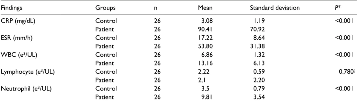
Table 1. Comparison of peripheral vein parameters between groups.
Findings Groups n Mean Standard deviation P*
CRP (mg/dL) Control 26 3.08 1.19 <0.001 Patient 26 90.41 70.92 ESR (mm/h) Control 26 17.22 8.64 <0.001 Patient 26 53.80 31.38 WBC (e3/UL) Control 26 6.86 1.32 <0.001 Patient 26 13.16 6.13
Lymphocyte (e3/UL) Control 26 2,22 0.59 0.780†
Patient 26 2,1 2.20
Neutrophil (e3/UL) Control 26 3.5 0.79 <0.001
Patient 26 9.81 3.54
*t test for independent groups.
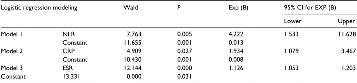
showed NLR OR as 4.22 (P = 0.005; 95% CI, 1.533–11.628). NLR in the septic group was found to be 4.22 times more than the control group. OR values of ESR and CRP were found to be 1.934 (P = 0.000; 95% CI, 1.079–3.467) and 1.126 (P = 0.027; 95% CI, 1.053–1.203), respectively. Based on these numbers, in the study group, NLR, ESR, and CRP values were 4.22, 1.93, and 1.13 times more than those of the control group, respectively (Table 3, Figure 3).
The NLR curve of the SA patients was observed to be over the reference line, and the area under the line was 0.896 (P <0.001; 95% CI, 0.796–0.996), which is very close to 1.
In the NLR validity calculation, the highest LR positive value was 11.89 and NLR was found to be 2.414. At this point, when the cutoff point for NLR was taken as 2.41, our method was observed to have a sensitivity and specificity of 88% and 93%, respectively.
Discussion
As it is known, SA is the inflammation of the syno-vial membrane and synosyno-vial fluid in joints caused by bacterial, viral, or fungal infections. Because of its highly vascular structure and absence of protec-tive basement membrane, microorganisms can eas-ily reach and colonize in synovial membranes by a hematogenous route.16
Septic arthritis is an important medical emer-gency with high morbidity. Epidemiological study is difficult to do for SA. For this reason incidence was given in variable ranges. While the incidence of SA in Europe is 4/100,000 per year, this rate is six times more common in eastern Europe and Australia. We could not find any report regarding incidence rate of SA in our country. Tarkowski et al. reported that the incidence in the general pop-ulation is 6/100,000.3 Furthermore there are too
few reports in the USA after 2000.17
That is why we believe that our study results containing 26 cases could provide a valuable con-tribution to the literature even it appears to be a small cohort.
Figure 2. Comparison of peripheral vein parameters between groups. Table 2. Joint fluid culture and Gram staining results of the
study group.
Application Patient group
Frequency %
Synovial joint aspirate culture
Culture negative 19 73.1 Coagulase negative Staphyloccus aureus 3 11.5 Gram-positive cocbacteria 2 7.7 Pseudomonas 2 7.7 Total 26 100.0
Synovial joint aspirate Gram staining
Positive 19 73.1
Negative 7 26.9
At the diagnosis stage of SA, time-consuming and high-cost tests are performed as a necessity. After the physical examination, increase of periph-eral blood WBC, ESR, and CRP levels are benefi-cial in diagnosis. However, espebenefi-cially in the diagnosis of suspicious cases of SA, if the case has an inflammatory joint disease like rheumatoid arthritis, the raise of the ESR is not helpful in dif-ferential diagnosis.18
In order to save the cartilage, the diagnosis of SA, which is characterized by suppurative inflam-mation, must be made as early as possible – a few hours after the onset of the symptoms – and then the surgical procedure must be carried out based on this diagnosis. NLR, which is calculated from complete blood count with differential, is an inex-pensive, easy to obtain, widely available marker of inflammation. So NLR can aid in the risk stratifi-cation of patients with various diseases in addition to the traditionally used markers. The NLR is reported to be increased in various inflammation-related diseases,19 but their clinical significance in
SA remains unclear.
For these reasons, in this study, we aimed to investigate the clinical significance of NLR value especially in patients who bear some features that cause some difficulties in differentiating and diag-nosing SA to start treatment as soon as possible.
It was previously reported that surgical interven-tion 48 h after the onset of the symptoms would be too late and lead to damage in the cartilage. Therefore, the most important factor in SA was reported to be early diagnosis.20,21
In the present study, surgery was performed in the first 6 h on average.
In the literature, it is reported that SA was a seri-ous disease and could be seen in all joints includ-ing the large, weight-bearinclud-ing, lower limb joints. Especially in the cases with non-gonococcal patho-gen, the disease is usually seen in a single joint.22
In our results, the most common joint that was involved was the knee (n = 23).
It was also reported in the literature that Gram staining and culture antibiograms in joint aspira-tions and crystal analyses must be performed as soon as possible.7,23 Further, along with ESR, CRP
increments, and a leucocyte count more than 50,000/mm3, and predominance of neutrophils in
synovial joint aspirate, supportive findings in the diagnosis were reported.23,24 Literature reported
that, based on the patients’ clinical findings, they were treated with wide spectrum antibiotic therapy even though the culture antibiograms of synovial samples resulted negative in more than 50% of the participants.25
In the present study culture results were nega-tive in 73.1% of the patients.
This is the first study in the literature supposing that NLR can support the diagnosis faster and more reliable compared to other inflammatory markers especially in uncertain SA cases.
In a previous study, no significant difference was found between healthy volunteers and ankylosing Table 3. Logistic regression models of the implicating factors in the study and control groups.
Logistic regression modeling Wald P Exp (B) 95% CI for EXP (B)
Lower Upper Model 1 NLR 7.763 0.005 4.222 1.533 11.628 Constant 11.655 0.001 0.013 Model 2 CRP 4.909 0.027 1.934 1.079 3.467 Constant 10.430 0.001 0.008 Model 3 ESR 12.144 0.000 1.126 1.053 1.203 Constant 13.331 0.000 0.031
Figure 3. The ROC curve showing the performance of NLR
spondylitis patients in terms of NLR values.26 In
another study, significantly higher NLR values were reported in ankylosing spondylitis group compared to the control.27 SA diagnosis is easily made when
patients’ synovial leukocyte count is over 50,000/mm3;
however, it has been difficult to make SA diagnosis in patients with a count less than 50,000/mm3.28 On
the other hand, synovial leucocyte count may not be increased over 50,000/mm3 in some other
con-ditions, such as corticosteroid or intravenous drug use, occurrence of malignant diseases, and in cases where immune system is deficient such as prematurity.
Further, polymorphonuclear leukocyte rate may generally be over 80% and there are studies in the literature reporting a PMNL increase in the syno-vial fluid following crystal accumulation with no rheumatoid arthritis infection.28–30
In addition, Gram staining and culturing of active pathogens in joint synovial fluid aspira-tions require selective media and techniques; the samples must therefore be transferred to the lab with detailed information, all of which is cumber-some and time-consuming. On the other hand, PCR technique, although it is not commonly used, may be utilized in some cases where Gram stain-ing is not effective, such as Neisseria gonorrhea arthritis. Other lab findings, such as the number of leukocytes, ESR, and CRP, are supporting evi-dence for the diagnosis. Recently, the level of procalcitonin in blood has been used in SA diag-nosis. Generally, CRP values in SA patients peak on day 1 and ESR values hit the maximum on days 3 and 5. These tests, like PCR technique, level of procalcitonin in blood, are expensive and time-consuming with respect to hemogram analy-sis. However, one of the most important thing in assessment of SA is immediate diagnosis and to start the proper treatment.17,31,32
All these findings are a result of NLR can be used as a differentiating marker for SA, transient arthritis, and other inflammatory arthritis. The results of research evaluating relationship between SA and NLR that was the first study we know in the literature showed that logistic regression analy-sis, which was used to monitor the changes in hemogram parameters, showed NLR OR as 4.22 (P = 0.005; 95% CI, 1.533–11.628). NLR in the septic group was found to be 4.22 times more than the control group. Based on these data, in the study
group, NLR, values were 4.22 times more than those of the control group. The NLR curve of the SA patients was observed to be over the reference line, and the area under the line was 0.896 (P <0.001; 95% CI, 0.796–0.996), which is very close to 1. In the NLR validity calculation, the highest LR positive value was 11.89 and NLR was found to be 2.414. At this point, when the cutoff point for NLR was taken as 2.41, our method was observed to have 88% sensitivity and 93% specificity. So, there was a statistical difference between groups regarding NLR (P = 0.005).
Especially in patients in whom it is difficult to diagnose, in order to begin treatment as soon as possible, cost-effective, short-term received results of the NLR parameter may be used in order to be able to support the clinical diagnosis.
The present study has several limitations. Biochemistry and hemogram devices in each health center from which the data were obtained may have had different calibration settings. Besides, the present study is a retrospective design, so measure-ment errors, if any, could not be controlled. Since measurement, weighing, and titration were all car-ried out at different times, there may have been variations in temperature, pressure, and relative humidity. It is therefore not possible to identify measurement errors in the analytical phase. Due to retrospective study and ethical inappropriateness, synovial joint sample could not be aspirated from the control group. Therefore, NLR in the periph-eral vein was compared between groups.
The specificity of NLR should always be looked at prospectively: in the first stage, the control group with joint interested other inflammatory events; and in the next step, patients diagnosed with dis-tant infection. According to the obtained results, NLR may be used as a marker to monitor disease progression and indicate a subclinical inflamma-tion in patients with SA.
Declaration of conflicting interests
The authors declared no potential conflicts of interest with respect to the research, authorship, and/or publication of this article.
Funding
This research received no specific grant from any fund-ing agency in the public, commercial, or not-for-profit sectors.
References
1. Mue D, Salihu M, Awonusi F et al. (2013) The epidemi-ology and outcome of acute septic arthritis: A hospital based study. Journal of the West African College of
Surgeons 3: 40–52.
2. Mankovecky MR and Roukis TS (2014) Arthroscopic synovectomy, irrigation, and debridement for treat-ment of septic ankle arthrosis: A systematic review and case series. Journal of Foot and Ankle Surgery 53: 615–619.
3. Tarkowski A (2006) Infection and musculoskel-etal conditions: Infectious arthritis. Best Practice &
Research: Clinical Rheumatology 20: 1029–1044.
4. Margaretten ME, Kohlwes J, Moore D et al. (2007) Does this adult patient have septic arthritis? Journal of
the American Medical Association 297: 1478–1488.
5. Montgomery CO, Siegel E, Blasier RD et al. (2013) Concurrent septic arthritis and osteomyelitis in chil-dren. Journal of Pediatric Orthopedics 33: 464–467. 6. Dlabach JA and Park AL (2008). Infectious arthritis.
In: Canale ST, Beaty JH, editors. Campbell’s
opera-tive orthopaedics. 11th ed. Philadelphia, PA: Mosby
Elsevier, pp. 723–750.
7. Tausche AK, Gehrisch S, Panzner I et al. (2013) A 3-day delay in synovial fluid crystal identification did not hinder the reliable detection of monosodium urate and calcium pyrophosphate crystals. Journal of
Clinical Rheumatology 19: 241–245.
8. Kim H, Kim J and Ihm C (2010) The usefulness of multiplex for the identification of bacteria in infection.
Journal of Clinical Laboratory Analysis 24: 175–181.
9. Gibson PH, Cuthbertson BH, Croal BL et al. (2010) Usefulness of neutrophil/lymphocyte ratio as predic-tor of new-onset atrial fibrillation after coronary artery bypass grafting. American Journal of Cardiology 105: 186–191.
10. Metz CE (1978) Basic principles of ROC analysis.
Seminars in Nuclear Medicine 8: 283–298.
11. Hanley JA and McNeil BJ (1982) The meaning and use of the area under a receiver operating characteris-tic (ROC) curve. Radiology 143: 29–36.
12. Knapp RG and Miller III MC (1992) Clinical Epidemiology and Biostatistics. Philadelphia, PA: Williams & Wilkins Press. Introductory Biostatistics. United States of America, Wiley & Sons Pres, 336–337.
13. Obuchowski NA and McClish D (1997) Sample size determination for diagnostic accuracy studies involving binormal ROC curve indices. Statistics in
Medicine 16: 1529–1542.
14. Zweih MH and Camphell G (1993) Receiver-operating characteristic (ROC) plots: A fundamental evalua-tion tool in clinical medicine. Clinical Chemistry 39: 561–577.
15. Mayer D (2004) Essential Evidence-based Medicine,
Dan Mayer Essential Medical Texts for Students and Trainees. 1st ed. Cambridge: Cambridge University
Press.
16. Ross JJ (2005) Septic arthritis. Infectious Disease
Clinics of North America 19: 799–817.
17. Lim SY, Pannikath D and Nugent K (2015) A retro-spective study of septic arthritis in a tertiary hospital in West Texas with high rates of methicillin-resist-ant Staphylococcus aureus infection. Rheumatology
International 35: 1251–1256.
18. Goodman SB, Chou LB and Schurman DJ (2001) Management of pyarthrosis. In: Chapman MW, edi-tor. Chapman’s orthopaedic surgery. Vol. 3. 3rd edn. Philadelphia, PA: Lippincott Williams & Wilkins, pp. 3561–3577.
19. Yilmaz H, Uyfun M, Yilmaz TS et al. (2014) Neutrophil-lymphocyte ratio may be superior to C-reactive protein for predicting the occurrence of postmenopausal osteoporosis. Endocrine Regulations 48: 25–33.
20. Smith RL, Schurman DJ, Kajiyama G et al. (1987) The effect of antibiotics on the destruction of car-tilage in experimental infectious arthritis. Journal
of Bone and Joint Surgery: American Volume 69:
1063–1068.
21. Daniel D, Akeson W, Amiel D et al. Lavage of sep-tic joints in rabbits: Effects of chondrolysis. Journal
of Bone and Joint Surgery: American Volume 58:
393–395.
22. Goldenberg DL and Reed JI (1985) Bacterial arthritis.
New England Journal of Medicine 312: 764–771.
23. Courtney P and Doherty M (2013) Joint aspiration and injection and synovial fluid analysis. Best Practice &
Research: Clinical Rheumatology 27: 137–169.
24. McCutchan HJ and Fisher RC (1990) Synovial leuko-cytosis in infectious arthritis. Clinical Orthopaedics
and Related Research 257: 226–230.
25. Dlabach JA and Park AL (1992) Infectious arthri-tis. In: Canale ST and Beaty JH, editors. Campbell’s
operative orthopaedics. Philadelphia, PA: Mosby
Year Book.
26. Boyraz I, Koç B, Boyacı A et al. (2014) Ratio of neutrophil/lymphocyte and platelet/lymphocyte in patient with ankylosing spondylitis that are treating with anti-TNF. International Journal of Clinical and
Experimental Medicine 7: 2912–2915.
27. Gokmen F, Akbal A, Resoglu H et al. (2015) Neutrophil-lymphocyte ratio connected to treatment options and inflammation markers of ankylosing spondylitis. Journal of Clinical Laboratory Analysis 29: 294–298.
28. McGillicuddy DC, Shah KH, Friedberg RP et al. (2007) How sensitive is the synovial fluid white blood
cell count in diagnosing septic arthritis? American
Journal of Emergency Medicine 25: 749–752.
29. Jones ST, Denton J, Holt PJ et al. (1993) Possible clearance of effete polymorphonuclear leucocytes from synovial fluid by cytophagocytic mononuclear cells: Implications for pathogenesis and chronicity in inflammatory arthritis. Annals of the Rheumatic
Diseases 52: 121–126.
30. Kung JW, Yablon C, Huang ES et al. (2012) Clinical and radiologic predictive factors of septic hip arthri-tis. American Journal of Roentgenology 199: 868– 872.
31. Muralidhar B, Rumore PM and Steinman CR (1994) Use of the polymerase chain reaction to study arthri-tis due to Neisseria gonorrhoeae. Arthriarthri-tis and
Rheumatism 37: 710–717.
32. Swan A, Amer H and Dieppe P (2002) The value of synovial fluid assays in the diagnosis of joint disease: a literature survey. Annals of the Rheumatic Diseases 61: 493–498.
33. Paosong S, Narongroeknawin P, Pakchotanon R et al. (2015) Serum procalcitonin as a diagnostic aid in patients with acute bacterial septic arthritis. International