Life-Threatening Respiratory Distress in a Total Laryngectomy
Patient: Aspirated Voice Prosthesis or Lung Tumor?
Selmin Karataylı Özgürsoy1, Ozan Özgürsoy2, Cabir Yüksel3, Gürsel Dursun2 1Department of Otorhinolaryngology, Ufuk University School of Medicine, Ankara, Turkey 2Department of Otorhinolaryngology, Ankara University School of Medicine, Ankara, Turkey 3Department of Thoracic Surgery, Ankara University School of Medicine, Ankara, Turkey Case Report
Address for Correspondence:
Selmin Karataylı Özgürsoy
E-mail: selminkrt@hotmail.com Received Date: 29.02.2016 Accepted Date: 12.04.2016
© Copyright 2016 by Official Journal of the Turkish Society of Otorhinolaryngology and Head and Neck Surgery Available online at www.turkarchotorhinolaryngol.org DOI: 10.5152/tao.2016.1592
131
Introduction
A laryngectomy patient with marked respiratory distress may pose a dilemma for the physician, par-ticularly when the patient has a tracheoesophageal puncture (TEP) (1). A missing TEP prosthesis (TEPP) represents a rare but threatening event (1). It may be coughed out, swallowed, or aspirated. The latter can be an emergency and may therefore require urgent surgical intervention. Moreover, stoma problems such as crust or excessive granu-lation tissue formation, recurrence of the primary cancer, a second primary cancer, and exacerbation of coexisting cardiopulmonary diseases may lead to respiratory disturbances in postlaryngectomy patients (2).
Most laryngectomy patients have poor baseline pulmonary functions because of long-term smok-ing that put them at a high risk for rapid decom-pensation(2). Therefore, early diagnosis of the underlying cause of respiratory distress and appro-priate management are crucial in these patients. Here we report an interesting case of a laryngec-tomy patient with two different clinical presenta-tions of life-threatening respiratory distress at the same time.
Case Report
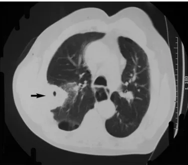
A 63 year-old male total laryngectomy patient with TEP was admitted to the emergency de-partment because of sudden-onset severe dyspnea. We figured out that the TEPP was not in place; however, the patient was not aware of the situation until he was asked about it. Chest X-rays did not reveal an overt foreign body but showed hyperin-flation of the right lung with complete collapse of the left lung and deviation of the trachea to the left (Figure 1). Anticipating that the patient aspirated his TEPP, he underwent flexible bronchoscopy by a thoracic surgeon. The Blom–Singer prosthesis (Inhealth Technologies; Carpinteria, CA, USA) was identified in the left main bronchus and re-moved with forceps through the use of an instru-ment port. Although there was a significant im-provement in the chest X-ray after the procedure (Figure 2), respiratory distress of the patient did not improve as much as we expected. This time, we noticed a hypodense area in the upper lobe of the right lung that was not clearly observed by any of us in the first chest X-ray (Figure 2). Next, computerized tomography (CT) scan of the chest revealed 6 cm mass in the upper lobe of the right lung with hypodense necrotized center,
Turkish Archives of Otorhinolaryngology
Türk Otorinolarengoloji Arşivi Turk Arch Otorhinolaryngol 2016; 54: 131-3
Abstract Laryngectomy patients usually have poor pulmonary functions due to long-term smoking. Their lungs can easily be decompensated. Hence, meticulous evalu-ation and timely management of severe respiratory distress in laryngectomy patients can be life savers. Here we present an interesting case of a laryngecto-my patient with two different clinical presentations of life-threatening respiratory distress at the same time (aspiration of voice prosthesis and a second primary
lung cancer). Marked or persistent respiratory distress in a laryngectomy patient deserves thorough clinical evaluation and may require urgent intervention. We consider that the presentation and course of respira-tory distress in our laryngectomy patient will provide an additional aspect for emergency room doctors and airway specialists dealing with such a patient.
Keywords: Laryngectomy, dyspnea, voice prosthesis,
which caused destruction in the 5th and 6th ribs, implying ma-lignancy (Figure 3). Bronchoscopy was repeated, and the lung mass was subjected to biopsy. Histological examination revealed a well-differentiated squamous cell carcinoma. Subsequent bone scintigraphy showed hyperactivity on the right 5th and 6th ribs. CT scans of the head, neck, and abdomen showed no evidence of locoregional recurrence or any other lesion. The patient was then referred to the oncology and thoracic surgery departments for further treatment.
A meticulous review of the patient’s medical records showed that he underwent total laryngectomy with TEP and selective left neck dissection (level 2-3-4) 19 months ago for T3NOM0 glottic squamous cell carcinoma with prominent paraglottic space invasion. Histological examination of the surgical
speci-men demonstrated medium-differentiated squamous cell laryn-geal carcinoma, and there was no lymph node involvement in any of the 29 lymph nodes in the neck dissection specimen. Pre-operative CT scan of the chest revealed milimetric parenchymal nodules in the right middle and inferior and left inferior lobes, which were attributed to be post-infectious and non-specific. There were also a few aorticopulmonary, precarinal, and par-aesophageal millimetric calcified lymph nodes, which were at-tributed to be specific post-infectious sequelae. He was followed up regularly, and his TEPP was replaced at first year visit. His last visit with us was approximately 4 months ago, with no sig-nificant problem. Our patient was also followed by his primary care physician, and chest X-rays were taken every 4–6 months in his hometown. According to his primary care medical records, there was no significant respiratory difficulty and no abnormal finding in his subsequent chest X-rays.
Discussion
The placement of voice prosthesis through TEP in a laryngecto-my patient was first introduced by Singer and Blom(3) in 1980 and has evolved as the most common technique of choice for the restoration of voice after total laryngectomy. TEPP has to be regularly replaced, and the lifetime of the prosthesis is around 5 months (1, 4). Spontaneous loss of prosthesis occurs at a rate of 1% (1, 4).
When TEPP is dislodged, there may be three possible explana-tions: 1) it may be coughed out, 2) it may fall into the esophageal side and get swallowed, or 3) it may fall into the tracheal side and get aspirated. All these possibilities require urgent medical care.
When TEPP is coughed out or swallowed, TEP has to be stent-ed or a new prosthesis should be put in place as soon as possi-ble. If stenting and/or replacement is not performed in a timely manner, TEP can easily get closed and the patient may require repeat TEP (5).
Turk Arch Otorhinolaryngol 2016; 54: 131-3 Karataylı Özgürsoy et al. Respiratory Distress in Laryngectomy Patient
132
Figure 1. Chest X-ray showing hyperinflation of the right lung with complete collapse of the left lung and deviation of the trachea to the left
Figure 3. CT scan of the chest. Arrow indicates a mass in the upper lobe of right the lung with a hypodense necrotized center
Figure 2. Chest X-ray showing radiological improvement after bronchoscopic removal of the aspirated prosthesis. Arrow indicates a hypodense area in the upper lobe of right the lung
The aspiration of TEPP has been reported to occur in 0.75%– 13% of patients (6). The most common location for aspirated TEPP is the upper right main stem bronchus and carina (1). Be-cause of the lethal consequences of aspirated TEPP, a laryngec-tomy patient with missing TEPP should always be evaluated for aspiration. If aspiration occurs, bronchoscopic removal is neces-sary (7). In some patients having no overt symptoms after the aspiration of TEPP, the situation may be noticeable at a routine follow-up visit (6). However, in our patient, aspirated TEPP led to severe respiratory distress, requiring urgent intervention. Contrary to the fact that aspirated foreign bodies usually lodge in the right lung or carina, TEPP was in the left main bronchus of our patient.
In a laryngectomy patient, stoma is an independent risk factor for foreign body aspiration (2). Moreover, extensive crust for-mation deep in the stoma may obstruct the airway. The aspi-ration of TEPP should be eliminated and managed first in a laryngectomy patient with a missing TEP. However, there may be other or additional possibilities causing respiratory problems in a laryngectomy patient, such as tracheal recurrence of the primary cancer or exacerbation of coexisting lung disease. Fur-thermore, there is a lifetime risk for a second primary cancer. In our patient, a second primary tumor was found incidentally, as described in the report. Because his primary care practitioner did not note any abnormal respiratory symptom or finding in chest X-rays, he has not undergone CT scan of the chest. Most likely, mild respiratory symptoms may have been attributed to chronic chest disease, and the lung mass may not be detected in X-rays, like the case of the first X-ray taken in the emergency room. CT scan of the chest would have provided much more in-formation. However, the necessity for CT scans of the chest for laryngectomy patients is still controversial worldwide, and it is not a routine practice at our institution. As more self-criticism, all our attention was focused on the left lung and the aspirat-ed prosthesis. We did not carefully assess the first X-ray and missed the suspicious area in the upper lobe of the right lung. Furthermore, we did not perform complete bronchoscopy while removing the prosthesis. If we would have observed the right lung during the first intervention, we could have noticed the tumor and performed a biopsy. Hence, a second bronchoscopy would not have been required.
Conclusion
Most laryngectomy patients are at a high risk for rapid pul-monary decompensation because of long-term smoking. We therefore believe that marked or persistent respiratory distress
in a laryngectomy patient deserves meticulous evaluation and timely management. We consider that the unique presentation and course of the respiratory distress in our laryngectomy pa-tient will notify emergency room doctors, airway specialists, and otolaryngologists when dealing with such a case.
Informed Consent: Written informed consent was obtained from
pa-tient who participated in this study.
Peer-review: Externally peer-reviewed.
Author Contributions: Concept - G.D., O.B.Ö.; Design - S.K.Ö.,
O.B.Ö.; Supervision - G.D., O.B.Ö.; Resources - S.K.Ö., C.Y.; Ma-terials - S.K.Ö., O.B.Ö., C.Y.; Data Collection and/or Processing - S.K.Ö., O.B.Ö., C.Y.; Analysis and/or Interpretation - C.Y., G.D., O.B.Ö., S.K.Ö.; Literature Search - S.K.Ö., O.B.Ö., C.Y.; Writing Manuscript - S.K.Ö., O.B.Ö., G.D., C.Y.; Critical Review - G.D., O.B.Ö.
Conflict of Interest: No conflict of interest was declared by the
au-thors.
Financial Disclosure: The authors declared that this study has
re-ceived no financial support.
References
1. Leuin SC, Deschler DG. The missing tracheoesophageal puncture prosthesis: evaluation and management. ENT J 2013; 92: E14-6. 2. Brenner MJ, Floyd L, Collins S. Role of computed tomography
and bronchoscopy in speech prosthesis aspiration. Ann Otol Rhi-nol Laryngol 2007; 116: 882-6. [CrossRef]
3. Singer MI, Blom ED. An endoscopic technique for restoration of voice after total laryngectomy. Ann Otol Rhinol Laryngol 1980; 89: 529-33. [CrossRef]
4. Op de Coul BM, Hilgers FJ, Balm AJ, Tan IB, van den Hoogen FJ, van Tinteren H. A decade of post laryngectomy vocal reha-bilitation in 318 patients: A single institution’s experience with consistent application of provox indwelling voice prostheses. Arch Otolaryngol Head Neck Surg 2000; 126: 1320-8. [CrossRef]
5. Hathurusinghe H, Uppal H, Shortridge R, Read C. Case of the month: Dislodged trachea-oesophageal valve: Importance of rapid replacement or stenting. Emerg Med J 2006; 23: 322-3.
[CrossRef]
6. Ostrovsky D, Netzer A, Goldenberg D, Joachims HZ, Golz A. Delayed diagnosis of tracheoesophageal prosthesis aspiration. Ann Otol Rhinol Laryngol 2004; 113: 828-9. [CrossRef]
7. Kilic D, Findikcioglu A, Bilen A, Habesoglu MA, Hurcan C, Hatipoglu MA. Delayed diagnosis of a foreign body: aspiration of voice prosthesis: case report. Turkiye Klinikleri J Med Sci 2007: 27: 471-3.
