Diagn Interv Radiol 2014; 20:511–512 © Turkish Society of Radiology 2014
LETTER TO THE EDITOR
511
Delayed failure of rectovaginal fistula
embolization with Amplatzer vascular plug 2
We read with great interest the paper published by Gun-eyli et al. (1) in Diagnostic and Interventional Radiology along with the other impressive papers regarding the use of Amplatzer® Vascular Plugs (AVPs, AGA Medical Corp., Golden Valley, Minnesota, USA) (1–3). We would like to share our experience about using the AVP for treating a rec-tovaginal fistula (RVF). Although initially successful, this treatment unfortunately failed in the long-term follow-up. A 47-year-old female patient was referred to our depart-ment for interventional radiologic treatdepart-ment of RVF, which occurred following a low anterior resection for T2N0M0 rectal cancer, performed 10 months ago in another cen-ter. On postoperative day 12 the patient suffered vaginal fecal discharge. Initial emergent loop diversion colostomy and primary surgical repair of the fistula six months lat-er, were both unsuccessful. Double-contrast barium enema (Prontobario Colon, Bracco, Milan, Italy) revealed passage of contrast and air from rectum to vagina consistent with RVF, and embolization of the fistula was planned (Fig. 1a). Under the guidance of a 5 F vertebral catheter (Cordis, Johnson & Johnson, Miami, Florida, USA) and an angled hydrophilic 0.035-inch radiofocus glidewire (Terumo, Som-erset, New Jersey, USA) the fistula was catheterized, and the wire accessed into the vaginal lumina. The glidewire was exchanged for a stiff Amplatz guidewire (Cook Medi-cal, Bloomington, Indiana, USA). An 8 F long sheath and a dilator (Shuttle, Cook Medical) were inserted over the ex-change wire into the fistula and advanced to the measured area. The dilator and the wire were removed, and angiog-raphy was performed through the guiding catheter to con-firm satisfactory location of the guiding catheter. An AVP 2 was introduced through the 8 F sheath. While the medial part of the plug was in the fistula, the distal and proxi-mal parts were in the vaginal and rectal walls, respective-ly (Fig. 1). The plug was deployed following confirmation of the device position. Postembolization angiography re-vealed occlusion of the fistula. The patient was discharged the next day. The control double-contrast barium enema examination at the first month revealed complete healing and the patient was free of symptoms (Fig. 2). However, the second control imaging performed at the third month, after relapsed complaints of the patient, revealed loss of the plug and reappearance of the fistula. In addition, there was a new blind-ended fistula towards the presacral space (Fig. 3). Although we could not demonstrate direct passage of contrast in to vagina, there was a late slight fluid discharge consistent with contrast material from the vagina.
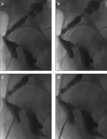
Figure 1. a–d. Lateral iodinated contrast study of the colon (a)
demonstrates the passage of contrast material from the rectum to vagina through a fistulous tract. Treatment begins with the passage of the wire through the fistula (b). Panel (c) shows catheterization of the fistula tract. Repeat colonography (d) confirms position before deployment of a 12 mm Amplatzer Vascular Plug in the rectovaginal fistula tract.
c a
d b
Figure 2. a,b. Lateral x-ray image (a) shows the plug in the
expected position in the first month follow-up of the patient. Control colonography (b) confirms the unchanged position of the plug and healed fistulous tract.
512 • November–December 2014 • Diagnostic and Interventional Radiology Kılıçkesmez et al. First-line treatment options for this
patient include palliative colostomy and surgical repair. An alternative method could be the use of an occlu-sion device designed for nonsurgical treatment of patients with acquired RVF, as reported by Lee et al. (4). Al-though this device is reported to have
a high success rate, there are certain limitations: each device should be con-structed according to the diameter and length of the fistula tract and deployed through a 24 F chest tube.
In this letter we present a case of ini-tially succesful nonsurgical technique for RVF treatment. To the best of our knowledge this is the first case in the literature reporting the use of an AVP in RVF. The late-onset failure and loss of AVP might be related to physiolog-ical rises in rectal pressure during the passage of stool and regional move-ments.
Since the introduction of AVPs for use in the cardiovascular system, vari-ous applications have been reported in different diseases and organ systems. Their applicability and effectiveness have been proved in numerous vascu-lar diseases along with reports confirm-ing successful use in esophagobronchi-al, gastrocolonic, and ureterovesical fistulas (1–3).
In conclusion, RVFs are rare compli-cations of various diseases or surgical procedures, and they should be treated immediately for distressing symptoms. The long-term failure of AVP must be kept in mind, when considering treat-ment options.
Conflict of interest disclosure
The authors declared no conflicts of interest. References
1. Guneyli S, Cinar C, Bozkaya H, Parildar M, Oran I. Applications of the Amplatzer Vascular Plug to various vascular lesions. Diagn Interv Radiol 2014; 20:155–159. [CrossRef]
2. Cil B, Peynircio¤lu B, Canyi¤it M, Geyik S, Çiftçi T. Peripheral vascular applications of the Amplatzer vascular plug. Diagn In-terv Radiol 2008; 14:35–39.
3. Young JA, Shimi SM, Alijani A, Patil PV, Bhat R. Occlusion of a neo-esopha-geal-bronchial fistula using the Amplatzer Vascular Plug 2. Diagn Interv Radiol 2013; 19:259–262.
4. Lee BH, Choe DH, Lee JH, et al. Device for occlusion of rectovaginal fistula: clinical tri-als. Radiology 1997; 203:65–69. [CrossRef]
Özgür Kılıçkesmez, Çağatay Andıç, Levent Oğuzkurt
From the Department of Interventional Radiolo-gy (Ö.K. [email protected]), İstanbul Training and Research Hospital, İstanbul, Turkey; Department of Interventional Radiology (Ç.A., L.O.), Başkent University School of Medicine, Adana Training and Medical Research Center, Adana, Turkey.
Received 20 April 2014; accepted 28 April 2014. Published online 11 July 2014
DOI 10.5152/dir.2014.14177. Figure 3. Contrast-injected lateral
colonography performed at the third month reveals the loss of the plug and a recently developed presacral blind-ended fistulous tract.