Contents lists available atScienceDirect
International Journal of Surgery
journal homepage:www.elsevier.com/locate/ijsuOriginal Research
The e
ffect of the cerclage wire in the treatment of subtrochanteric femur
fracture with the long proximal femoral nail: A review of 52 cases
Bekir Eray Kilinc
a,∗, Yunus Oc
b, Adnan Kara
c, Ramazan Erden Erturer
daGolhisar State Hospital, Turkey
bSisli Hamidiye Etfal Training and Research Hospital, Halaskargazi Cad., Etfal Sk, 34371,Şişli, İstanbul, Turkey cMedipol University Medical Faculty, Göztepe Mahallesi, Metin Sk. No:4, 34214, Bağcılar, İstanbul, Turkey dİstinye University Medical Faculty, Esenkent Mahallesi, Süleyman Demirel Cd, 34510, Esenyurt, İstanbul, Turkey
A R T I C L E I N F O Keywords:
Subtrochanteric femur fracture Long proximal femoral nail PFNA
Cerclage wire
A B S T R A C T
Introduction: To present the effect of the cerclage fixation, which was performed for the purpose of preserving the alignment obtained by open reduction, on the long-term clinical and radiological results of subtrochanteric fractures.
Materials and methods: The inclusion criterias were at least 2 years of follow-up, no severe cognitive impairment, and to be able to walk independently prior to fracture. Patients with transverse or short oblique subtrochanteric hip fractures (AO/OTA class 32-A3.1), patients underwent previous femoral or hip operation for the same side and those with segmental fractures, bilateral fractures and pathological fractures were not included in the study. The clinical and radiological results of the patients were evaluated. The clinical evaluations were performed with Palmer and Parker Mobility Score (PPM), Lower Extremity Functional Score (LEFS), visual analogue score (VAS) and return to pre-injury activity status.
The elapsed time between the trauma and the surgery date, the duration of surgery the estimated amount of bleeding, and the length of hospital stay of patients were assessed.
Results: Thirty-two of the 52 patients were female and 20 were male. The mean age of females was 77.80 ± 9.75 years and the mean age of the males was 79.18 ± 6.50 years. The mean follow-up period of the patients was 62.25 ± 34.68 months. The mean time until the patients' surgery was 6.18 ± 3.32 days. The mean duration of surgery was 52.6 ± 13.8 min. The mean amount of bleeding was 176 ± 90 ml. The mean length of patients' hospital stay was 6.2 ± 3.2 days. The postoperative mean duration of union was found to be 3.8 ± 1.6 months. The mean value of varus/valgus angulation in coronal plane measurements was 0.52°. No complication was observed in any patient during the cerclage application. The mean number of wire was 1.3. LEFS difference was statistically significant. PPM decline was not statistically significant.
Conclusion: Open reduction and the use of cerclage did not produce a negative effect in terms of fracture union. The generation of medial support by anatomical reduction of the fracture prevents the implant failure and provide a basis for union.
1. Introduction
Subtrochanteric fractures of the femur are defined as fractures in-volving the area between the trochanter minor and isthmus of the femur. Although these fractures are usually seen in young patients who go through high-energy trauma, it may also occur as a result of simple falls in elderly patients due to reduced bone quality [1].
The subtrochanteric region of the femur is exposed to a large variety of stress that comes up as a result of bending movements and com-pressive forces generated by body weight and the hip muscles. Surgery of the subtrochanteric femoral fractures can be challenging because of
the instability of bone fragments and the stretching and pulling forces of the muscles. Strong abductor muscles, iliopsoas and adductor mus-cles cause a displacement of the proximal fragment and resist the anatomical reduction of the fracture. This may lead to non-union, malunion in fractures, and mechanical failure of implants [2,3].
The use of numerous intra- and extramedullary implants has been described for internalfixation of the subtrochanteric fractures [4–7]. In order to achieve a stablefixation, many authors suggest treating the subtrochanteric fractures with intramedullary implants which are bio-mechanically superior. However, depending on the displacement, pro-viding and maintaining an appropriate alignment prior to implant
https://doi.org/10.1016/j.ijsu.2018.06.035
Received 20 November 2017; Received in revised form 30 April 2018; Accepted 25 June 2018
∗Corresponding author.
E-mail addresses:[email protected](B.E. Kilinc),[email protected](Y. Oc),[email protected](A. Kara),[email protected](R.E. Erturer).
Available online 28 June 2018
1743-9191/ © 2018 IJS Publishing Group Ltd. Published by Elsevier Ltd. All rights reserved.
placement may be difficult for displaced subtrochanteric fractures. Moreover, the risk of non-union due to the varus of fracture alignment was also reported to be higher in the literature [6,7].
A possible approach to perform a stable osteosynthesis with an in-tramedullary implant in subtrochanteric fractures is to reduce the medial hinge anatomically. The displacement of fracture fragments due to used forces by strong muscles may not usually allow the closed re-duction of the fracture.
The aim of this study is to present the effect of the cerclage fixation, which was performed for the purpose of preserving the alignment ob-tained by open reduction, on the long-term clinical and radiological results of subtrochanteric fractures.
2. Material and methods
A retrospective analysis was performed using the data of patients treated with long hip nails (long PFN [300–420 mm]; Synthes1, Oberdorf, Switzerland) and cerclage due to subtrochanteric fracture between January 2008 and December 2014. The Orthopedic Trauma Association (AO/OTA) classification was used to classify the inter-trochanteric fractures (31A3), including the subinter-trochanteric region fractures (32A-B-C) and the reverse oblique fractures extending to the subtrochanteric region [8]. The inclusion criteria for participation in the study were determined for patients to have at least 2 years of follow-up, no severe cognitive impairment, and to be able to walk in-dependently prior to fracture. Patients with transverse or short oblique subtrochanteric hip fractures (AO/OTA class 32-A3.1), patients under-went previous femoral or hip operation for the same side and those with segmental fractures, bilateral fractures and pathological fractures were not included in the study. The study included 52 patients with appro-priate follow-up duration and data.
The work has been reported in line with the STROCSS criteria [9]. The clinical and radiological results of the patients at the last follow-up were evaluated. The clinical evaluations were performed with Palmer and Parker Mobility Score (PPM) [10], Lower Extremity Func-tional Score (LEFS), visual analogue score (VAS) and return to pre-in-jury activity status.
The elapsed time (days) between the trauma and the surgery date, the duration of surgery (minutes), the estimated amount of bleeding (milliliters), and the length of hospital stay (days) of patients were as-sessed. The union was evaluated by a single surgeon with standard orthogonal radiographs taken during the follow-up of patients. The presence of union was defined as the presence of bridging callus for-mation at least on three cortices in two orthogonal projections [11]. The angulations in the coronal plane were evaluated. Our study was performed by obtaining the approval of the ethics committee.
In statistical analyses, Statistical Package for Social Sciences (SPSS) software (version 21.0,SPSS Inc., Chicago, IL, USA) was used. The paired Student's t-test was used to compare the preoperative and postoperative outcomes. Statistical significance was accepted at p < 0.05.
Data of the radiological and clinical evaluations in preoperative and follow-up periods were compared statistically.
2.1. Surgical technique
After an appropriate anesthesia, the patients were placed in supine position on the traction table, the other extremity was placed in hemilithotomy position while the fractured extremity was taken in traction boot, allowing the appropriate lateral imaging of the fluoro-scopy and distal locking. The reduction of the fracture was evaluated with antero-posterior (AP) and lateral imaging under fluoroscopy by performing traction. In patients in whom an adequate closed reduction of the fracture could not be obtained, the skin was opened by a 5-cm small incision that was made from the lateral thigh in the fracture area underfluoroscopy control.
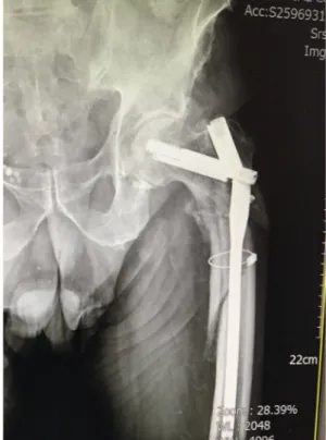
Later, the fascia lata was opened longitudinally and blunt dissection of the vastus lateralisfibers was performed. Then, an open reduction without periosteal dissection was performed with the help of a reduc-tion clamp (Verbrugge bone holding clamp) and cerclage-assisted (Dall Miles)fixation of the fracture was obtained. Care was exercised to the localization to prevent the applied wire from affecting the insertion point. A longitudinal incision of approximately 3 cm was made towards the proximal side after the type of the trochanter major was palpated by the surgeon. The appropriate point for the nail insertion, which was slightly at the medial, was determined according to the type of tro-chanter major, and the long PFNA was implanted into the anatomically reduced femur. After ensuring that the nail was fully inserted, the clamp was removed (Figs. 1 and 2). The implantation of the PFNA was per-formed with a smooth placement of the screws by obtaining a standard tip-apex distance (TAD) of 20 mm. The distal locking of the nail was Fig. 1. Aplication of the long PFNA and cerclage wire antero-posterior view.
performed with a free-hand technique underfluoroscopy.
No bone graft was used in any of the cases. On the first post-operative day, active hip and knee flexion-extension exercises were started for the operated side of the patient. On the second postoperative day, the patients were walked with the help of a walker by thefinger-tip contact. If no reduction loss was detected in the radiographs taken on the postoperative sixth week, the patients were asked to walk by bearing weight as much as they could tolerate. They were walked, bearing full weight on the eighth and twelfth weeks. The patients were called up for routine follow-up visits after the postoperative 6th week, 3rd month, 6th month, 1st year and 2 nd year.
3. Results
Thirty-two of the 52 patients, who participated in the study, 32 were female and 20 were male. The mean age of females was 77.80 ± 9.75 years (range, 46–94) and the mean age of the males was 79.18 ± 6.50 years (range, 23–92) (Table 1). The mean follow-up period of the patients was 62.25 ± 34.68 months. The mean time until the patients' surgery was 6.18 ± 3.32 days. The mean duration of surgery was 52.6 ± 13.8 min. The mean amount of bleeding was 176 ± 90 ml (range, 100–320). The mean length of patients' hospital stay was 6.2 ± 3.2 days (range, 4–21) (Table 2).
Falling down from standing height was the most common type of injury, accounting for 90% of the total number of cases whereas the remaining part consisted of motor vehicle injuries.
Complete fracture union was detected in all of the patients. It was found that the union times prolonged from 32 A to C according to the type of AO/OTA fracture classification. The postoperative mean dura-tion of union was found to be 3.8 ± 1.6 (range, 3–7) months. The ratio of union was 100%. The mean value of varus/valgus angulation in coronal plane measurements was 0.52° (range, varus 4°-valgus 7°). No complication was observed in any patient during the cerclage applica-tion. The mean number of wire was 1.3 (range, 1–3) (Table 2).
One patient developed implant brekage despite a good reduction (Figs. 3 and 4), and revision surgery was performed. Two patients de-veloped fracture around the implant due to falling down after the union of the fracture. At the same time, fracture reduction with a single wire had been preferred in these patients. 3 of the patients developed su-perficial surgical infection that healed with antibiotics and dressing. Debridement was performed in two of the patients due to severe wound drainage and they were treated with appropriate antibiotherapy ac-cording to culture result. The duration of union was observed to be
longer in these patients than in other patients. At the same time, the number of wires used in these patients was 3. No sign of infection was found at the last follow-up of these patients.
One of the patients was identified to have a iatrogenic non-displaced fracture of the trochanter major during reaming. The union of the fracture occurred with conservative treatment (Table 3). All of the patients could return to their previous residence after the treatment period in the hospital. Nine patients were walking with a single cane support, while 43 patients did not need any equipment to walk. Table 1
Demographic profile of study patients. Characteristic Sex Female n: 32 Male n: 20 Age Female 77.8 ± 9.75 Male 79.18 ± 6.5 Table 2 Perioperative measures. Characteristic Average
Follow-up time 62.25 ± 34.68 month
Injury-operation time 6.18 ± 3.32 day
Operation time 52.6 ± 13.8 min
Estimated blood loss 176 ± 90 ml
Day of stay 6.2. ± 3.2 day
Union time 3.8 ± 1.6 month
Cable number 1.3
Fig. 3. Callus formation of the healing.
The preoperative mean score of the patients, who were evaluated to have a maximum score of 80 and a minimum score of 0 according to LEFS, was 64.1, whereas the postoperative mean score declined to 56.8, and this 7.3-point difference was not found to be statistically sig-nificant. The patients' ability to walk was evaluated using a 3-item PPM score with a maximum score of 9. The mean preoperative score of 7.2 was found to be 6.4 postoperatively. This decline was not statistically significant (Table 4).
According to VAS in which the maximum pain was evaluated to be 10 and no pain was evaluated to be 0, 38 of the patients had no pain due to femoral fracture. 6 patients reported 7/10 of pain during long-distance walking.
4. Discussion
The treatment of unstable subtrochanteric fractures of the femur continues to be challenging for orthopedic surgeons. Subtrochanteric fractures tend to slide toward the varus deformity due to disintegration of the medial cortical support. The fragments in the subtrochanteric fracture of the femur may be deformed by the muscular forces causing flexion, abduction, and external rotation of the proximal fragment. Strong abductor muscles, iliopsoas and adductor muscles deteriorate the alignment of the proximal fragment and put up resistance to the anatomical reduction of the fracture. Forces acting in the proximal femur (gluteus medius and minimus, psoas, and short external rotators) create a characteristic pattern of fracture that results in aflexed, ab-ducted, and externally rotated proximal fragment, and a shortened and adducted distal fragment (hamstrings and adductors).
The nearly anatomical reduction of the fracture and the optimal placement of the fracture fragments are of great importance for good outcomes and reducing the risk of complications. Especially if AO 32A-C 1–3.1 type long oblique and spiral fractures are desired to be treated with closed reduction and intramedullary nails, they may be technically difficult surgical procedures [12,13]. Some studies argue that the periosteal bloodflow should not be disrupted by open reduction tech-niques to treat these fractures, and line up with the biologicalfixation which is an ideal technique [14–18]. Extramedullaryfixation with plate screws has potential disadvantages such as large surgical dissection, severe soft tissue injury and blood loss. Therefore, it leads to a failure in fracture healing and implanting. Furthermore, the eccentric plating is prone to implant fatigue-related implant failure due to its mechanical load-sharing effect [2,3,19–21]. Due to the superior mechanical per-formance of intramedullary implants, it has gained popularity in the treatment of displaced subtrochanteric fractures in adults [3,22–28]. In
the case of unstable subtrochanteric fractures, the advantage of load sharing with smaller bending moments of intramedullary devices al-lows early weight-bearing and prevents the excessive collapse [29]. Biomechanically, the posteromedial wall and the intramedullary nail that resists excessive collapse are a better choice for the fixation of unstable fractures.
After temporary reduction andfixation with long PFN, the fracture fragments may still appear to be relatively 5–10 mm displaced from each other. And this may lead to a long delay in radiographically union of the fracture [30,31]. Due to certain unstable biomechanical forces acting on the implant around the hip joint, implant failure may occur in PFNA.
These deformities may make thefixation difficult by significantly altering the orbits of the nail insertion point [32]. The determination of the nail insertion point that is tried to be found before obtaining re-duction takes a long time and increases the exposure to radiation. At the same time, the minimally medialized nail insertion point may lead to a slide towards fossa piriformis, and the lateralized point may lead tro-chanter major to displace towards the lateral side during reaming. A 52.6-min mean duration of surgery in our case series, which was lower than the previously reported mean duration of subtrochanteric fracture surgery, indicates that the wire application did not prolong the duration of the surgery [19,21]. We think that the anatomical reduction of the fracture performed through a small incision and the wire application was the key point in accurate determination of the nail insertion point and success of the operation in our study. This anatomical reduction also helps the load sharing between the bone and the implant. Sup-porting the lateral wall with the use of wire and narrowing the me-dullary canal diameter and increasing the rigidity of the bone implant structure prevented complications and increased the stability of the surgicalfixation. With the minimally invasive technique we performed, we minimized the fracture union-related complications, preserving the original hematoma of the fracture as much as possible and not losing the potential of fracture healing which has been emphasized to be important in fracture healing. This technique shows an excellent frac-ture healing with a low rate of complications associated with fracfrac-ture healing.
An important part of the complications that may occur after PFNA surgery depends on the implant failure. Mechanical complications specified in the literature include withdrawal of the screws, implant cut-out,“Z” effect and “reverse Z” effect or broken implant. Cut-out complication in subtrochanteric fractures of the femur treated with long PFN or gamma nail has been commonly reported in the literature [19,21]. In our case series, only one patient developed this complica-tion. We associated this condition that we almost had no cut-out complication with the importance we attached to the placement of the lag screw (closeness to the femoral calculus in the lower part of the femoral neck and 10 mm distance to the articular cartilage in the AP image; the exact middle location of the femoral neck in the lateral image).
The load transfer has been demonstrated to be depended on the medial wall support while using intramedullary nail in the treatment of subtrochanteric fractures. Medial buttress effect has been specified to be the key element in stable osteosynthesis [33]. The medial wall is under pressure in axial loads applied to the proximal femur whereas, the lateral wall is under the influence of tensile forces [34–36]. In cases where anatomical reduction cannot be obtained, medial load transfer also lacks and provides a basis for mechanical failure. Our opinion is that anatomical reduction should be obtained prior to nail placement for the biomechanical superiority of the intramedullary nail to arise. The medial wall of the wire used in our cases was reconstructed to provide the medial buttress effect, thus the varus bending force in the fracture line was eliminated, preventing the lateralization of the ana-tomical.
Bone-implant stability is important in the treatment of fracture for early mobilization until the period of bone union in the treatment Table 3 Complication of patients. Complication n Infection Superficial 3 Deep 2 Nonunion 0 Implant related Cut-out 1 Breakage 0 Z effect 0 Revision Surgery 1 Trokanteric fracture 1 Table 4
Functional score analysis.
Outcome Score Pre-op Post-op Px
LEFS 64.1 56.8 0.48
course. The increase in the distance between the displaced medial fracture fragments leads to an implant failure by increasing the stress on the implant used and the duration of fracture union. It also lengthens the time for postoperative weight-bearing. Allowing the patients to bear weight in the early-term thanks to the biomechanical stability that we increased with the use of wire supports our satisfaction with the clinical results.
Based on our experience, we recommend the use of wire with in-tramedullary nails in the treatment of suitable subtrochanteric fracture types. The intraoperative use of wire also reduces the risk of varus-type union by providing the anatomical reduction of the fracture prior to nail insertion and by straightening the intramedullary canal.
Various histological studies of the femoral periosteum showed that the concept of periosteal arteries that provide a longitudinal blood supply to the large regions of the femur was controversial. These studies showed that the periosteal circulation had a longitudinal extension, and that the use of wire produced a negligible effect on the periosteal cir-culation [37–40]. Based on the high union rates in our study, we have found that the optimal use of wire did not produce a negative effect on fracture healing which supported the previous studies. Codesia and et al. reported that cerclage wiring was associated with quicker healing and better functional results, without increased complications [41]. This study was conducted in osteoporotic femur fractures. In our study our we included younger population as well and our results also showed that cerclage wire application allowed for better fracture reduction of subtrochanteric fractures, when they could not be reduced by closed methods to the surgeons' satisfaction. When compared with that study our operation time average showed that cerclage wire application did not prolong the surgical time and length of stay at the hospital. Both study showed that cerclage wiring was associated with quicker healing and better functional results, without increased complications.
The fact that our study was retrospective and there was no control group can be seen as a limitation. Meanwhile, age, gender of our patient population and the energy source leading to a fracture were not homogenized. Despite these limitations, we believe that we are suc-cessful in highlighting the effect of the use of wire on healing of the subtrochanteric femoral fractures since a larger number of cases than the previous studies increases the reliability and effectiveness of our study.
5. Conclusion
Intramedullaryfixation with PFNA nail performed after the anato-mical reduction obtained by a limited open reduction and wire cerclage is a safe and effective treatment option in cases of subtrochanteric fractures of the femur where closed anatomical reduction could not be performed. A limited open reduction and the use of wire did not pro-duce a negative effect in terms of fracture union. The generation of medial support by anatomical reduction of the fracture prevents the implant failure and provide a basis for union.
Ethical approval
Ethical approval was given by Medipol University with the number 2017/23443178.
Sources of funding
There is no sources of funding for this study. Author contribution
Bekir Eray Kilinc: Study design, data collection and analysis, manuscript writing.
Yunus Oc: Data collection and analysis. Adnan Kara: Surgery performed, data analysis.
Ramazan Erden Erturer: manuscript writing. Conflicts of interest
There is no conflict of interest for this study. Research registration
researchregistry3294. Guarantor
Bekir Eray Kilinc and Adnan Kara are the guarantor of this study. Acknowledgement
The author(s) declare(s) that there is no conflict of interest re-garding the publication of this paper.
References
[1] J.W. Nieves, J.P. Bilezikian, J.M. Lane, T.A. Einhorn, Y. Wang, et al., Fragility fractures of the hip and femur: incidence and patient characteristics, Osteoporos. Int. 21 (2010) 399–408.
[2] P.L. Broos, P. Reynders, The use of the unreamed AO femoral intramedullary nail with spiral blade in nonpathologic fractures of the femur: experiences with eighty consecutive cases, J. Orthop. Trauma 16 (2002) 150–154.
[3] G.J. Haidukewych, D.J. Berry, Non-union of fractures of the subtrochanteric region of the femur, Clin. Orthop. Relat. Res. 419 (2004) 185–188.
[4] T.K. Hotz, R. Zellweger, K.P. Kach, Minimal invasive treatment of proximal femur fractures with the long gamma nail: indication, technique, results, J. Trauma 47 (1999) 942–945.
[5] K.A. Siebenrock, U. Muller, R. Ganz, Indirect reduction with a condylar blade plate for osteosynthesis of subtrochanteric fractures, Injury 29 (3) (1998) 7–15. [6] P. Tornetta III, Subtrochanteric femur fracture, J. Orthop. Trauma 16 (2002)
280–283.
[7] G. Blatter, M. Janssen, Treatment of subtrochanteric fractures of the femur: re-duction on the traction table andfixation with dynamic condylar screw, Arch. Orthop. Trauma Surg. 113 (1994) 138–141.
[8] J.L. Marsh, T.F. Slongo, J. Agel, J.S. Broderick, W. Creevey, et al., Fracture and dislocation classification compendium—2007: orthopaedic Trauma Association classification, database and outcomes committee, J. Orthop. Trauma 21 (2007) 1–133.
[9] R.A. Agha, M.R. Borrelli, M. Vella-Baldacchino, R. Thavayogan, D.P. Orgill, for the STROCSS Group, The STROCSS statement: strengthening the reporting of cohort studies in surgery, Int. J. Surg. 46 (2017) 198–202.
[10] M.J. Parker, C.R. Palmer, A new mobility score for predicting mortality after hip fracture, J. Bone Joint Surg. Br. 75 (5) (1993) 797–798.
[11] L.A. Corrales, S. Morshed, M. Bhandari, T. Miclau, Variability in the assessment of fracture-healing in orthopaedic trauma studies, J Bone Joint Surg Am 90 (2008) 1862–1868.
[12] R.E. Zickel, An intramedullaryfixation device for the proximal part of the femur. None years experience, J Bone Joint Surg [Am] 58-A (1976) 866–872. [13] C.L. Loizou, I. McNamara, K. Ahmed, G.A. Pryor, M.J. Parker, Classification of
subtrochanteric femoral fractures, Injury 41 (7) (2010) 922–928.
[14] S.M. Perren, Evolution of the internalfixation of long bone fractures. The scientific basis of biological internalfixation: choosing a new balance between stability and biology, J Bone Joint Surg [Br] 84-B (2002) 1093–1110.
[15] S.M. Perren, The concept of biological plating using the limited contact-dynamic compression plate (LC-DCP), Sci. Backgr.Des Appl Injury 22 (1) (1991) 1–41. [16] R.U. Velasco, T.H. Comfort, Analysis of treatment problems in subtrochanteric
fractures of the femur, J. Trauma 18 (7) (1978) 513–523.
[17] S.V. Vaidya, D.B. Dholakia, A. Chatterjee, The use of a dynamic condylar screw and biological reduction techniques for subtrochanteric femur fracture, Injury 34 (2) (2003) 123–128.
[18] V.S. Nikolaou, A. Papathanasopoulos, P.V. Giannoudis, What's new in the man-agement of proximal femoral fractures? Injury 39 (12) (2008) 1309–1318. [19] M. Ramakrishnan, S.S. Prasad, R.W. Parkinson, J.C. Kaye, Management of
sub-trochanteric femoral fractures and metastases using long proximal femoral nail, Injury 35 (2004) 184–190.
[20] K. Lunsjo, L. Ceder, K.G. Thorngren, B. Skytting, J. Tidermark, et al., Extramedullaryfixation of 569 unstable intertrochanteric fractures, Acta Orthop. Scand. 72 (2001) 133–140.
[21] O. Borens, M. Wettstein, C. Kombot, F. Chevalley, E. Mouhsine, et al., Long gamma nail in the treatment of subtrotrochanteric fractures, Arch. Orthop. Trauma Surg. 124 (7) (2004) 443–447.
[22] J.O. Anglen, J.N. Weinstein, American board of orthopaedic surgery research committee. Nail or platefixation of intertrochanteric hip fractures: changing pat-tern of practice. A review of the American board of orthopaedic surgery database, J
Bone Joint Surg Am 90 (2008) 700–707.
[23] D.T. Chou, A.M. Taylor, C. Boulton, C.G. Moran, Reverse oblique intertrochanteric femoral fractures treated with the intramedullary hip screw (IMHS), Injury 43 (6) (2012) 817–821.
[24] L. Celebi, M. Can, H.H. Muratli, M.F. Yagmurlu, H.Y. Yuksel, A. Bicimoğlu, Indirect reduction and biological internalfixation of comminuted subtrochanteric fractures of the femur, Injury 37 (2006) 740–750.
[25] W.M. Ricci, J. Schwappach, M. Tucker, K. Coupe, A. Brandt, R. Sanders, R. Leighton, Trochanteric versus piriformis entry portal for the treatment of femoral shaft fractures, J. Orthop. Trauma 20 (2006) 663–667.
[26] C.S. Roberts, A. Nawab, M. Wang, M.J. Voor, D. Seligson, Second generation in-tramedullary nailing of subtrochanteric femur fractures: a biomechanical study of fracture site motion, J. Orthop. Trauma 16 (2002) 231–238.
[27] C.M. Robinson, S. Houshian, L.A. Khan, Trochanteric-entry long cephalomedullary nailing of subtrochanteric fractures caused by low-energy trauma, J Bone Joint Surg Am. 87 (2005) 2217–2226.
[28] S.J. Morgan, Fractures of the hip, in: J.R. Lieberman (Ed.), AAOS Comprehensive Orthopaedic Review, American Academy of Orthopaedic Surgeons, Rosemont, IL, 2009, pp. 597–609.
[29] H.W. Jones, P. Johnston, M. Parker, Are short femoral nails superior to the sliding hip screw? A meta-analysis of 24 studies involving 3279 fractures, Int. Orthop. 30 (2) (2006) 69–78.
[30] J.S. de Vries, P. Kloen, O. Borens, R.K. Marti, D.L. Helfet, Treatment of sub-trochanteric nonunions, Injury 37 (2) (2006) 203–211.
[31] P. Persiani, G. Noia, C. de Cristo, J. Graci, M.D. Gurzì, C. Villani, A study of 44 patients with subtrochanteric fractures treated using long nail and cerclage cables, Musculoskelet Surg 99 (2015) 225–230.
[32] R.F. Ostrum, A. Marcantonio, R. Margurger, A critical analysis of the eccentric
starting point for trochanteric intramedullary femoral nailing, J. Orthop. Trauma 19 (2005) 681–686.
[33] N. Mahomed, I. Harrington, J. Kellam, G. Maistrelli, T. Hearn, J. Vroemen, Biomechanical analysis of the Gamma nail and sliding hip screw, Clin. Orthop. Relat. Res. 304 (1994) 280–288.
[34] J.C. Koch, The laws of bone architecture, Am. J. Anat. 21 (1917) 177–298. [35] S.F. Rosenblum, J.D. Zuckerman, F.J. Kummer, B.S. Tam, A biomechanical
eva-luation of the Gamma nail, J Bone Joint Surg Br 74 (3) (1992) 352–357. [36] J. Mingo-Robinet, M. Torres-Torres, M. Moreno-Barrero, J.A. Alonso, S.
García-González, Minimally invasive clamp assisted reduction and cephalomedullary nailing without cerclage cables for subtrochanteric femur fractures in the elderly: surgical technique and results, Injury 46 (6) (2015) 1036–1041.
[37] A. Nather, H.J.C. Ong, Z. Aziz, Structure of bone, in: A. Nather (Ed.), Bone Grafts and Bone Substitutes. Basic Science and Clinical Applications, World Scientific Publishing Company, 2005, p. 16.
[38] U.E. Pazzaglia, T. Congiu, M. Raspanti, F. Ranchetti, D. Quacci, Anatomy of the intracortical canal system. Scanning electron microscopy study in rabbit femur, Clin. Orthop. Relat. Res. 467 (2009) 2446–2456.
[39] W.M. Gadegone, B. Shivashankar, V. Lokhande, Y. Salphale, Augmentation of proximal femoral nail in unstable trochanteric fractures, SICOT-J (2017) 3. [40] S.G. Kulkarni, S.S. Babhulkar, S.M. Kulkarni, G.S. Kulkarni, M.S. Kulkarni, R. Patil,
Augmentation of intramedullary nailing in unstable intertrochanteric fractures using cerclage wire and lag screws: a comparative study, Injury 48 (2) (2017) 18–22.
[41] P. Codesido, A. Mejía, J. Riego, C. Ojeda-Thies, Subtrochanteric fractures in elderly people treated with intramedullaryfixation: quality of life and complications fol-lowing open reduction and cerclage wiring versus closed reduction, Arch. Orthop. Trauma Surg. 137 (8) (2017) 1077–1085.