Case Report
203 Urology Journal Vol 5 No 3 Summer 2008
Radical Retropubic Prostatectomy as a Solo
Therapy for Treatment of Adult Rhabdomyosarcoma
Cuneyd Sevinc,1 Haluk Akpinar,1 Ilter Tufek,1 Can Obek,2 Ali Riza Kural3
Urol J. 2008;5:203-5. www.uj.unrc.ir Keywords: prostate neoplasms,
rhabdomyosarcoma, prostatectomy, treatment protocol
1Department of Urology, Istanbul Bilim University, Istanbul, Turkey 2Department of Urology, Yeditepe
University, Istanbul, Turkey 3Department of Urology, Medical School of Cerrahpasa, University of Istanbul, Istanbul, Turkey Corresponding Author: Cuneyd Sevinc, MD Hakki Yeten St, No 8/5 Fulya
Besiktas, Istanbul, Turkey Tel: +90 533 557 5696 Fax: +90 212 219 0987 E-mail: [email protected] Received September 2007 Accepted March 2008
INTRODUCTION
Incidence of prostaterhabdomyosarcoma in adults is not yet known. Together with other sarcoma subtypes they cover less than 0.1% of the primary prostate malignancies.(1) We report an adult
patient diagnosed with localized rhabdomyosarcoma treated with radical prostatectomy alone.
CASE REPORT
A 57-year-old man presented with obstructive urinary symptoms for 6 months. Physical examination did not reveal any abnormality except for the slightly enlarged prostate. Prostate volume was estimated to be 55 mL, and serum level of prostate-specific
antigen (PSA) was 5.4 ng/mL. Eight-quadrant transrectal biopsy revealed no malignancy. Transurethral prostate resection was done and pathologic examination revealed benign prostatic hyperplasia. Postoperative period was uneventful. Seven months thereafter, he was admitted with urinary retention. On urethrocystoscopy, proliferative tissue
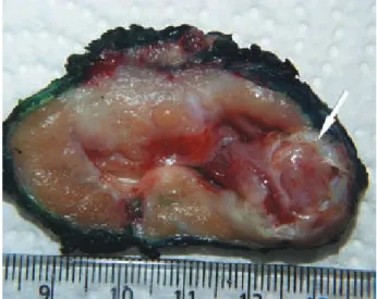
obstructing the prostatic urethra was seen and resected. Histopathological evaluation of the 6-mL resected material revealed mesenchymal malignant cells including rhabdomyoblasts in the subepithelial region of all resected specimens. These mesenchymal cells showed positive staining for desmin and myoglobin. There was no evidence of local or distant invasion of the tumor on thoracic and abdominal computed tomography. Bilateral pelvic lymph node dissection and radical prostatectomy were performed. The tumor was measured 2.8 × 2.2 × 1.5 cm and was located between the right peripheric and transitional zones (Figure 1). Surgical margins
Figure 1. Macroscopic view of prostate rhabdomyosarcoma
(arrow). The tumor is extended through the prostatic urethra. The capsule is intact and the surgical margin is negative for tumoral tissue.
Treatment of Adult Rhabdomyosarcoma—Sevinc et al
204 Urology Journal Vol 5 No 3 Summer 2008
were negative. Histopathological evaluation of the radical prostatectomy specimen confirmed the diagnosis of embryonal rhabdomyosarcoma (Figures 2 and 3). In addition to the sarcomatous tissue, a 3-mm tumor was detected which was prostate adenocarcinoma with a Gleason score of 6 (3 + 3).
The recovery was uneventful and no adjuvant therapy was instituted. Under the surveillance with magnetic resonance imaging and regular measurement of serum PSA, no evidence of recurrence has been found during the 54-month follow-up period.
DISCUSSION
There are limited reports of adult prostate
rhabdomyosarcoma in the literature. Rapidly progressing obstructive lower urinary tract symptoms are the major complaint as in our patient.(2) The age of onset may be less than what
is expected for benign prostatic hyperplasia or prostate carcinoma. Diagnosis of prostate rhabdomyosarcoma is challenging and is usually made by transrectal ultrasonography-guided biopsy or transurethral resection of the prostate.(1)
Digital rectal examination suggests no specific finding. Serum PSA may be within normal limits because of the nonepithelial origin of the disease. However, due to concomitant adenocarcinoma, PSA may be elevated, too, as in the present case. At the initial presentation, digital rectal examination and transrectal ultrasonography-guided biopsy revealed no evidence of malignancy in our patient. Definite diagnosis was made only after the second resection of the prostate performed because of the urinary retention. There is no pathognomonic radiological finding for prostate rhabdomyosarcoma. Extensive local invasion of the bladder neck and the trigone may be accompanied by some changes related to ureteral obstruction.(3) Ultrasonography and
computed tomography are not valuable for early detection of the disease. Magnetic resonance imaging has been the study of choice for most sarcomas, but may not be helpful for prostatic lesions.(4)
Histopathological evaluation of
rhabdomyosarcoma usually reveals variable differentiation along the myogenesis pathway and may appear as strap cells or myotubes that sometimes contain muscle
cross-striations. As what we observed in our case, rhabdomyosarcoma cells may demonstrate positive immunohistochemical results for muscle-specific markers such as myoglobin, actin, and desmin.(5)
Embryonal rhabdomyosarcoma, mainly its botryoides subtype, is the usual presentation of urogenital rhabdomyosarcoma in infants and toddlers. They respond well to radiation and chemotherapy.(6) In contrast, adults usually
present with nonembryonal subtypes which tend to be widely disseminated. The results are poor and despite good initial responses to
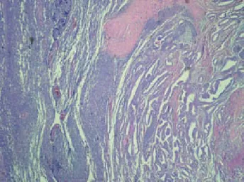
Figure 2. Microscopic evaluation of prostate rhabdomyosarcoma.
Tumoral tissue is seen on the right side, while normal prostatic ducts are on the left (hematoxylin-eosin, × 40).
Figure 3. Cytoplasmic staining with desmin antibody in tumoral
Treatment of Adult Rhabdomyosarcoma—Sevinc et al
Urology Journal Vol 5 No 3 Summer 2008 205
chemotherapy, they eventually die of their disease.(7) Localized presentation and favorable
prognosis of our case may be due to his histological subtype.
Although it is difficult to compare data from different series because of varying documentation and very small number of patients with prostate rhabdomyosarcoma, the overall survival
for adults is discouraging. The largest group of patients reported recently by Dotan and colleagues included 131 patients with urogenital sarcoma. Within this group, 21 of the tumors were originated from prostate and only 9 (6.8%) were rhabdomyosarcomas. This small subgroup of patients had the worst survival rates. At initial presentation, 9% of the prostate sarcomas were smaller than 5 cm, 10% were low grade, and 38% were free of metastasis. However, only 24% of them were free of disease at long-term follow-up.(8)
Treatment of prostatic rhabdomyosarcoma is multidisciplinary. The main aim of surgery for rhabdomyosarcoma is to completely remove the tumor with negative surgical margins while preserving all functional tissue. If feasible, it should be performed even in case of documented metastasis. However, definitive surgery is usually delayed until chemotherapy and/or radiotherapy has caused the shrinkage of large tumors that are not initially resectable.(9) Radical prostatectomy
alone may be an alternative only when the sarcoma is small and confined to the prostate. Quinlan and colleagues reported a prostate sarcoma treated with radical prostatectomy, hemicystectomy, and ureteroureterostomy.(10)
They instituted no adjuvant treatment and their patient was healthy on the 6th year of follow-up. In another study, Sakura and coworkers reported a 19-year-old man treated with radical prostatectomy without any additional therapy. On follow-up, he developed bilateral metastases to the obturator lymph nodes that led to
chemotherapy and pelvic radiation.(11) To our
knowledge, our patient is the only one so far treated alone with radical prostatectomy with an uneventful long-term follow-up.
CONFLICT OF INTEREST
None declared.
REFERENCES
1. Sexton WJ, Lance RE, Reyes AO, Pisters PW, Tu SM, Pisters LL. Adult prostate sarcoma: the M. D. Anderson Cancer Center Experience. J Urol. 2001;166:521-5.
2. Cheville JC, Dundore PA, Nascimento AG, et al. Leiomyosarcoma of the prostate. Report of 23 cases. Cancer. 1995;76:1422-7.
3. King DG, Finney RP. Embryonal rhabdomyosarcoma of the prostate. J Urol. 1977;117:88-90.
4. Nabi G, Dinda AK, Dogra PN. Primary embryonal rhabdomyosarcoma of prostate in adults: diagnosis and management. Int Urol Nephrol. 2002;34:531-4. 5. Pappo AS, Shapiro DN, Crist WM.
Rhabdomyosarcoma. Biology and treatment. Pediatr Clin North Am. 1997;44:953-72.
6. Crist WM, Garnsey L, Beltangady MS, et al. Prognosis in children with rhabdomyosarcoma: a report of the intergroup rhabdomyosarcoma studies I and II. Intergroup Rhabdomyosarcoma Committee. J Clin Oncol. 1990;8:443-52.
7. Russo P. Urologic sarcoma in adults. Memorial Sloan-Kettering Cancer Center experience based on a prospective database between 1982 and 1989. Urol Clin North Am. 1991;18:581-8.
8. Dotan ZA, Tal R, Golijanin D, et al. Adult genitourinary sarcoma: the 25-year Memorial Sloan-Kettering experience. J Urol. 2006;176:2033-8.
9. Ferrer FA, Isakoff M, Koyle MA. Bladder/prostate rhabdomyosarcoma: past, present and future. J Urol. 2006;176:1283-91.
10. Quinlan DM, Stutzman RE, Peters CA, Walsh PC. Unilateral nerve-sparing radical prostatectomy and hemicystectomy in management of prostate sarcoma. Urology. 1993;41:308-10.
11. Sakura M, Tsukamoto T, Yonese J, Ishikawa Y, Aoki N, Fukui I. Successful therapy of a malignant phyllodes tumor of the prostate after postoperative local failure. Urology. 2006;67:845.e11-3.
Case Report
206 Urology Journal Vol 5 No 3 Summer 2008
Spermatocele Presenting as Acute Scrotum
Atsuya Hikosaka, Yutaka Iwase
Urol J. 2008;5:206-8. www.uj.unrc.ir Keywords: spermatocele,
epididymis, acute scrotum, torsion abnormality
Department of Urology, Toyota Kosei Hospital, Toyota, Japan Corresponding Author: Atsuya Hikosaka, MD Department of Urology, Toyota Kosei Hospital, 500-1, Ibobara, Josui-cho, Toyota, Aichi 470-0396, Japan Tel: +81 565 43 5000 Fax: +81 565 43 5100 E-mail: [email protected]
INTRODUCTION
Spermatocele, a retention cyst of the scrotum which is or has been in communication with the semen-carrying system,(1) is a
relatively common clinical entity. It presents typically as an intrascrotal paratesticular mass, but it usually has few subjective symptoms. Here, we report a case of spermatocele manifested with acute scrotum due to its unique feature.
CASE REPORT
A 25-year-old man presented to our hospital complaining of persistent left scrotal pain with a sudden onset 12 hours earlier. He had no history of scrotal injury or vasectomy. Physical examination noticed a thumb-head-sized soft subcutaneous ovoid mass with severe tenderness above the left
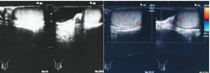
testicle, but bilateral testes were normally palpable. Laboratory findings were unremarkable. Doppler ultrasonography showed a simple cystic mass adjacent to the upper pole of the left testis and normal appearance of the both testes. However, slight decrease of blood flow in the left testis was suggested (Figure 1). Due to these confusing findings and perpetual pain, immediate surgical exploration was performed, which revealed a cystic lesion with a short stalk arising from the head of the left epididymis (Figure 2). The cyst was filled with yellowish turbid fluid and was twisted about 180 degrees. Histologically, the inner surface of the cyst wall was lined with columnar epithelial cells with cilia (Figure 3, Left). On cytological examination, the fluid in the cyst included spermatozoa (Figure 3,
Figure 1. Left, Scrotal ultrasonography demonstrates a cystic lesion adjacent to the left testicle. Right, Doppler ultrasonography