Original Article
Effects of melatonin and 5-methoxytryptophol on
synovial inflammation in the zymosan-induced
rheumatoid arthritis in rats
Gokce Savtekin1, Mustafa Senol Tuzum1, Lokman Onur Uyanik1, Aysa Ayali1, Ayliz Velioğlu Öğünç2, Şule Çetinel3, Ertuğrul Kılıç4, Ahmet Özer Şehirli5,6
1Near East University, Faculty of Dentistry, Department of Oral and Maxillofacial Surgery, Mersin 10, Turkey; 2Marmara University, Vocational School of Health Related Professions, Turkey; 3Marmara University, School of Medicine, Department of Histology & Embryology, Turkey; 4Medipol University, School of Medicine, Department of Physiology, Turkey; 5Marmara University, Faculty of Pharmacy, Department of Pharmacology, Turkey; 6Near East University, Faculty of Dentistry, Turkey
Received December 30, 2015; Accepted March 24, 2016; Epub April 15, 2016; Published April 30, 2016
Abstract: The aim of this study is the investigation of synoviocytes, cytokines and metalloproteinases in the syno-vial inflammation model which is created by zymosan application to temporomandibular joint (TMJ) and effects of pineal hormones melatonin (MEL) and 5-methoxytryptophol (5-MTX) on these parameters. 200-250 g Wistar albino rats of both sexes were used for modeling arthritis in this study. Arthritis model was created by intraarticularly (i.a.) injecting 2 mg zymosan in 40 ml saline into the left temporamandibular joint of the rats while the sham group was created by only injecting 40 ml saline solution (intraarticulary). MEL and 5-MTX administration was made intraperi-toneal before zymosan injection. 6 hours after zymosan or saline administration of MEL and 5-MTX the synovial fluid was collected from the animals and the synovial membrane was collected for histological assessment. The administration of zymosan increased the release of IL-1β and TNFα and the activity of metalloproteinases (MMM-9) and metalloproteinases-2 (MMP-2) and with this administration values got closer to the sham group. The histologi-cal evaluation showed significant increase in the intensity of synoviocytes that arose in the inflammation was found to subside. In conclusion, MEL and 5-MTX reducing inflammation in arthritis suggests these agents might constitute a new therapeutic principle clinically.
Keywords: Zymosan, synovial membrane, melatonin, 5-methoxytriptophol
Introduction
Temporomandibular joint (TMJ) disorder is a condition that results in a high degree of pain in speech, eating and other daily activities [1]. The most common of these disorders which is rheu-matoid arthritis (RA) is musculo-skeletal dis-comfort which affects a large portion of the population and can be seen at any age [2, 3]. Rheumatoid arthritis (RA) is a chronic inflam-matory autoimmune disease and the condition is characterized by particularly inflammatory cell infiltration of the synovial membrane [4]. Excessive release of inflammatory mediators that are produced by inflammatory cells, con-tributes to joint destruction. Mediators such as tumor necrosis factor-alpha (TNF-α) and inter-lokin1-beta (IL-1β) are associated with the pa-
thogenesis of arthritis and causes an increase in the secretion of proteolytic enzymes such as matrix synovial metalopreoteinase-2 (MMP-2) and metalopreoteinase-9 (MMP-9) in stromal cells [5, 6]. These enzymes degrade specific peptide bonds of the extracellular matrix pro-teins and help expression of inflammatory cells. Zymosan which we used in our study to create the arthritis is a polysaccharide that is capable of severe and erosive synovitis that can be pro-duced from yeast cell walls. The intraarticular injection of this agent has been shown to cause inflammation by enhancing the activation of cytokines such as TNF-α and IL-1β and proteo-lytic enzymes [7].
Melatonin (N-acetyl-5-methoxytryptamine) (M- EL), the disintegration product of serotonin,
mainly released from the pineal gland especial-ly in the dark, plays a role in many physiological processes such as circadian rhythm, immune function and sexual behavior [8]. In earlier stud-ies antioxidant and anti-inflammatory effects of these agents has been shown [9, 10]. It creates anti-inflammatory effect by inhibiting the adhe-sion molecules stimulated by NF-κβ, reducing the migration of leukocytes from endothelial cells and thus preventing the PMN leukocytes from aggregating in the inflamed area [11, 12]. Consequently, it is stated that MEL is reported to inhibit tissue damage by inhibiting the gen-eration of proteolytic enzymes and cytokines that are caused by the release of both direct and indirect free radicals during inflammatory events [13].
5-Methoxytryptophol (5-MTX), another pineal indole created by the decomposition of sero-tonin, plays a role in the regulation of many bio-logical activities such as pro or anti-gonado-tropic effects of biological rhythms and their reproductive behavior [14]. Features demon-strate an adverse effect on the light dark cycle with MEL. Since the secretion of MEL is high in the dark, while 5 MTX is secreted in light [15]. Despite a lack of studies with 5-MTX, antioxi-dant and immunomodulatory effects were ob- served [16, 17]. Studies in recent years focused on the evaluation of superiority or differences of the effects of MEL and 5-MTX [18, 19]. The recent study, assess the effects of these two pineal indoles on bone marrow formation re- vealed 5-MTX has more protective effects with its antioxidant and anti-inflammatory effects compared with MEL [20].
In this study we aimed to compare the effects of MEL and 5-MTX and to examine whether there is a synergistic effect of the combined used of MEL and 5-MTX on synovial inflamma-tion model that created with zymosan appli- cation.
Materials and methods
200-250 g Wistar albino rats of both sexes were used. Animals were acclimated in the lab-oratory conditions two weeks prior to the exper-iment (20°C ± 2, 12 h light/12 hours dark). [This study has been done with the approval of Medipol University Animal Ethics Committee (Ref No. 38328770-46)].
Groups and experimental protocol
Our study comprised of 5 groups with 8 rats per group. The synovial inflammation rat model was set up as previously described [1].
Sham group: The rats in this group were
admin-istered saline intraarticularly (i.a.) to the TMJ under ketamine anesthesia (100 mg/kg, 40 ml).
Synovial inflammation (SI) group: 2 mg
zymo-san 40 ml was dissolved in saline and adminis-tered intraarticularly (i.a.) into the TMJ.
SI + melatonin group: 10 mg/kg
intraperitone-ally MEL injection 15 minutes before zymosan administration.
SI + 5-methoxytryptophol group: 30 minutes
before injection of zymosan (i.a.), 5 mg/kg 5-methoxytryptophol intraperitoneally (i.p.) was injected.
SI + 5-methoxytryptophol + melatonin group: 5
mg/kg 5-MTX intraperitoneally 30 minutes before zymosan and 10 mg/kg melatonin intra-peritoneally (i.p.) 15 minutes before zymosan were injected.
Synovial Fluid Collection, done on anesthetized rats by zymosan injection (i.a). 0.05 ml of EDTA in neutral buffered PBS used to wash TMJ cav-ity. Washing done twice with pumping and aspi-rating technique and then synovial fluid was collected and the rats were decapitated under anesthesia after 6 hours [1]. In synovial fluid, tumor necrosis factor-alpha (TNF-α), interleukin 1-beta (IL-1β) levels and matrix metalloprotein-ase (MMP-2 and MMP-9) activities were mea-sured [21].
Histological studies
Light microscopy: Synovial membrane was ex-
cised after sacrificing animals. Then specimens were fixed in 10% neutral buffered formalin for 24 h, followed by demineralization in 10% ED- TA, embedded in paraffin, and 5-6 μm section- ed along the synovial membrane. Synovial me- mbrane sections were evaluated under light microscopy at 400×. For the specimens pro-cessed for routine hematoxylin-eosin (H&E) staining, histological analysis considered to the synoviocytes influx in the synovial membrane [1].
Examinations of Synovial Fluid
TNF-α, IL-1β, MMP-2, MMP-9 analyses: In
syno-vial fluid the analysis and measurements of TNF-α (ELISA, BioSource Europe S.A. Catalog No. KRC 3014 Nivelles, Belgium) IL-1β (ELISA BioSource Catalog No. KRC0011, Nivelles, Belgium) MMP-2 (ELISA, Catalog No. Abnova KAO393, Taoyuan, Taiwan), MMP-9 (ELISA, Ca- talog No. Abnova KAO398, Taoyuan, Taiwan) were done considering appropriate criteria according to the instructions.
Statistical methods
One-way analysis of variance (ANOVA) based on statistical evaluation and as advanced analysis Tukey’s test was performed. Where P values were less than 0.05, were considered signifi- cant.
Results
In SI group, TNF-α, IL-1β levels belonging to the synovial fluid were significantly higher than the control group (Table 1). In contrast to SI group increase in TNF-α, IL-1β levels in melatonin (MEL), 5-methoxytryptophol (5-MTX) and MEL+ 5-MTX administrated groups was significantly lower. In only MEL and only 5-MTX administered groups, TNF-α and IL-1β levels remained higher than the control group.
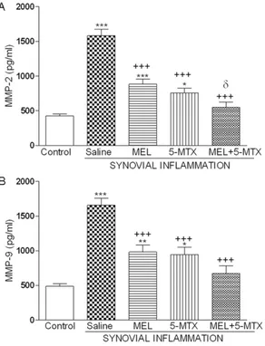
Upon the assessment of the synovial fluid’s MMP-2 and MMP-9 activities, SI administered group was found to be significantly higher com-pared to the control group. In contrast, melato-nin (MEL), 5-methoxytryptophol (5-MTX) admin-istered groups has significantly dropped and was closer to control group values. Between the combined application and single applica-tions, only MEL administered group showed sig-nificant reduction in their MMP-2 levels and other parameters did not show any variance (Figure 1).
The histological examination of the tissue made synovial tissue synoviocytes in sham group,
Table 1. Tumor necrosis factor-α (TNF-α), interleukin-1β (IL-1β) values of all groups of synovial fluid in synovial inflammation (SI) model on rats created by zymosan
SHAM SI SI-MEL SI-5-MTX SI-MEL + 5-MTX TNF-α (pg/ml) 152 ± 11 254 ± 17*** 179 ± 15+ 171 ± 11++ 150 ± 11++
IL-1β (pg/ml) 51.8 ± 9.9 136.8 ± 14.8*** 68.2 ± 9.1++ 70.3 ± 11.5++ 55.6 ± 9.6+++
***P<0.001 comparisons according to the control group, +P<0.05, ++P<0.01, +++P<0.001 comparisons according to SI group.
Figure 1. Activities of matrix metalopreoteinases (MMP-2, MMP-9) belonging to the synovial fluids of all groups in the (SI) model which was created by sy-novial inflammation with zymosan administration on rats. MEL: Melatonin, 5-MTX: 5-methoxytryptophol.
**P<0.01, ***P<0.001 according to the control group; +P<0.05, ++P<0.01, +++P<0.001 comparisons
accord-ing to SI group; P<0.05 comparisons accordaccord-ing to MEL group.
Figure 2. Sham group: The synoviocytes (arrowheads) observed with the spread morphology in the synovial membrane, synovia (*). Image magnification X400.
worldwide. In particular, it does not have a per-fect cure despite many studies done on the subject and many applications administered in the treatment of synovial inflammation due to temporomandibular joint diseases [22].
Different mechanisms are suggested to explain temporomandibular joint disorders character-ized by pain, tenderness and limitation of the jaw movement. Many mediators such as pro-teolytic enzymes, synoviocytes, monocytes/ma- crophages, platelets, complement and coagu-lation systems, cytokines are thought to be involved in the formation of injury [23]. These mediators are collected mainly in the synovial area and cause structural damage on the membrane.
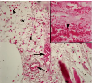
Figure 3. Saline administered synovial inflammation (SI) group: Intense congestion of the capillaries (ar-rows) and synoviocytes (arrowheads), intense syno-via (*). Image magnification X400.
Figure 4. SI + 5-methoxytryptophol (5-MTX) group: Capillary congestion and synoviocytes intensity (ar-rowheads) moderate regression. Image magnifica-tion X400.
exhibits a smooth appearance (Figure 2) while SI group synoviocytes showed damage charac-terized by an increase in synoviocytes density and capillary congestion (Figure 3). SI + 5-MTX group showed the congestion decreased while cell density remain decreased (Figure 4). In SI + MEL group decreased congestion and a sig- nificant decrease in cell density was observed (Figure 5). SI + MEL + MTX group showed appar- ent reduction in density of synoviocytes (Figure 6).
Discussion
Inflammation associated with arthritis is a seri-ous health problem that affects many people
Figure 5. SI + melatonin (MEL) group: Capillary con-gestion and fairly significant regression in synovio-cytes intensity (arrowheads), synovial cell density (*). Image magnification X400.
Figure 6. SI + 5-MTX + MEL: Synovium (*) signifi-cantly reduced synoviocytes intensity (arrowheads). Image magnification X400.
In our study, zymosan which is a polysaccha-ride synthesized from yeast cell walls has been shown to promote the formation of the synovi-tis by causing an increase in cell migration in the vascular permeability with mononuclear cell infiltration [24-26]. Especially in zymosan inflammation models 0.25, 0.5, 1 or 2 mg doses tried and the effects in different times were evaluated [1]. Both literature findings and our preliminary study shows the 2 mg intervals zymosan injection causes intense inflamma-tion after 6 hours.
Cytokines, secreted by immune cells, play a major role in hematopoiesis, cell division and differentiation control, tissue repair, and inflam-mation. Synovial inflammation leads to release of the pro-inflammatory mediators such as TNF-α, IL-1β from synovial cells and an increase of the biosynthesis of adhesion molecules that mediate leukocyte-endothelial cell adhesion [27]. In a recent study, it is stated that synovio-cytes infiltration in the synovial fluid TNF-α and IL-1β levels are increased [28]. A clinic study of 31 arthritis patients suggests increased cyto-kines, TNF-α and IL-1β over inflammation by activating the collagen synthesis in fibroblast type 1 by activating metalloproteases [29]. In accordance with these literatures our study showed in zymosan caused inflammation also there was an increase in cytokines, TNF-α and IL-1β in synovial fluid. As is known, increase in pro-inflammatory cytokines indicate an inflam-matory response by the organism in which inflammation is important starting causing tis-sue damage [30]. The positive effects of thera-peutic agents on tissue parameters of our study given the levels of cytokines in synovial fluid of these agents in the synovial inflamma-tion is significantly suppressed by supporting the important role of these cytokines. There- fore, the agents used in synovial inflammation treatment having inhibitory properties on cyto-kines expression will increase the success of treatment. The present study showed that MEL prevents the inflammation caused by IL-1β on mesenchymal stem cells and contributes to bone regeneration [23]. In the arthritis model in which the antioxidant and anti-inflammat- ory effect of MEL is studied has shown that these agents decrease levels of TNF-α and IL- 1β [12].
The study has shown that antioxidant effects of 5-MTX which is another pineal indole, IL-6 is
reduced which is a pro-inflammatory cytokines and increased IL-2 levels in the serum which is an inflammatory cytokines with an immune-modulator effect despite the studies about inflammations show a lack [31]. However, there aren’t enough studies in the literature on the effects of 5-MTX on TNF-α and IL-1β. When all these studies are considered our findings con-form to the literature, showing that MEL and 5-MTX’s inhibitory effects on pro-inflammatory cytokines helps the tissue protective effects. Matrix metalloproteinases (MMPs) are a family of zinc-dependent metalloendopeptidases, co- nsists of 5 subgroups namely; collagenase, st- romelysin, gelatinase, membrane-type, and ot- hers and be expressed in epithelial, mesenchy-mal, and haematopoietic cells [32]. In experi-mental and clinical studies, inflammation and gelatinase subfamily consists of connective ti- ssue diseases in MMP-2 (gelatinase A) and MMP-9 (gelatinase B) shows the important role played [33]. The present study showed the increased activation of this proteolytic enzyme in experimental inflammation both created with zymosan and macrophage migration inhibitory factor to investigate the activity of MMP-2 [34]. On the other hand, the study suggest that the expression of MMP-9 increased in synovio-cytes, macrophages and master cells in the experimental peritonitis model created with zymosan [35]. Therefore, these MMPs show activity by destroying the extracellular matrix proteins by breaking down specific peptide bonds in various cell and tissues and play a role in pathological conditions such as rheumatoid arthritis, tumor invasion, apoptosis and angio-genesis [36]. Our results from the study are consistent with the literature. Zymosan applica-tion resulted in synovial fluid MMP-2 and MMP-9 activation increasing significantly com-pared with the control group and with MEL and 5-MTX administration improved significantly. MEL’s effects on expressions of MMP-2 and MMP-9 has been studied in, several models of inflammation such as spinal cord injury [37], colitis [38], cerebral ischemia [39]. Antioxidant and anti-inflammatory properties of MEL in these models was shown that the protective effect is created by reducing the expression of MMP-9 and MMP-2. However, effects of both MEL and 5-MTX’s on MMP-2 and MMP-9 activa-tion has not been studied in any arthritis model. But so far effects of 5-MTX on proteolytic
enzymes has not been studied before, it is a first with our research.
In conclusion: In our experimental study, pro-inflammatory cytokines and proteolytic enzy- mes increased and also that change observed in biochemical parameters accompanied by increase in synoviocytes and so the advert of tissue damage was observed. Pineal hormones melatonin and 5-methoxytryptophol, which we used for treatment has shown to significantly reduce this damage. To our knowledge this is the first study investigating the anti-inflamma-tion effects of two pineal hormones in synovial inflammation and these two pineal hormones are normally responsible for the regulation of the circadian rhythm, effects are considered; 5-MTX that is released in brightness and MEL that is released in darkness. On the other hand this study being the first to research the effects of 5-MTX on cytokines levels and proteolytic enzymes is of importance in terms of contribut-ing to the literature. Despite the fact more extensive and comparative clinical and experi-mental studies are needed for these agents to be used in the clinics our results suggest that MEL and 5-MTX’s plays an important role on synovial inflammation repair thus we believe that further studies will shed light on later. Acknowledgements
This research supported by the Near East Uni- versity Centre of Excellence Research Foun- dation (www.neu.edu.tr) with the project num-ber CE021-2015.
Disclosure of conflict of interest None.
Address correspondence to: Dr. Gokce Savtekin, De- partment of Oral and Maxillofacial Surgery, Faculty of Dentistry, Near East University, Nicosia-Cyprus, Mersin 10, Turkey. Tel: 00 90 533 8494997; Fax: 00 90 392 6802050; E-mail: [email protected]
References
[1] Chaves HV, Ribeiro Rde A, de Souza AM, Ro-drigues e Silva AA, Gomes AS, Vale ML, Bezer-ra MM, Brito GA. Experimental Model of Zymo-san-Induced Arthritis in the Rat Temporom- andibular Joint: Role of Nitric Oxide and Neu-trophils. Journal of Biomedicine and Biotech-nology. J Biomed Biotechnol 2011; 2011: 707985.
[2] Carlos FP, de Paula Alves da Silva M, de Lemos Vasconcelos Silva Melo E, Costa MS, Zamuner SR. Protective effect of low-level laser therapy (LLLT) on acute zymosan-induced arthritis. La-sers Med Sci 2014; 29: 757-763.
[3] Nonose N, Pereira JA, Machado PR, Rodrigues MR, Sato DT, Martinez CA. Oral administration of curcumin (Curcuma longa) can attenuate the neutrophil inflammatory response in zymo-san-induced arthritis in rats. Acta Cir Bras 2014; 29: 727-34.
[4] Ji J, Dou H, Li X, Song Y, Li X, Li E, Tan R and Hou Y. Novel benzenediamine derivative FC99 ameliorates zymosan-induced arthritis by in-hibiting RORgt expression and Th17 cell differ-entiation. Acta Biochim Biophys Sin 2014; 46: 829-836.
[5] Ed H. Rheumatoid arthritis: pathophysiology and implications for therapy. N Engl J Med 1990; 322: 1277-1289.
[6] Nielsen RH, Christiansen C, Stolina M, Karsdal MA. Oestrogen exhibits type II collagen protec-tive effects and attenuates collagen-induced arthritis in rats. Clin Exp Imun 2008; 152: 21-27.
[7] da Conceição Rivanor RL, Chaves HV, do Val DR, de Freitas AR, Lemos JC, Rodrigues JA, Pereira KM, de Araújo IW, Bezerra MM, Benev-ides NM. A lectin from the green seaweed Caulerpa cupressoides reduces mechanical hyper-nociception and inflammation in the rat temporomandibular joint during zymosan-in-duced arthritis. Int Immunopharmacol 2014; 21: 34-43.
[8] Rudra DS, Pal U, Maiti NC, Reiter RJ and Swarnakar S. Melatonin inhibits matrix metal-loproteinase-9 activity by binding to its active site. J Pineal Res 2013; 54: 398-405.
[9] Şehirli AÖ, Koyun D, Tetik Ş, Özsavcı D, Yiğiner Ö, Çetinel Ş, Tok OE, Kaya Z, Akkiprik M, Kılıç E, Şener G. Melatonin protects against ischemic heart failure in rats. J Pineal Res 2013; 55: 138-48.
[10] Borges Lda S, Dermargos A, da Silva Junior EP, Weimann E, Lambertucci RH and Hatanaka E. Melatonin decreases muscular oxidative stress and inflammation induced by strenuous exercise and stimulates growth factor synthe-sis. J Pineal Res 2015; 58: 166-72.
[11] Reiter RJ, Calvo JR, Karbownık M, Qi W and Tan DX. Melatonin and its relation to the immune system and inflammation. Ann N Y Acad Sci 2000; 917: 376-86.
[12] Forrest CM, Mackay GM, Stoy N, Stone TW and Darlington LG. Inflammatory status and kyn-urenine metabolism in rheumatoid arthritis treated with melatonin. Br J Clin Pharmacol 2007; 64: 517-526.
[13] Cuzzocrea S and Reiter RJ. Pharmacological action of melatonin in shock, inflammation and ischemia/reperfusion injury. Eur J Phar-macol 2001; 426: 1-10.
[14] Ouzir M, Bouhaddou N, Khalki H and Lakhdar-Ghazal N. Physiological and pharmacological properties of 5-methoxytryptophol. Expert Rev Endocrinol Metab 2013; 8: 355-364.
[15] Lewczuk B, Ziółkowska N, Prusik M and Przyb-ylska-Gornowicz B. Diurnal profiles of melato-nin synthesis-related ındoles, catecholamines and their metabolites in the duck pineal organ. Int J Mol Sci 2014; 15: 12604-12630. [16] Lissoni P, Pittalis S, Rovelli F, Zecchini S, Casati
M, Tremolada M and Pelizzoni F. Immunomod-ulatory properties of a pineal indole hormone other than melatonin, the 5-methoxytrypto-phol. J Biol Regul Homeost Agents 1996; 10: 27-30.
[17] Rodriguez-Naranjo MI, Moyá ML, Cantos-Villar E and Garcia-Parrilla MC. Comparative evalua-tion of the antioxidant activity of melatonin and related indoles. J Food Comp Anal 2012; 28: 16-22.
[18] Wang H and Ng TB. Hypotensive activity of the pineal indoleamine hormones melatonin, 5- methoxytryptophol and 5-methoxytryptamine. Pharmacol Toxicol 2000; 86: 125-8.
[19] Ng TB, Liu F and Zhao L. Antioxidative and free scavenging activities of pineal indoles. J Neu-ral Transm 2000; 107: 1243-1251.
[20] Satué M, Ramis JM, Arriero MM and Monjo M. A new role for 5-methoxytryptophol on bone cells function in vitro. J Cell Biochem 2015; 116: 551-8.
[21] Holwegner C, Reinhardt AL, Schmid MJ, Marx DB and Reinhardte RA. Impact of local steroid or statin treatment of experimental temporo-mandibular joint arthritis on bone growth in young rats. Am J Orthod Dentofacial Orthop 2015; 147: 80-8.
[22] Wang XD, Zhang JN, Gan YH and Zhou YH. Cur-rent understanding of pathogenesis and treat-ment of tmj osteoarthritis. J Dent Res 2015; 94: 666-73.
[23] Liu X, Gong Y, Xiong K, Ye Y, Xiong Y, Zhuang Z, Luo Y, Jiang Q and He F. Melatonin mediates protective effects on inflammatory response induced by interleukin-1 beta in human mes-enchymal stem cells. J Pineal Res 2013; 55: 14-25.
[24] Keystone EC, Schorlemmer HU, Pope C and Al-lison AC. Zymosan-induced arthritis: a model of chronic proliferative arthritis following acti-vation of the alternative pathway of comple-ment. Arthritis Rheum 1977; 20: 1396-1401. [25] Gegout P, Gillet P, Chevrier D, Guingamp C, Ter-lain B and Netter P. Characterization of zymo-san-induced arthritis in the rat: effects on joint
inflammation and cartilage metabolism. Life Sci 1994; 55: 321-6.
[26] Rocha FA, Aragão AG Jr, Oliveira RC, Pompeu MM, Vale MR, Ribeiro RA. Periarthritis pro-motes gait disturbance in zymosan-induced arthritis in rats. Inflamm Res 1999; 48: 485-90.
[27] Wahl SM, McCartney-Francis N, Chan J, Di-onne R, Ta L and Orenstein JM. Nitric oxide in experimental joint ınflammation. Cells Tissues Organs 2003; 174: 26-33.
[28] Belenska-Todorova L, Gyurkovska V and Iva-novska N. How complement activation influ-ences the development of chronic synovitis in a mouse model of rheumatoid arthritis. Scand J Rheumatol 2015; 1-10.
[29] Suzuki T, Segami N, Nishimura M and Nojima T. Co-expression of interleukin-1β and tumor necrosis factor a in synovial tissues and syno-vial fluids of temporomandibular joint with in-ternal derangement: comparision with histo-logical grading of synovial inflammation. J Oral Pathol Med 2002; 549-57.
[30] Güven O, Tekin U, Salmanoğlu B and Kaymak E. Tumor necrosis factor-alpha levels in the sy-novial fluid of patients with temporomandibu-lar joint internal derangement. J Craniomaxil-lofac Surg 2015; 43: 102-5.
[31] Lissoni P, Messina G and Rovelli F. Cancer as the main aging factor for humans: the funda-mental role of 5-methoxy-tryptamine in rever-sal of cancer-induced aging processes in meta-bolic and immune reactions by non-melatonin pineal hormones. Curr Aging Sci 2012; 5: 231-5.
[32] Xue M, March L, Sambrook PN and Jackson CJ. Differential regulation of matrix metallopro-teinase 2 and matrix metalloprometallopro-teinase 9 by activated protein C: relevance to inflammation in rheumatoid arthritis. Arthritis Rheum 2007; 56: 2864-2874.
[33] Clegg PD, Burket RM, Coughlans AR, Rlggs CM and Carter SD. Characterisation of equine ma-trix metalloproteinase 2 and 9; and identifica-tion of the cellular sources of these enzymes in joints. Equine Vet J 1997; 29: 335-342. [34] Pakozdi A, Amin MA, Haas CS, Martinez RJ,
Haines GK, Santos LL, Morand EF, David JR and Koch AE. Macrophage migration inhibitory factor: a mediator of matrix metalloprotein-ase-2 production in rheumatoid arthritis. Ar-thritis Res Ther 2006; 8: R132.
[35] Kolaczkowska E, Grzybek W, Rooijen N, Picca-rd H, Plytycz B, Arnold B and Opdenakker G. Neutrophil elastase activity compensates for a genetic lack of matrix metalloproteinase-9 (MMP-9) in leukocyte infiltration in a model of experimental peritonitis. J Leukoc Biol 2009; 85: 374-381.
7144 Int J Clin Exp Med 2016;9(4):7137-7144
[36] Cunnane G, FitzGerald O, Hummel KM, Youssef PP, Gay RE, Gay S and Bresnihan B. Synovial tissue protease gene expression and joint ero-sions in early rheumatoid arthritis. Arthritis Rheum 2001; 44: 1744-1753.
[37] Esposito E, Genovese T, Caminiti R, Bramanti P, Meli R and Cuzzocrea S. Melatonin regulates matrix metalloproteinases after traumatic ex-perimental spinal cord injury. J Pineal Res 2008; 45: 149-156.
[38] Esposito E, Mazzon E, Riccardi L, Caminiti R, Meli R and Cuzzocrea S. Matrix metalloprotein-ase-9 and metalloproteinase-2 activity and pression is reduced by melatonin during ex-perimental colitis. J Pineal Res 2008; 45: 166-173.
[39] Kim SJ and Lee SR. Protective effect of melato-nin against transient global cerebral ischemia-induced neuronal cell damage via inhibition of matrix metalloproteinase-9. Life Sci 2014; 94: 8-16.