Introduction
Cutaneous leishmaniasis (CL) is a zoonotic disease caused by Leishmania species, spread by vector sandflies (Phlebotomus spp) with an obligatory intracellular protozoon andwhich leaves a scar on the skin [1]. The occurrence of CL disease and the healing pro-cess are determined by the host immune
system, the virulence of the parasite and the characteristics of the vector [2,3]. In addition, the continuity of anthroponotic focus especi-ally in unplanned urbanisation with no in-frastructure has been found to contribute to causing a positive effect as a global risk factor for CL disease [4]. Recently, an increase in the number of cases of CL disease in Turkey
The Sociodemographic, Living and Environmental
Characteristics of Patients with Cutaneous Leishmaniasis
Mustafa Aksoy,1MD, Yavuz Yeşilova,2*MD, Hacer Altun Sürücü,1MD, Nurettin Ardıç,3MD, Abdullah Yeşilova,4MDAddress:1Faculty of Medicine, Harran University College School, Department of Dermatology, Sanliurfa, 2Special
Lokman Physician Van Hospital, Dermatology Clinic, Van, 3Gulhane Military Medical Academy, Department of
Microbiology, Ankara, 4Yuzuncu Yıl University College School, Department of Biostatistics, Van, Turkey E-mail: [email protected]
*Corresponding Author: Dr.Yavuz Yeşilova, Special Lokman Physician Hospital Department of Dermatology,Van
Published: J Turk Acad Dermatol 2017; 11 (1): 17111a1.
This article is available from: http://www.jtad.org/2017/1/jtad17111a1.pdf
Keywords: Cutaneous leishmaniasis, phlebotomus, socio-economic living
Abstract
Background: Cutaneous leishmaniasis is a parasitic skin disease caused by various leishmaniasis species. Material and Methods: In this study, the clinical, sociodemographic, living and environmental characteristics of 4048 cutaneous leshmaniasis patients are presented. A retrospective evaluation was made of 4048 cutaneous leishmaniasis patients in terms of gender, lesion diameter (mm), number of lesions, duration of lesions (weeks) and living conditions, then statistical analysis was applied.
Results: The cutaneous leismaniasis patients comprised 52.47% female and 47.53% males with a mean age of 16.48±0.23 years. The mean duration of the disease was 9.62±0.33 weeks, lesion diameter was 12.197±0.10 mm and mean number of lesions was 1.75±0.02. The number of people in the patient’s family was generally 5-10 and the number of rooms in the house was 2-3 for 70.63% of patients. The vast majority of the cutaneous leishmaniasis patients, 96.96%, lived in a lowland plains area and no patient lived in wetlands. The types of houses were of concrete construction in 94.94% and stone in 1.61% and 1.41% lived in an apartment block. Animals were reported to be kept in the living area of 17.02% of patients and of those, 2.30% had a WC.
Conclusion: The determination of an excessive number of Phlebotomus in areas of low socio-economic living conditions and the high incidence of cases in these areas has revealed a direct, positive relationship between cutaneous leishmaniasis disease and the socio-economic conditions of the patients.
has been determined [5,6]. According to the Ministry of Health, approximately 45% of the cases of CL in Turkey in the last 20 years, in the province of Şanliurfa in particular, have been seen to originate in areas of highly un-planned residences without sufficient infras-tructure [7].
Two types of sandfly species (Phlebotomus spp) are responsible for the spread of CL di-sease; the vector sandfly Lutzomyia for New World CL seen on the American continent and the vector sandfly Phlebotomus (Phlebo-tomus papatasi and Phlebo(Phlebo-tomus sergenti) for Old World CL seen in the Mediterranean region [8]. While Phlebotomus sergenti is ac-cepted as the vector for Leishmania tropica, Phlebotomus papatasi is accepted as the vec-tor for Leishmania major [9,10,11]. The agent often causing CL disease seen in the province of Şanliurfa has been shown to be Leishma-nia tropica [8,10,12,13,14,15]. The aim of this study was to make a retrospective exa-mination of the clinical and socio-demograp-hic characteristics and living environments of 4048 CL patients recorded in Turkey at Şan-liurfa Public Health Centre and Harran Uni-versity Medical Faculty Dermatology Clinic.
Material and Methods
A retrospective evaluation was made of the records of 4048 CL patients registered at Şanliurfa Public Health Centre and Harran University Medical Faculty Dermatology Cli-nic between 1998 and 2004. Approval for the study was granted by the Local Ethics Com-mittee. An examination was made of the age
(years) of the CL patients, gender, lesion dia-meter (mm), number of lesions, duration of lesions (months) and living conditions. The data were statistically evaluated.
Diagnosis of CL disease was made from clini-cal appearance of the patient, and positive demonstration of Leishmania with laboratory tests (parasites-amastigotes-in skin smears). When there were difficulties in diagnosis, la-boratory tests such as polymerase chain re-action and immune fluorescent antibody techniques were of benefit in the evaluation of the histopathological results.
Statistical evaluation
In the statistical evaluation of the data, the SAS 9.12 statistical software program was used. For examination of the variables in the study, firstly descriptive statistical calculati-ons were made. Then, to establish differences between the characteristics of the groups such as age, gender, duration of the disease, lesion type, localization and diameter, the proportions test was used. A value of p<0.05 was accepted as statistically significant.
Results
General characteristics of the CL patients
The study group comprised a total of 4048 pati-ents; 2124 (52.47%) females and 1924 (47.53%) males. The mean age of the patients was 16.48±0.23 years. The mean duration of the di-sease was 9.62±0.33 weeks (range, 1-500 months). Mean lesion diameter was found to be 12.197±0.10 mm (range, 1-80mm) and the mean
Table 1. General Features of Patients with Cutaneous Leishmaniasis Patients
General features Frequency
Age (years)(n=4048) 16.48±0.23 Female 2124(52.47%) Male 1924(47.53%) Duration (months) (n=3996) 9.62±0.33 Size (mm) (n=4048) 12.197±0.10 Number (n=4000) 1.75±0.02
number of lesions was 1.75±0.02 (range, 1-10) (Table 1).
The number of rooms in the houses where CL patients lived and the number of family members
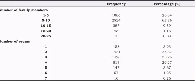
The number of rooms in the house where CL pati-ents lived and the number of people living there are shown in (Table 2.) The number of family members of CL patients ranged from 1 to 25 and generally (62.36%) the number of people living to-gether in the family was 5-10. It was noticeable that 1-5 children in the families of the patients was at a low rate (26.84%) in CL patients. In respect of the number of rooms in the house, 70.63% of the patients lived in houses with 2 or 3 rooms.
Living environments of the CL patients
The vast majority of the CL patients lived in flat plains areas. A very low rate (0.82%) lived in mo-untainous areas and only 0.05% lived in forested areas. No patient lived in wetlands or surrounding areas (Table 3). The houses where the CL patients lived were concrete type houses in 94.94% of cases, stone houses in 1.61% and flats in apart-ment blocks in 1.41%. Only 3.06% of the houses lived in by CL patients were made of mud bricks. None of the patients lived in wooden houses or tents (Table 3). This section should include a con-cise textual description of the data presented in tables and/or figures. The Results and Discussion may be combined if by doing so, space is saved or the logical sequence of the manuscript is impro-ved. Excessive repetition of table or figure contents shouldbe avoided.
Discussion
Old World CL shows a wide geographic distribution, being seen in many countries, primarily Afghanistan, Algeria, Iran and Syria [16]. CL disease is generally seen during childhood and the prevalence decreases with increasing age [17]. The number of lesions in CL disease is usually limited to 1 or 2 [18]. and the duration of the disease has be en reported as mean 12.71 months [19]. Clinically, the disease starts with a spot a few millimetres in diameter and continues with papulonodular or ulcerative lesions growing over time. These lesions later heal leaving permanent scar tissue when not treated [18,20]. The Phlebotomus which play a role in the spread of the disease are found during the day in places such as piles of stones, cracks in walls, tree hollows and animal shelters. At dusk they emerge from these hiding places and the female Phlebotomus takes in amastigotes when sucking blood from sick humans or other mammals. In the intestines of the Phlebotomus, the ingested amastigotes become promastigotes, rapidly reproduce and mature. While sucking blood at the next feeding of the Phlebotomus, these parasites in promastigote form spread the disease by innoculating non-infected skin. The parasites entering the host macrophages return to amastigote form and reproduce within the cells, which results in the death of the host macrophage cells and new cells are infected by dissemination into the
Table 2. The Number of Family Members and the Number of Rooms in the Home of Cutaneous Leishmaniasis Pati-ents
Frequency Percentage (%) Number of family members
1-5 1086 26.84 5-10 2524 62.36 10-15 387 9.59 15-20 48 1.13 20-25 3 0.08 Number of rooms 1 158 3.93 2 1431 35.37 3 1426 35.25 4 819 20.27 5 147 3.67 6 57 1.25 7 10 0.26
surroundings. In terms of infection, the time of highest risk when sandflies are most active together with warming of the air is from dusk to dawn [21, 22, 23, 24, 25]. In the summer months when the density of Phlebotomus increases, in open areas known as tents by people in villages, people sleep uncovered without mosquito nets, so it is inevitable that fly bites in uncovered areas such as the head and upper extremities will result in CL disease. In the sampling of homes on the outskirts of Şanliurfa, reasons such as the general condition of the house, there being a garden, the house not having plastered walls, keeping animals in part of the house, there being a barn, keeping pigeons, the toilet being outside the house and poor hygiene conditions have been determined to make a positive contribution to the sandfly population [26]. Especially when the temperature of animal shelters is higher than the surroundings, the spread and population density of these species can be
affected negatively. This can be explained by the placing of light traps in outdoor open areas in July which catch a relatively higher number of adult flies [7,26].
In the current study, there was plaster on the walls of 96.4% of the houses lived in by CL patients. The toilets used by the household were outside the house for the majority (97.70%) of the CL patients and most of the houses were single-storey concrete houses (94.94%). In 17.02% of the places lived in by the CL patients, there were animal shelters. The rates of those living in apartment blocks (1.41%) or in houses with stone walls (1.61%) were very low. None of the patients were living in tents. The results of the current study are in contrast with those of Toprak et al as a high proportion of the houses lived in by the CL patients had plastered walls and the rate of animal shelters in the living accommodation was very low. However, the finding that a high proportion of the houses where the CL patients
Living conditions of CL patients Frequency Percentage (%)
Plastered walls in the house (n=4048) No 3924 96.94
Yes 124 306 Mountainous area (n=4048) No 4015 99.18 Wetlands (n=4048) Yes 33 0.82 No 123 3.04 Plain/lowlands (n=4048) Yes 3925 96.96 No 4048 10.000 Forest (n=4048) No 4046 99.95 Yes 2 0.05 Tent (n=4048) No 4048 100.00 Wooden house (n=4048) No 4048 100.00 No 3991 98.59
Flat in apartment block (n=4048) Yes 57 1.41
No 205 5.06
Concrete house (n=4048) Yes 3843 94.94
No 3983 98.39
Stone house (n=4048) Yes 65 1.61
No 3359 82.98
Animals in the house (n=4048) Yes 689 17.02
No 93 2.30
Outside WC (n=4048) Yes 3955 97.70
lived had an outside toilet supports the findings of Toprak et al.
Phlebotomus are generally found in flat lowland areas [27]. This species has been seen to adapt well to drought and is not found at high altitude due to the drop in temperature [28]. In the current study, the vast majority of the CL patients lived in the lowland areas (96.96%). The rate of those living in mountainous (0.82%) or forested (0.05%) areas was very low and none of the CL patients lived in areas such as wetlands. This finding that generally the patients lived in lowlands and very few lived in mountainous areas supports the data in literature.
As a much greater number of Phlebotomus were collected in neighbourhoods with a low socio-economic living standard, and at the same time most CL patients comprised those who lived in these types of houses in these areas, a direct positive relationship has been shown between disease-sandfly density and socio-economic structure [7,26]. Another result of the current study was that CL disease is related to the number of family members and the number of rooms in the accommodation. The number of family members of the patients in the current study ranged from 1 to 25 but generally (59.2% of the CL patients) they were living in familes of 5-8 individuals. The living areas of the CL patients generally (70.63%) had 2-3 rooms. The high number of children and low number of rooms in the home of the CL patients in this study can be accepted as an indicator of low socio-economic level.
Socio-economic, political and environmental factors have a decisive role in the epidemiology of CL disease in the province of Şanliurfa. In CL disease it has been determined that animals were kept in houses where people were living and the lavatories used by the household were generally in outside areas. At the same time, especially in the outlying areas of Şanliurfa where CL patients were more concentrated, residental areas had developed rapidly and in an unplanned way without infrastructure such as sewerage systems, thus creating an
unsuitable living environment but one which is suitable for Phlebotomus.
In the struggle against CL disease, besides effective medical treatment for patients, the implementation of environmental rehabilitation of the slum areas of cities where patients are concentrated to improve the socio-economic levels of the patients and the application of insecticides effective against Phlebotomus, will be able to make a significant reduction in the number of patients. In conclusion, CL dsease should not be considered as a purely medical disease. Especially in countries where the disease is seen at high rates, evaluation is required by a committee comprised of dermatologists, parasitologists, biologists and environmental health specialists.
References
1. Turhanoglu M, Alp Erdal S, Bayindir Bilman F. A nine-year evaluation of cutaneous leishmaniasis pa-tients in Diyarbakir Training and Research Hospital, Turkey. Mikrobiyol Bul 2014; 48: 335-340 PMID: 24819271
2. Klaus SN, Frankenburg S, Ingber A. Epidemiology of cutaneous leishmaniasis. Clin Dermatol 1999; 17: 257-260. PMID: 10384863
3. Salman SM, Rubeiz NG, Kibbi A. Cutaneous leishma-niasis: clinical features and diagnosis. Clin Dermatol 1999; 17: 291-296. PMID: 10384868
4. WHO. Urbanization: an increasing risk factor for le-ishmaniasis. Wkly Epidemiol Rec 2002; 77: 365-370. PMID: 12428426
5. Rodriguez-Barraquer I, Gongora R, Prager M, et al. Etiologic agent of an epidemic of cutaneous leishma-niasis in Tolima, Colombia. Am J Trop Med Hyg 2008; 78: 276-282. PMID: 18256429
6. Campbell-Lendrum D, Dujardin JP, Martinez E, et al. Domestic and peridomestic transmission of American cutaneous leishmaniasis: changing epidemiological patterns present new control opportunities. Mem Inst Oswaldo Cruz 2001; 96: 159-162. PMID: 11285490 7. Gürel MS, Yeşilova Y, Olgen MK, Özbel Y. Cutaneous
leishmaniasis in Turkey. Turkiye Parazitol Derg 2012; 36: 121-129. PMID: 22801920
8. Killick-Kendrick R, Tang Y, Killick-Kendrick et al. Phlebotomine of Kenya III. Ann Trop Med Parasitol 1994; 88: 183-196. PMID: 8067814
9. Anis E, Leventhal A, Elkana Y, Wilamowski A, Pener H. Cutaneous leishmaniasis in Israel in the era of changing environment. Public Health Rev 2001; 29: 37-47 PMID: 11780715.
10. Ozbel Y, Turgay N, Ozensoy S, et al. Epidemiology, di-agnosis and control of leishmaniasis in the Mediter-ranean region. Ann Trop Med Parasitol 1995; 89: 89-93. PMID: 8745931
11. Guilvard E, Rioux JA, Gallego M, et al. Leishmania tropica in Morocco. III--The vector of Phlebotomus
sergenti. Apropos of 89 isolates. Ann Parasitol Hum Comp 1991; 66: 96-99. PMID: 1776784
12. Alptekin D, Kasap M, Lüleyap U, , Kasap lH , Aksoy S , Wilson ML. Sand flies (Diptera: Pyschodidae) as-sociated with epidemic cutaneous leishmaniasis in sanlıurfa, Turkey. J Med Entomol 1999; 36 277-281 PMID: 10337097
13. Pazarbasi A, Alptekin D, Luleyap HU, Kasap M, Kasap H. Use of enzyme-linked immunosorbent assay for detec-tion of natural leishmania infections in phle-botomine sand flies from southeastern Turkey. J Med Entomol 2006; 43: 248-251. PMID: 16619606 14. Svobodová M, Alten B, Zídková L, et al. Cutaneous
le-ishmaniasis caused by Leishmania in-fantum trans-mitted by Phlebotomus tobbi. Int J Parasitol 2009; 39: 251-256. PMID: 18761342
15. Toprak S, Ozer N. Sand fly species of Sanlıurfa pro-vince in Turkey. Med Vet Entomol 2005; 19: 107-110. PMID: 15752185
16. Zeyrek FY, Gürses G, Uluca N, et al. Is the agent of cutaneous leishmaniasis in Sanliurfa changing? First cases of Leishmania major. Turkiye Parazitol Derg 2014; 38: 270-274 PMID: 25732888
17. Alvar J, Velez ID, Bern C. et al. Leishmaniasis world-wide and global estimates of its incidence. PLoS One 2012; 7: 35671. PMID: 22693548
18. Gurel MS, Ulukanligil M, Ozbilge H. Cutaneous leish-maniasis in Sanliurfa: epidemiologic and clinical fea-tures of the last four years (1997-2000). Int J Dermatol 2002; 41: 32-37. PMID: 11895511 19. Abdellatif MZ, El-Mabrouk K, Ewis AA. An
epidemio-logical study of cutaneous leishmaniasis in Al-jabal Al-gharbi, Libya. Kore J Parasitol 2013; 51: 75-84 PMID: 23467624
20. Zaraa I, Ishak F, Kort R, et all. Childhood and adult cutaneous leishmaniasis in Tunisia. Int J Dermatol 2010; 49: 790-793. PMID: 20618499
21. Balasegaram M, Ritmeijer K, Lima MA, et al. Liposo-mal amphotericin B as a treatment for human leish-maniasis. Expert Opin Emerg Drugs 2012; 17: 493-510. PMID: 23167833
22. Murray HW, Berman JD, Davies CR, Saravia NG. Av-dances in leishmaniasis. Lancet 2005; 366: 1561-1577. PMID: 16257344
23. Kafetzis DA. An overview of pediatric leishmaniasis. J Postgrad Med 2003; 49: 31-38. PMID: 12865569 24. Markle WH, Makhoul K. Cutaneous leishmaniasis:
Recognition and treatment. Am Fam Physician 2004; 15: 1455-1460. PMID: 15053410
25. Samady JA, Schwartz RA. Old World cutaneousleish-maniasis. Int J Dermatol 1997; 36: 161-166. PMID: 9158994
26. Desjeux P. Leishmaniasis. Public health aspects and control. Clin Dermatol 1996; 14: 417-423. PMID: 8889319
27. Toprak S, Ozer N. Distribution of sand fly (Diptera: Psychodidae) species and efficiency of capturing met-hods in Sanliurfa province, Turkey. J Med Entomol 2007; 44: 23-28. PMID: 17294917
28. Fryauff D, Hanafi H. Demonstration of hybridization between Phlebotomus papatasi (Scopoli) and Phlebo-tomus bergeroti (Parrot). Parassitologia 1991; 33: 237-243. PMID: 1841213