T.C.
BIRUNI UNIVERSITY
INSTITUTE OF GRADUATE EDUCATION
DEPARTMENT OF MOLECULAR BIOLOGY AND GENETICS MOLECULAR AND MEDICAL GENETICS GRADUATE PROGRAM
KLK GENE EDITING USING CRISPR TECHNOLOGY IN
PROSTATE CANCER CELL LINES FOR CANCER THERAPY
Nasim KHERAD
ADVISOR
Asst. Prof. Dr. Meryem ALAGÖZ
T.C.
BIRUNI UNIVERSITY
INSTITUTE OF GRADUATE EDUCATION
DEPARTMENT OF MOLECULAR BIOLOGY AND GENETICS MOLECULAR AND MEDICAL GENETICS GRADUATE PROGRAM
KLK GENE EDITING USING CRISPR TECHNOLOGY IN
PROSTATE CANCER CELL LINES FOR CANCER THERAPY
Nasim KHERAD
ADVISOR
Asst. Prof. Dr. Meryem ALAGÖZ
iii
DECLARATION
I declare that I have designed and performed all experiments in the current study entitled “Klk Gene Editing Using Crispr Technology In Prostate Cancer Cell Lines For Cancer Therapy” according to good scientific practices. I obtained all the information contained in this thesis under academic and ethical rules. All the information I have used from secondary literature has been respectively referenced. I also declare that, I have not violated any patents and copyrights during the preparation and writing of this thesis.
iv
ACKNOWLEDGEMENTS
I wish to express my sincere gratitude to my advisor Assistant Professor Dr. Meryem Alagoz As, for her great help on my master’s thesis.
Also, I thank lab assistant Ezgi Guner for her help in experiments and technical support, and Assistant Professor Dr. Meryem Alagoz for samples for the experiments. I also thank to my supportive mother, my dearest father, my kind brother, and my encouraging friends for their positive motivation.
v
TABLE OF CONTENTS
Page
THESIS APPROVAL PAGE ...
DECLARATION ... iii
ACKNOWLEDGEMENTS ... iv
TABLE OF CONTENTS ... v
ABBREVIATIONS ... ix
LIST OF TABLES ... xi
LIST OF FIGURES ... xii
TURKISH ABSTRACT and KEYWORDS ... xiii
ABSTRACT and KEYWORDS ... xiv
1. INTRODUCTION AND PURPOSE ... 1
2. GENERAL INFORMATION ... 3
2.1. Epidemiology of Prostate Cancer... 4
2.1.1. microRNAs in Prostate Cancer ... 5
2.1.2. KLKs in Prostate Cancer ... 6
2.2. External Factors ... 7
2.3. CRISPR ... 8
3. MATERIALS and METHOD ... 11
3.1. Competent Cell Preparation ... 11
3.1.1. Materials ... 11
3.1.2. Method ... 11
3.2. Bacterial Transformation ... 11
3.3. Plasmid DNA Extraction (MiniPrep) ... 12
3.3.1. Material ... 12
3.3.2. Method ... 13
3.4. Gel Electrophoresis ... 14
3.4.1. Material ... 14
3.4.2. Method ... 14
3.5. Cell culture: Growing Cells ... 15
3.6. Cell Passaging (Subculturing) ... 15
vi
3.6.2. Method ... 16
3.7. Preparation of Cell Pellet ... 16
3.7.1. Material ... 16
3.7.2. Method ... 16
3.8. Cryopreservation of Cell Culture ... 17
3.8.1. Materials ... 17
3.8.2. Method ... 17
3.9. Thawing Frozen Cell Lines ... 17
3.9.1. Material ... 17
3.9.2. Method ... 18
3.10. Genomic DNA Purification from Mammalian Cells ... 18
3.10.1. Material ... 18
3.10.2. Method ... 18
3.11. Primer Design For Pcr and Sanger Sequencing ... 19
3.12. PCR Amplification ... 19
3.12.1 Material ... 19
3.12.2. Method ... 19
3.13. Sanger Sequencing Workflow... 20
3.14. PCR clean-up ... 21 3.14.1. Material ... 21 3.14.2. Method ... 21 3.15. Cycle Sequencing ... 22 3.15.1 Material ... 22 3.15.2. Method ... 22
3.16. Purification of sequencing products ... 24
3.16.1. Material ... 24
3.16.2. Method ... 24
3.17. Capillary Electrophoresis and Data Analysis ... 25
3.18. Computational Analysis of Protein Structure and Severity of Damage Caused by Mutation ... 25
3.19. Construction of the CRISPR system ... 25
3.20. Mammalian cell Transfection with DNAfectin Reagent ... 26
3.21.1. Material ... 26
vii
3.23. Antibiotic Selection ... 28
3.23.1. Material ... 28
3.23.2. Method ... 28
3.24. Drug Resistance Assay ... 29
3.25. Cell Counting ... 29
3.25.1. Material ... 29
3.25.2. Method ... 29
3.26. Cell Viability Assay ... 30
3.26.1. Material ... 30
3.26.2. Method ... 31
3.27. MTT Assay ... 33
3.27.1. Material ... 33
3.27.2. Method ... 33
3.28. Colony Formation Assay... 35
3.28.1. Material ... 35 3.28.2. Method ... 35 4. RESULTS ... 38 4.1. Bacterial Transformation ... 38 4.2. Gel Electrophoresis ... 39 4.3. CRISPR Construct ... 41
4.4. Sanger Sequencing for KLK5 Mutation Analysis; ... 42
4.5. Computational Analysis of Protein Structure and Severity of Damage Caused by Mutation ... 45
4.5.1. KLK5 protein domain structure ... 45
4.5.2. 3D protein structure conformation ... 46
4.6. Antibiotic Selection ... 46
4.7. Cytotoxicity Assay ... 48
4.7.1. Cell viability Assay Using Trypan Blue ... 48
4.7.2. MTT Analysis ... 50
4.7.3. Colony Formation Assay (CFA) ... 52
5. DISCUSSION, CONCLUSION AND RECOMMENDATION ... 57
5.1. Conclusion ... 62
6. REFERENCES ... 64
viii 8. PLAGIARISM REPORT ... 75
ix
ABBREVIATIONS
Abbreviation/Symbols Definition/Full form
PC Prostate Cancer
KLK Kallikrein
CRISPR/Cas9 Clustered Regularly Interspaced Short
Palindromic Repeats/Cas9
MTT
3-(4,5-Dimethylthiazol-2-yl)-2,5-diphenyltetrazolium bromidefor
PARP Poly (ADP-ribose) polymerase
TA transit amplifying
CB committed basal
PAP prostatic acid phosphate
BPH benign prostatic hyperplasia
RNASEL ribonuclease L
BRCA breast cancer susceptibility gene
AR androgen receptor
E.coli Escherichia coli
LB Lucia-Bertani
SOB super optimal broth
MgCl2 Magnesium chloride
CaCl2 Calcium chloride
DNA Deoxyribonucleic Acid
SOC Super Optimal broth
MgSO4 Magnesium sulphate
EDTA Ethylenediaminetetraacetic acid
TBE Tris-borate-EDTA
UV ultraviolet
EMEM Eagle's minimal essential medium
FBS Fetal Bovine Serum
DMSO Dimethyl sulfoxide
CO2 Carbon dioxide
x
GC% guanine-cytosine content
PCR Polymerase chain reaction
ddH2O Double distilled water
Dntp deoxyribonucleotide triphosphate
SAP shrimp alkaline phosphatase
gRNA Guide Ribonucleic Acid
PAM protospacer adjacent motif
sgRNA Short Guide Ribonucleic Acid
CFA Colony formation assay
Kb Kilobyte
DSB Double strand break
NHEJ Non-homologous end-joining
οC Degree of Celcius
MW Molecular weight
OD Optical density
µG Microgram
ug/umole Microgram per micromole
uM or µM Micromolar
3D Three dimension
xi
LIST OF TABLES
Table No Name Of Table Page
Table 3.1 : Cell Culture Conditions ... 15
Table 3.2. PCR Amplification Components ... 20
Table 3.3: PCR Conditions ... 20
Table 3.4. Components of Cycle Sequencing in Sanger Sequencing ... 22
Table 3.5. Cycle Sequencing Condition ... 23
Table 3.6. Cycle Sequencing Reaction Components ... 23
Table 3.7. Cycle Sequencing Incubation Condition... 24
Table 3.8. Components for Sequencing Products’ Purification ... 25
Table 3.9. Reagent Quantities for Different Culture Vessels in Mammalian Cell Transfection... 27
Table 3.10. Preparation of Drug Solution ... 31
Table 3.11. Cell Numbers and Single Drug Concentration ... 32
Table 3.12. Cell Numbers and Single Drug Concentration ... 32
Table 3.13. MTT Assay Doxorubicin Concentration Per Well ... 34
Table 3.14. MTT Assay Doxorubicin and PARP Inhibitor Concentration Per Well . 34 Table 3.15. Cell Number and Single Drug Concentration in CFA ... 36
Table 3.16. Cell Number and Double Drug Concentration in CFA... 37
xii
LIST OF FIGURES
Figure No Name Of Figure Page
Figure 4.1. Gel Electrophoresis of Competent Cells... 40
Figure 4.2. Antibiotic Resistance Screening on Plasmids... 41
Figure 4.3. Lentiviral Vector Map ... 42
Figure 4.4. Schematic Representation of Sanger Sequencing Analysis for KLK5 Gene ... 44
Figure 4.5. Schematic Representation of KLK5 Protein Domain. The figure was taken from ensembl.org ... 45
Figure 4.6. Schematic Representation of 3D Protein Structure of KLK5 ... 46
Figure 4.7. Polyphen2 analysis KLK5 Amino Acid Substitution ... 46
Figure 4.8 . Agarose Gel Electrophoresis of Transfected DU145 Cell Line and Wild DU145 Cell Line ... 47
Figure 4.9. Schematic Representation of Cell Viability Percentage in PC3 Cell ... 48
Figure 4.10. Schematic Representation of Cell Viability Percentage in DU145 Wild Cell Lines ... 49
Figure 4.11. Schematic Representation of Cell Viability Percentage in Knockout ... 49
Figure 4.12. Schematic Representation of MTT Analysis in PC3 Cell Lines ... 50
Figure 4.13. Schematic Representation of MTT Analysis in Wild Type DU145 ... 51
Figure 4.14. Schematic Representation of MTT Analysis in Knockout DU145 ... 51
Figure 4.15. CFA for PC3 at Different Single and Double Drug Concentration. ... 52
Figure 4.16. Schematic Representation of CFA Effect on PC3 Tumorgenesis ... 53
Figure 4.17. Colony Formation Assay for Wild Type DU145 ... 54
Figure 4.18. Schematic Representation of CFA Effect on Wild Type DU145 Tumorgenesis ... 55
Figure 4.19. CFA for Knockout DU145 ... 55
Figure 4.20. Schematic Representation of CFA Effect on Wild Type DU145 Tumorgenesis ... 56
xiii
TURKISH ABSTRACT and KEYWORDS
Kherad, N. (2020). Kanser terapisi için Klk Gen Düzenlenmesini Crispr Teknolojisi Kullanilarak Prostat Kanseri Hücre Hatlarinda Yapilmasi. Yüksek Lisans Tezi, Biruni Üniversitesi Lisansüstü Eğitim Enstitüsü, İstanbul.
Erkeklerde, kansere bağlı ölümlerde prostat kanseri ikinci sırada yer almaktadır. Genetik mutasyon, etnik köken, yaş, diyet vb. gibi iç ve dış faktörlerin, kanserin ortaya çıkmasında ki nedensel faktörlerden olduğu gözlemlenmiştir. Kallikrein (KLK) gen ailesi tarafından kodlanan serin proteazlar PC kanseri üzerinde dikkate değer etkiler gösterirler ve KLK3,KLK2 gibi bazı çeşitleri PC varlığında androjene bağlı kanda tespit edildiği için biyo marker olarak tanınmaktadır. Bununla birlikte, androjen bağımlılığı ve düşük özgüllük nedeniyle, tam olarak güvenilir değillerdir. Bu çalışmada, mutasyonun PC de tümör oluşumuna etkisini gözlemlemek için androjenden bağımsız PC3 ve DU145 PC hücre hatlarında KLK5 gen mutasyonuna odaklandık. Gen düzenleme teknolojisi, uzun vadede bilim adamlarının en az yan etki ile kanser tedavisi araştırmalarına faydalı olabilecek yeni bir yol sağladı.Bu çalışmada KLK geninde tespit edilen ve Sanger dizilemeyle doğrulanan mutasyon c.245C>A (p.A82D) CRISPR/Cas9 teknolojisi kullanılarak nakavt edilmiştir.Bu çalışmada, gelecekte kanser tedavisinde CRISPR sisteminin umut veren sonuçlarını göstemek için, yapılan gen düzenlemesi Doxorubicin ve PARP inhibitörü kullanılarak tripan mavisi ile hücre canlılığı, MTT testi ve koloni oluşumu gibi çeşitli sitotoksisite deneyleri ile değerlendirilmiştir.
xiv
ABSTRACT and KEYWORDS
Kherad, N. (2020). Klk Gene Editing Using Crispr Technology In Prostate Cancer Cell Lines For Cancer Therapy. Master Thesis, Biruni University, Institute of Graduate Education, İstanbul.
Prostate cancer (PC) has been accounted for as the second leading cause of death by cancer in men. Internal and external factors, such as genetic mutation, ethnicity, age, diet, etc., are observed to have been the causative elements for cancer to occur. Serine proteases, that are encoded by Kallikrein (KLK) gene family, show a remarkable impact on PC and some are introduced as PC biomarkers, such as KLK3 and KLK2, as both androgen-dependent are detected in the bloodstream when PC occurs. However, due to androgen dependence and low specificity, they are not quite reliable. In this study, we focused on KLK5 gene mutation in different PC androgen-independent cell lines, PC3, and DU145 cell lines to observe the effect of mutation of PC tumorigenesis. To monitor the effect of the mutation, the KLK5 gene’s detected mutation point was knocked out using a genome-editing tool. Gene-editing technology has opened a new path to the scientists’ investigation for designing the method that can benefit cancer treatment with the lowest side effects in the long term. In this study, CRISPR/Cas9 gene-editing technology was used to knock out c.245C>A (p.A82D) mutation in the KLK gene which was detected and confirmed by Sanger sequencing. To evaluate the performed gene editing, various cytotoxicity assays, such as cell viability with trypan blue, MTT assay, and colony formation was carried using Doxorubicin and PARP inhibitor which showed a promising result of CRISPR system as a future prostate cancer treatment.
1
1. INTRODUCTION AND PURPOSE
Prostate cancer (PC) has been posing a huge threat to men because of heterogeneous factors and carcinogenic chemicals (N.J. Maitland 2013). The First report of Prostate cancer occurrence belongs to ancient times. However, the first treatment method that developed more than a century, is the development of prostatectomy, a surgical procedure with which prostate glands are removed (Capasso LL. 2005). PC has been reported as a slow and fast life-threatening tumour. Therefore, scientists focused on finding a better solution to detect PC at early stages to avoid the surgical approach.
Prostate-specific antigen (PSA) blood test has been commonly used to determine the manifestation of PC by the presence of kallikrein-related serine proteases, which are found in prostate secretion and the case of prostate glands’ functional abnormality, in the bloodstream. And therefore, the test facilitates early stages of prostate cancer’s diagnosis (Lilja H. Eet.al. 2008). However PSA dropdown in a mild and acute case of prostatitis and other prostate-related problems makes the antigen unspecific and difficult to consider as a PC biomarker (Guess HA. et.al. 1993).
Human serine proteases are members of human tissue kallikrein (KLK) family with 15 homologues serine proteases (KLK1-KLK15) that are encoded by the largest clustered human genome located on chromosome 19 (Borgoño CA.et.al. 2004). KLK2, KLK4, KLK5, KLK14, and KLK15 have shown to be the key element in KLK3 activation whose function includes digestion of seminogelins I and II, and fibronectin (Michael IP et.al. 2006, Emami N et.al. 2008)
Over the past few decades, various therapeutic methods have been developed to reduce the mortality rate of PC; drug treatment such as bicalutamide (Gravina GL et.al 2010), application short interfering RNA (siRNA) with chemotherapy (Izquierdo M. 2005), transfecting LNCaP and PC3 cell lines with RNA-aptamer (Marangoni K et.al. 2015), and many other methods has been used. Nevertheless, drawbacks, toxicity, cost, and PC recurrence due to short-term effects were observed.
2 Genome editing technologies are considered to be the most efficient tools to assist therapeutic approaches. Many studies are being carried out to design new methods that can improve gene editing function, their effectiveness, with minimum off-target effects. Clustered regularly interspaced palindromic repeats associated protein Cas9 (CRISPR/CAS9) provide has shaken up the genome editing in disease treatment, including cancers. The great capability of CRISPR/CAS9 in the treatment of broad ranges of rare and neurodegenerative diseases has been demonstrated. Use of CRISPR-Cas9 editing system is performed in ex-vivo models of somatic and pluripotent stem cells as wells as in-vivo animal models that can shed light on developing an approach that can be used in gene alteration and correction.
Before the discovery of gene editing tools, prostate cancer therapy complications such as drug resistance eluded physicians and scientists as they could not identify the causes of drug effects. The CRISPR-Cas9 ability of insertion and deletion of particular gene sequence on target DNA has given scientists and medical professions a remarkable solution in identifying drug-resistance genes by exposing drug-resistant cells to CRISPR/Cas9 gRNAs, which individually knockout single gene at the time in each cell (Scott, A. 2018). Therefore, the result of the experiment can improve drug therapy by introducing other more effective drugs for successful therapy (Scott, A. 2018).
This study aimed to introduce the effect of CRISPR knockout on the KLK5 gene in PC cell lines which is reported to be down-regulated in PC. Moreover, the experiments focused on monitoring the effect of KLK5 knockout on PC cells mutagenesis, proliferation, and viability rate under drug combination therapy which is hoped to be the positive approach for a better and less-aggressive therapy in a long term.
3
2. GENERAL INFORMATION
Prostate cancer (PC)is considered the second most common cancer among the male population and it has been posing a huge threat to men due to its high prevalence as 3.8% of death in men was caused by PC in 2018 (Ferlay J EM et.al 2019). Cancer begins in the prostate gland, located inferior to the bladder, which is a part of the male reproductive system and responsible for semen production in the male body (Robinson EJ et.al. 1998). Almost 98% of prostate cancers are reported to have been originated in glandular origin (Greenberg R. 2003). This organ is made of epithelial cell clusters which are arranged in the form of the basal layer and contains stem cells, transit-amplifying (TA) cells, and committed basal (CB) cells as the most dominant cell population in this structure (Robinson EJ et.al. 1998). Based on histological analysis of prostate organ, stem cells undergo division and form TA cells which are further divided by mitosis and differentiate into CB cells, the only cell type in prostate capable of DNA synthesis and proliferation, which are finally transformed into differentiating luminal cells in human (Greenberg R. 2003). Prostate cancer showed lack, deformation, and abnormal cell number of the basal layer in the prostate gland (M.M. Shen. et.al 2010).
Varieties of internal and external factors including family history, hormonal dysregulation, age, ethnicity, and infections (Patel, A.R.et.al 2009) has made the design of particular treatment difficult and ineffective. Age is of significance in occurrence and death by PC as the average age is calculated to be 66 at the time of diagnosis (Ferlay J EM et.al 2019). Ethnic difference is another element as the rate of PC in African-American men shows twice the rate of death caused by PC compared to white men (Ferlay J EM et.al 2019). Moreover, external factors such as social, environmental, and dietary factors are significantly effective in the cause of PC (Chan JM.et.al 2005, Willis MS.et.al 2003). Prostate cancer has existed since ancient times. However, the development of prostatectomy, a surgical mean for prostate cancer treatment, has been achieved for more than a century (Capasso LL. 2005). PC has been categorized as heterogeneous and it is observed as a slow-growing and fatal fast-growing form. Many studies have been researched investigating genes and genetic impairments involving in PC, which include the epidemiology of the disease while evaluating all the risk factors causing the disease.
4 2.1. Epidemiology of Prostate Cancer
Recent statistics show that the rate of PC is 7.1% of all cancers in males. However, the percentage correlates with the geographical location (Bray F.et.al 2018). The incident rate of PC elevates as the age of individuals is higher and it is 60%. Various methods were designed to identify the most convenient methods to diagnose PC at an early age. Stem cells characterization, collected by biopsy of patients, were carried out using CD44+ α2β1hi CD133+ markers as the phenotype which unfortunately was not specific for only cancer stem cells (CSCs) (G.D. Richardson.et.al 2004). Additionally, PAP (prostatic acid phosphate) was found to not be fully special for prostate cancer diagnosis since it was found to be elevated in benign prostatic hyperplasia (BPH) and other organ’s malfunctions such as liver (Demichelis F.et.al 2015). Years of studies and analysis have introduced a more convenient way for diagnosis of the disease. Prostate-specific antigen (PSA) blood test is the test performed to determine the presence of kallikrein-related serine protease, an enzyme which is normally found in prostate secretion, but found in the bloodstream due to functional and histological anomaly of the prostate, and helps to diagnose prostate cancer in early stages for further treatment (Lilja H.et.al 2008).
Genetic factors are considered to have approximately a 5% contribution to the occurrence of the disease (Ferris-i-Tortajada J.et.al. 2011, Sridhar G.et.al. 2010). Still, the percentage elevates when causative alleles are passed on to the individuals. Added to the genetic factors, the loci and their rate of susceptibility has a slight contribution to the cause of the PC. Linkage study of PC introduces 7 loci that are estimated to be the most sensitive loci whose distortion or damage can lead to PC as these loci harbor the genes that maintain the sustainability of immune system mechanism and cell cycle that can eliminate abnormally-grown cells (Malathi K.et.al. 2007, Eeles RA .et.al. 2008, Schlaberg R.et.al 2009). Cell-death programming and antiviral activity are regulated by the enzyme known as ribonuclease L (RNASEL) which is encoded by the HPC1 gene on chromosome 1q 24-25 (Chen H.et.al. 2003, Malathi K.et.al. 2007). Impairment of some genes has shown to be the causative factor in retrovirus activities that is likely to cause PC (Schlaberg R.et.al 2009). An aggressive and familial form of PC was demonstrated to be closely associated with BRCA1, BRCA2, and PLAB2 genes (Gallagher DJ.et.al 2010). Besides generic mutations, distortion of the X
5 chromosome in Xq26.3-q27.3 causes hereditary prostate cancer, as the X chromosome carries a gene that encodes androgen receptor (AR) (Xu J.et.al. 2000).
2.1.1. microRNAs in Prostate Cancer
microRNAs, also known as miRNAs, are small, non-coding endogenous RNA molecules that are found in eukaryotes as well as some viruses and function in RNA regulations and diagnosis of various diseases because of their circulation in body fluids (Wahid F.et.al. 2010). Recent studies on miRNA transcripts have demonstrated their significance in prostate cancer as biomarkers. MicroRNA-21, also known as miR-21, mammalian microRNA encoded by MIR21 gene located on chromosome 17, has been observed to suppress PTEN (tumor suppressor gene) in various cancers such as colorectal and breast cancer (Sheng WZ.et.al and Fragni M.et.al. 2016) but no indication of its direct effect on prostate cancer. Experiment on the effect of miR-21 in prostate cancer cells (PC3 cell lines) by transfecting them miR-21 to show its effect on prostate cancer cells. In the presence of matured miR-21, which is generally conserved in mammalian cells, prostate cancer cells evade immune defense due to suppression of PTEN by miR-21 and cause cell proliferation and metastasis (Yang Y.et.al. 2017). Therefore, miR-21 has been introduced as a prostate cancer biomarker, however, needs further studies are required to detect possible epigenetic influence on cancer cells.
Up-regulation of miR-21 led to the down-regulation of miR-15 and mirR-16 as well as overexpression of TGF-β signaling due to suppression of SMAD7 (Mothers against decapentaplegic homolog 7 leads to degradation of TGF-β and inhibits hedgehog signaling). which leads to AR hyper-activation and generation of the bone lesion in prostate cancer (Bonci D.et.al. 2016). Moreover, the impact of miR-221 and miR-222 on tumorgenesis and cancer progression in prostate cancer was seen (Yang, X.et.al. 2014). Furthermore, when PC3 cells are induced with miR-221 and miR-222 inhibitors in-vivo and in-vitro, their proliferation dropped down. This result indicates miR-221/222 increasing expression in prostate cancer and its metastasis (Yang, X.et.al. 2014). Other studies revealed elevation of other different miRNAs by examining prostate cancer patients against controls which could be advantageous in the diagnosis of prostate cancer.
6 Other miRNAs expressed a rather significant change in their level in response to tumorigenesis. To demonstrate, researchers analyzed miR-152-3p in LNCaP and PC3 in vitro compared to the bloodstream. The result indicated a low miR-152-3p level of tumour cells compared to healthy control cells. However, the impact of high-level miR-152-3p caused elevated proliferation and metastasis which shows presenting the external effect of miR-152-3p on tumorigenesis (Matin F.at.al 2018).
2.1.2. KLKs in Prostate Cancer
To this date, 15 members of tissue kallikrein (KLK1) and the kallikrein-related peptidases (KLK2–KLK15) with either trypsin or chymotrypsin-like activities have been identified and categorized (Lawrence MG.et.al. 2010, Emami N.et.al, 2007). KLK2 and KLK3 are found only in prostate and KLK3 (Prostate-specific antigen (PSA) is used as the biomarker of prostate cancer diagnosis (Borgoño CA.et.al. 2004, Shaw JL.et.al. 2007). The discovery of PSA dates back to 1979 when it was found in prostatic tissues (Wang MC.et.al. 1979). Apart from contributing to cancer diagnosis, KLK3 is the main modulator of sperm status in terms of semen liquefaction and coagulation (Lilja H.et.al. 1987). KLK3 also contributes to male infertility, as a degrading factor of semenogelin (Lilja H.et.al. 1987). Increased expression of KLK2, KLK3/PSA, and KLK4 in prostate cancer has been correlated with over-expression of KLK mediated proteolytic cascades that increases the rate of mutagenesis (Avgeris M.et.al. 2011, Herrala AM.et.al. 2001, Pollak M. 2008, Lawrence MG.at.al. 2010). Additionally, a study on DU145 cell lines shows their increased proliferation rate which is caused by KLK2- and KLK4 protease-activated receptors (PARs) (Mize GJ.et.al. 2008).
Moreover, the epithelial-mesenchymal transition (EMT) of prostate cancer cells have been observed to be strengthened by KLK3, KLK4, and KLK7 which could be a potential indicator of KLK3, KLK4, and KLK7 role in prostate cancer mutagenesis (Veveris-Lowe TL.et.al. 2005, Whitbread AK.et.al. 2006)
7 2.2. External Factors
Apart from genetic factors, external factors play a crucial role in PC occurrence. Many studies refer to the role of geographical location to be a significant element in the development of prostate cancer. Studies on Chinese and African immigrants in the USA showed a higher prevalence of PC compared to the ones living in their homeland, which could indicate the importance of lifestyle (Chu LW.et.al. 2011). Furthermore, dietary intake has shown to impart the prevalence rate of PC. Multiple studies found the effect of hight percentage of fat intake (meat, beaf, etc) to increase the rate of PC development and this could be due to effect of the saturated fat on insulin growth factor and higher proliferation rate, higher leukotrienes and prostaglandins, and increasing carcinogenicity of prostate cancer cells by androgen stimulations (Aronson WJ.et.al. 2010, Pauwels EK. 2011). The investigations on people of less fatty diets indicated that high-fat levels increase oxidative stress which can cause pre-oxidation leading to DNA damage (Lloyd JC.et.al. 2013). Another study shows that food and oils that are rich in omega 6 can increase proliferation by producing pro-inflammatory prostaglandins which enhances the malignancy of PC. On the other hand, a rich amount of Omega 3 can reduce the chance of the cancer cells' proliferation and eventually cause malignancy (Berquin IM.et.al. 2007). The amount of meat in a man's diet presents remarkable effect on the occurrence of PC as men with lower intake of meat shower lower rate of PC compared to the ones with higher intake of meat especially if the meat is cooked at great temperature (Major JM.et.al. 2011, Rohrmann S.et.al. 2007). This could be due to DNA damage which happens by the free radicals that are formed with lipid peroxidation caused by high temperature (Sinha R.et.al. 2009). The type of the consumed has shown to have a significant effect on the prevalence of PC. Consumption of calcium and calcium-rich products, such as dairy food increases the risk of PC (Wilson KM.et.al. 2015). Additionally, men with a rich diet of crucifers e.g. Broccoli have a low risk of PC over their lifespan which could be due to their antioxidants (Stram DO.et.al. 2006). Other things such as green tea, soy, and tomato have shown to decrease the chance of PC in males (103-119). The level of exposure to sunlight was shown to have a direct relationship with the risk of prostate cancer, as lack of vitamin D can lead to a higher risk of PC (Erdman JW.et.al. 2009). Further studies on race presented the effect of high melanin concentration on vitamin D deficiency, as African-American males showed a higher prevalence of PC which
8 remarks on inhibitory role of vitamin D on cell proliferation and invasion as well as apoptosis stimulator (Bhatia V .et.al. 2009, Chen TC.et.al. 2003).
2.3. CRISPR
Since the discovery of the CRISPR system, there have been remarkable achievements in the field of gene therapy. CRISPR system, an adaptive prokaryotic immune system acts as a bacterial defense mechanism against the foreign genome invasion and prevents destructive impacts of mobile genetic elements (MGE) which are delivered by phages and plasmids. CRISPR/Cas9 has shown to boost the host immune system using the invader’s genetic material to protect the host from further invasion. The protection mechanism is completed with the acquisition of "spacer" sequences’ by Cas (CRISPR-associated CAS) proteins. Cas proteins are guided to the exogenous spacer sequences of foreign nucleic acids by crRNA (CRISPR-associated RNA). The mechanism involves spacers' identification and anchoring by Cas proteins which is to provide a vindicative shield against further invasion. The CRISPR existence was found in 1987 by Yoshizumi Ishino et al., whose initial target, which was the iap gene, was cloned with a portion of CRISPR array (Y Ishino.et.al. 1987). The discovery incepted a new path in the gene treatment of challenging disorders. Following the discovery, further analysis of prokaryotes, such as Archaeoglobus fulgidus, showed other constituents of the CRISPR system including non-messenger RNA sequencing, transcription of DNA repeats loci to small RNAs (DNA repeats are the target DNA sequences that are acquired and preserved in CRISPR loci), (Barrangou R.et.al. 2007) and cas gene family (cas gene family are associated with CRISPR loci during immunity mechanism) (Tang TH et al..2002). Moreover, identification of specific spacer sequences from the viral genome described how bacterial systems show phenotypic resistance against the phage (Hefferin ML.et.al. 2002).
Classification of CRISPR/CAS systems are in 2 classes, with each class comprising various types (Type I-V) based on their flanking cas genes and location of the target on foreign DNAs (Sergey Shmakov et al. 2017) has given valuable information to design phage-resistance strains and phylogenetic classification of bacteria. During the phage/plasmid invasion, CRISPR-Cas functions in three
phases-9 adaptation, expression, and interference. Each stage expresses particular characteristics that result in anti-plasmid or antiviral immunity (Sergey Shmakov et al. 2016). The adaptation stage consists of an integration process by which the Invader-derived spacers (known as spacer sequence) merge with CRISPR array. In the next step, the CRISPR loci are transcribed into trans-activating RNA (crRNA) that contains spacer sequences. Consequently, cas endonuclease manifests itself and uses the spacer sequences as a guide to cleave the invader genome (Jeffry D.et.al. 2014)
The functional characteristics CRISPR-CAS system is defined by cas1 and cas2 genes. Recent taxonomic studies have classified the CRISPR-CAS system into three types based on the particular marker proteins; cas 3 (type I), cas 9 (type II), and Csm/Cmr (type III) (Makarova KS et al. 2011). Recent studies have introduced class IV and V CRISPR editing system (Makarova KS et al. 2015). A general classification of the CRISPR system is based on genes that express the functional proteins and factors (Cascade, Csm, Cmr complex, or Cas 9).
To accomplish drug design, animal models and human cell lines are the most ideal elements to test specific drug toxicity and efficacy before its applications on humans. However, animal models, as well as in-vitro analysis of human cell lines, may not provide a conclusive result on the effectiveness of a particular drug. CRISPR-Cas9 has provided means to modify cells that can represent human models for different types of cancers. ID8 (mouse ovarian surface epithelium) cells, that were obtained from ID8 mouse with ovarian cancer and the TP53 and BRCA2 genes knocked out by CRISPR/Cas9, demonstrated characteristics of human ovarian cancer cell lines (; high-grade serous carcinoma) (Walton, J.et.al. 2016). HGSCs show inhibited the action of TP53 and BRCA genes, loss of ability to form Rad51 foci ( help in DSB repair), and sensitivity to PARP inhibition (Cruz, C.et.al. 2018). CRISPR-Cas9 has assisted in designing strong models that are likely to be used in measuring drug safety and eliminating drug resistance in human diseases.
In addition to drug design, CRISPR-Cas9 has been indicated as a promising gene-editing tool in cancer therapy. The dCas9 (mutated Cas9 without endonuclease activity, with added transcriptional activators on dCas9 or gRNA) is used to target specific genes by either activating or knocking them out when it is fused to
10 transcriptional activation or inhibition (Chen, B.et.al. 2013). Epigenome editing is another approach against cancer, with which dCas9 is tethered to histone modifiers involved in DNA methylation to disturb cancer mechanism, as DNA methylation is observed to have been affected in many cancers (Klann, T.S..et.al. 2017).
To find out the effect of the CRISPR-Cas9 system on cancers, such as Leukaemia, the human myeloid leukemia cell line, K562, without mutation on Isocitrate dehydrogenase (IDH2) gene underwent DSB and point mutation on IDH2 R140Q locus after being transfected by a plasmid that carries sgRNA and CRISPR-Cas9. Following such mutation, the gene repair was carried out by another transfection using pBS-SK(+) vector with CRISPR-Cas9 and fluorescent-template DNA followed by sgRNA to check the gene correction rate on point-mutated cells (Cruz, C.et.al. 2018). The result showed a high level of H3K9me2, H3K27me2, and H3K4me3 that indicated hypermethylation of chromatin in mutated cells129. In other studies, CRISPR-Cas9 was used as a tracking device to find out an effect of IL4 (Interleukin 4)-induced Stat6 activation on the elimination of leukemia cells by using lentiviral vectors that carry Cas9 and sgRNA for Stat6 (Signal transducer and activator of transcription) (Chen, B.et.al. 2013). The following experiment presented that IL4 and its antileukemic effect depended on AML cells to activate stat6 (Chen, B.et.al. 2013) which restate the efficiency of the CRISPR-Cas9 system as a therapeutic and diagnostic tool in various diseases. Several researchers have been carried out in cancer therapies using the CRISPR system. The 2CT-CRISPR assay is used to identify the genes that cause resistance to immune cells (Klann, T.S..et.al. 2017). The test consists of 2 types of target cells and effector cells such as melanoma cells and CD8+ T cells respectively. The assay aims to find out the factors mediating the growth of melanoma cells in response to an immune system and to design the efficient therapeutic ways in immunotherapy against cancer cells which were performed in-vivo on C57BL/6J mice (Klann, T.S..et.al. 2017).
11
3. MATERIALS and METHOD
3.1. Competent Cell Preparation
3.1.1. Materials
1. Single colony of E.coli cells
2. LB (Lucia-Bertani) medium (1000 ml volume); 10gr tryptone, 5g yeast extract, 10 gr sodium chloride. The medium was sterilized by autoclaving
3. SOB (super optimal broth) medium (1000 ml volume); 20 gr tryptone, 5 gr yeast extract, 0.5 gr sodium chloride. Sterilization was performed by autoclaving and 5 ml of 2M MgCl2 was added.
4. 0.1M CaCl2
5. LB plates with ampicillin; 10 gr tryptone, 5 gr yeast extract, 10 gr sodium chloride, 15 gr bacteriological agar. The medium was sterilized by incubating and after cooling down, 50 mg/ml ampicillin was added to avoid contamination.
6. plasmid DNA
7. 50-ml polypropylene tubes 8.15-ml polypropylene tubes
3.1.2. Method
E.coli was used as candidate bacteria to prepare competent cells before plasmid transformation. First, E.coli cells were grown on LB (Lucia-Bertani) agar medium and were harvested after 48-72 hours growth while they were in the logarithmic phase of growth. To make sure the growth phase is adequate, we checked cell viability not to be more than 1×10⁸ viable cells/ml.
3.2. Bacterial Transformation
To transfer the CRISPR system and yield plasmid DNA, E.coli bacteria were transformed with the plasmids that contained the CRISPR product (vector). After incubating E.coli cells for 24-48 hours at 37οC, a single colony of 2-3 mm diameter was picked and transferred into 50-ml of LB medium into a sterile 300-ml flask. Then,
12 the flask was incubated at 37οC overnight in a rotary shaking incubator with optimum shaking of 50-60 cycles/min. The next day, cells were transferred into 50-ml prechilled, sterile polypropylene tubes and left on ice for 10 minutes. Next, the suspension was centrifuged at 2400g for 10 minutes at 4οC. After centrifugation, the supernatant was discarded and the pellet was suspended in 2.5 ml of an ice-cold solution of 80 mM CaCl2 50mM MgCl2 and stored in ice for 10 minutes. The procedure was repeated twice to increase the recovery yield of cells.
Next, repeat the above procedure and resuspend the palette in 2 ml of the ice-cold 0.1M CaCl2 an equal volume f 50% glycerol into polypropylene tube. Next, the cells were frozen at -70οC. To increase the transformation efficiency, we used the stored cells no later than 24 hours.
1-5 µl of DNA (usually 10pg-100 ng) was transferred in 10-ml test tubes and was put in ice. Following this, the competent cells were thawed by warming them with hand and 20-50 µl of the suspension was added to the DNA tubes and mixed gently by flicking the bottom of the tube with your finger a few times. Next, the mixture was placed on ice for 30 minutes. Without shaking the tubes, they were given a heat shock by putting them into a water bath at 42οC for 2 minutes. Then the tubes were transferred to ice immediately and held for 1-2 minutes. Then, 1 ml of LB or SOB medium was added and placed in a rotary shaker for 45 mins-1 hour for allowing the bacteria to acquire antibiotic resistance marker from the plasmid. The suspension was transferred on LB ampicillin medium with 20mM MgSO4 and spread over the surface of the medium and left in room temperature until the suspension is adsorbed to the medium. Then, the plates were inverted and incubated at 37 οC for 12-20 hours until the colonies appeared. To ensure DNA transformation was successful, E.coli colonies were collected from the plates and DNA purification was carried out.
3.3. Plasmid DNA Extraction (MiniPrep)
3.3.1. Material
1. E.coli cells
13 3. Freezer 4. Micropipette 5. Microcentrifuge tube 6. Water bath 7. Collection tube 8. Centrifuge machine 3.3.2. Method
To find out whether DNA transformation was successful in the plasmid, the DNA of E.coli cells were purified. To extract the DNA, Zymo Research Quick-DNA Universal Kit was used. The pellet was removed from the freezer and washed with 200 μl PBS. Next, 20 μl proteinase K was added to 200 μl of DNA sample and mixed well by pipetting up and down. The mixture was transferred into a microcentrifuge tube and the tubes were incubated in a water bath set at 55 οC for 10 minutes until a clear mixture was observed. Next, 1 volume (420 μl) of genomic binding buffer was added to the digested sample and were transferred into Zymo-Spin IIC-XL Column in a collection tube. Then, the columns were centrifuged at 12000 x g for a minute and flow through in a collection tube was discarded. Then, 400 μl of DNA pre-wash buffer was added into a column and centrifuged at 12000 x g for another minute. The flow-through in the collection tube was discarded and 700 μl of g-DNA wash buffer was added into a column and centrifuged at 12000 x g for another minute. Next, 200 μl g-DNA wash buffer was added to each column and centrifuged at 12000 x g for another minute. The collection tube was discarded and the columns were put in a clean sterile microcentrifuge tube. Then 40-50 μl of elution buffer was added into the columns and they were incubated for 5 minutes at room temperature and then centrifuged at 12000 x g for 1 minute. The columns were discarded and the microcentrifuge tubes containing extracted DNA were stored at -20 oC for further analysis.
14 3.4. Gel Electrophoresis
3.4.1. Material
1. E.coli cells
2. Gel electrophoresis chamber 3. Agarose gel
4. TBE buffer; Tris base, Boric acid, EDTA, Distilled water 5. Microwave 6. 500 microwave flask 7. Ethidium bromide 8. Casting tray 9. Comb 10 DNA ladder 11. Loading dye 12. Paraffin paper 13. Micropipette 14. UV light 3.4.2. Method
To confirm the DNA purification of E.coli cells, gel electrophoresis was run.0.7 % gel was prepared by mixing 0.35 gr of agarose gell in 50 ml of 1X TBE buffer in microwave flask and then the mixture was put in microwave for about 2 minutes until a clear solution was seen. (To prepare 10X TBE buffer, 11.6 gr Tris base, 5.5 gr Boric acid, and 0.93 gr EDTA was added in 100 ml distilled water and mixed properly until a clear solution was observed). The solution was cooled down at room temperature until it reached 50 οC and then 2 µl of ethidium bromide (is a dye that binds to DNA and helps DNA to be observed under ultraviolet (UV) light was added to the solution. The solution was transferred into a casting tray with a comb put in before pouring the gel. The tray was settled until the gel was solidified. Then the gel-containing tray was moved into the electrophoresis chamber and 1xTBE buffer was poured in the chamber until it covered over the gel. Then, in the first hole, 2 µl of 1 kb DNA ladder was loaded as control. In the next holes, the sample that was mixed with
15 loading dye (5 µl DNA sample+ 1 µl of loading dye on paraffin paper) was loaded into the holes using a micropipette. Finally, the gel was run at 70 V for 45-60 minutes. The gel was carefully removed and moved to a tray and observed under UV light.
3.5. Cell culture: Growing Cells
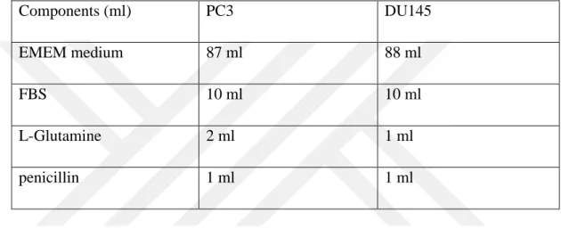
For further cell culturing protocol, the growth medium component percentage was adjusted based on the cell line type as follows;
Table 3.1 : Cell Culture Conditions
Components (ml) PC3 DU145
EMEM medium 87 ml 88 ml
FBS 10 ml 10 ml
L-Glutamine 2 ml 1 ml
penicillin 1 ml 1 ml
3.6. Cell Passaging (Subculturing)
3.6.1. Material
1. Cell Lines (PC3 and DU145) 2. CO2 incubator
3. PBS
4. Trypsin-EDTA
5. EMEM/DMEM medium containing FBS, Glutamine, penicillin 6. Culturing flask
7. Centrifuge tube 8. Centrifuge Machine
16
3.6.2. Method
To enhance cell growth, cells were passaged when they were 70-80% confluent. For sub-culturing, the flasks containing grown cells were removed from the incubator and the medium was discarded by pipetting out. Then, cells were washed with 1 ml of PBS to remove disturbing material that can cause contamination. Then cells went through trypsinization by adding 1 ml trypsin-EDTA to each flask and then the flasks were incubated at 37 ο C for about 2-3 minutes. The time of incubation can be adjusted by checking the detachment of the cells from the flask bottom observing under a microscope. Trypsinized cells start floating into trypsin-EDTA. 4 ml of fresh growth medium was added to each flask to deactivate the trypsin function to avoid cell death. Then, the whole medium containing cells was pipetted out and transferred into 10-ml centrifuge tubes and centrifuged at 1000-1200 rpm for 5 minutes. The supernatant was gently discarded and the pellet was transferred into a new growth medium (EMEM+10 FBS+ 1-2% L-Glutamine+ 1% penicillin) and incubated at 37 ο C.
3.7. Preparation of Cell Pellet
3.7.1. Material
1. Cell Lines culture 2. PBS
3. Trypsin-EDTA 4. Culturing flask
5. EMEM/DMEM medium containing FBS, Glutamine, penicillin 6. Centrifuge tube
7. Centrifuge machine 8. CO2 incubator
3.7.2. Method
For further experiments, cells were preserved at a low temperature by making pellets. To make pellets, the growth medium was removed and cells were washed by
17 1 ml PBS to remove harmful substances. 1 ml of trypsin-EDTA was added for trypsinization step and the flasks were incubated at 37 ο C. For 2-3 minutes. Then fresh growth medium was added to deactivate trypsin reaction and the whole culture was transferred into a 10-ml centrifuge tube and then, centrifuged at 1000-1200 rpm for minutes, the supernatant was discarded.
3.8. Cryopreservation of Cell Culture
3.8.1. Materials
1. Cell pellet 2. 10% DMSO 3. Cryotube
4. Cryogenic freezer
5. Freezing container (Mr.Frosty) 6. Liquid nitrogen tank
3.8.2. Method
Prepared cell pellets in the above technique were preserved by freezing technique in which cells were suspended in 10% DMSO growth medium and were transferred into cryotube. Then, the tubes were kept in a cryogenic freezer where the temperature reduced gradually to -30℃ followed by lowering the temperature to -60℃ overnight. Then, for a longer preservation period, cryotubes were transferred to a freezing container to be kept in a liquid nitrogen tank where the temperature drops to -130 οC.
3.9. Thawing Frozen Cell Lines
3.9.1. Material
1. PC3 and DU145 cell lines 2. Water bath
3. EMEM medium containing 10% fetal bovine serum (FBS), 1% L-Glutamine, and 1% penicillin
18 4. CO2 incubator
3.9.2. Method
PC3 and DU145 cell lines that were obtained in a frozen form, were warm in a water bath which was set at 37οC for about 3-5 mins and then cultured to the growth EMEM medium containing 10% fetal bovine serum (FBS), 1% L-Glutamine, and 1% penicillin and were grown in a CO2 incubator at 37 οC.
3.10. Genomic DNA Purification from Mammalian Cells
3.10.1. Material
1. PC3 and DU145 cells lines
2. Zymo Research Quick-DNA mini-prep Kit 3. Freezer 4. Micropipette 5. Microcentrifuge tube 6. Water bath 7. Collection tube 3.10.2. Method
To carry out Sanger sequencing, PC3 and DU145 cell lines underwent DNA isolation to extract the DNA for further analysis. For the DNA isolation method, Zymo Research Quick-DNA Universal Kit was used. The pellet was removed from the freezer and washed with 200 μl PBS. Next, 20 μl proteinase K was added to 200 μl of DNA sample and mixed well by pipetting up and down. The mixture was transferred into a microcentrifuge tube and the tubes were incubated in a water bath set at 55 οC for 10 minutes until a clear mixture was observed. Next, 1 volume (420 μl) of genomic binding buffer was added to the digested sample and were transferred into Zymo-Spin IIC-XL Column in a collection tube. Then, the columns were centrifuged at 12000 x g for a minute and flow through in a collection tube was discarded. Then, 400 μl of DNA pre-wash buffer was added into a column and centrifuged at 12000 x g for another minute. The flow-through in the collection tube was discarded and 700 μl of g-DNA
19 wash buffer was added into a column and centrifuged at 12000 x g for another minute. Next, 200 μl g-DNA wash buffer was added to each column and centrifuged at 12000 x g for another minute. The collection tube was discarded and the columns were put in a clean sterile microcentrifuge tube. Then 40-50 μl of elution buffer was added into the columns and they were incubated for 5 minutes at room temperature and then centrifuged at 12000 x g for 1 minute. The columns were discarded and the microcentrifuge tubes containing extracted DNA were stored at -20 o C for further analysis.
3.11. Primer Design For Pcr and Sanger Sequencing
To design the desired primers for Sanger sequencing and other analysis, Primer3 software was used and the following criteria were taken into account while selecting the primers; length should be 20 bps, and GC% should be between 50-6-%, and to increase primer efficiency, the primers with G or C 3’ end was selected.
3.12. PCR Amplification
3.12.1 Material
1. ddH2O
2. Master Mix; Taq polymerase enzyme, dNTPs, 1.5 5mM MgCl2, and reaction buffer 3. Vortex
4. Eppendorf reaction tube 5. Thermal Cycler
3.12.2. Method
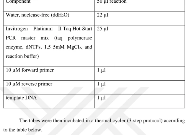
All components were mixed and centrifuged for 30 sec-1 min before use. 22 µl ddH2O and 25 µl of the master mix were added to 200 µl PCR eppendorf reaction tube. The components were mixed and vortexed gently. Next, 1 µl of the DNA template, 1 µl of forward primer, and 1 µl of reverse primer were added and gently mixed.
20 Table 3.2. PCR Amplification Components
Component 50 µl reaction
Water, nuclease-free (ddH2O) 22 µl Invitrogen Platinum II Taq Hot-Start
PCR master mix (taq polymerase enzyme, dNTPs, 1.5 5mM MgCl2, and reaction buffer) 25 µl 10 µM forward primer 1 µl 10 µM reverse primer 1 µl template DNA 1 µl
The tubes were then incubated in a thermal cycler (3-step protocol) according to the table below.
Table 3.3: PCR Conditions
step 3-step protocol 2-step protocol
Temp time Temp time
Initial denaturation 94οC 2 min 94οC 2 min
25035 PCR cycles
Daneutre Anneal 15 sec 98 οC 5 sec
Anneal 60 οC 15 sec 60 οC 15 sec
68 οC 60 οC 15 sec
hold 4 οC hold 4 οC hold
3.13. Sanger Sequencing Workflow
To ensure KLK5 gene mutation in cell lines, Sanger Sequencing was carried out by the Genetiks company. Primer design for PCR (Polymerase Chain Reaction)
21 and Sanger sequencing was carried out using ThermoFisher primer design tool ( Invitrogen™ Primer Designer™ Tool) by uploading KLK5 gene sequence and the following criteria were takeninto account while selecting the desired primer; primer length (between 18-22 bps), primer’s GC content (50-55%), GC-lock on the 3’ end of the primer, melting temperature (50-55οC), no poly base regions in the primer’s sequence, no 4 or bases complement either direction of primer.
After selecting the primers, the sequences were sent to the company for designing, where they were checked by mass spectroscopy to check the success rate to more than 95%. Invitrogen Platinum II Taq Hot-Start DNA Polymerase was used in the PCR protocol.
3.14. PCR clean-up
3.14.1. Material
1. ExoSAP-IT Express Reagent 2. Freezer
3. Ice 4. Vortex
5. Eppendorf reaction tube 6. Centrifuge machine 7. Thermal cycler
3.14.2. Method
PCR reactions were cleaned up using ExoSAP-IT Express Reagent (a mixture of exonuclease I and shrimp alkaline phosphatase (SAP) that removes excess primers and dNTPs after a PCR reaction). The method was as follows; the ExoSAP-IT Express Reagent was removed from -20οC freezer and transferred on the ice during the whole procedure. 5 µl of the above PCR product was mixed with 2 µl of the reagent and vortexed gently to mix them properly. Next, the tubes had a quick spin to help the content precipitate to the bottom of the tube. Then the tubes were incubated in a thermal cycler at 37οC for 4 minutes (to degrade the primers nucleotides that are still left in the PCR product) and 80οC for 1 minute to inactivate ExoSAP-IT Express
22 Reagent. Then the tubes were held at 4 ο C and transferred to ice.
3.15. Cycle Sequencing
3.15.1 Material
1. Genomic DNA
2. Forward and reverse primers 3. BigDye Direct PCR master mix 4. ddH2O
5. Eppendorf reaction tube 6. Thermal cycler
7. Micropipette
3.15.2. Method
The sequencing was carried out by Applied Biosystems BigDye Direct Cycle Sequencing Kit. To prepare the reaction for PCR, the following components were added as it is shown in the table below;
Table 3.4. Components of Cycle Sequencing in Sanger Sequencing
Component Volume
Genomic DNA (4ng/ µl) 1 µl
M13-tailed PCR primers (0.8 µ each) (it included both forward and reverse primers)
1.5 µl
BigDye Direct PCR master mix 5 µl
ddH2O 2.5 µl
Total volume for each PCR reaction 10 µl
23 span briefly. Then, the tubes were run in a thermal cycler with the following time and temperature panel;
Table 3.5. Cycle Sequencing Condition
Stage Verity thermal cyclers 9700 thermal cycler
Temp time Temp time
Hold 95 οC 10 min 96 οC 5 min
Cycle (35 cycles) 96 οC 3 sec 94 οC 30 sec
62 οC 15 sec 62 οC 45 sec
68 οC 30 sec 68 οC 45 sec
hold 72 οC 2 min 72 οC 2 min
hold 4 οC ∞ 4 οC ∞
In the next step, the cycle sequencing reactions were prepared according to the company’s manual based on the table below;
Table 3.6. Cycle Sequencing Reaction Components
Component Volume
BigDye Direct sequencing master mix 2 μl One sequencing primer
BigDye Direct M13 forward primer or
BigDye Direct M13 forward primer
1 μl
The total volume to add to each sequencing reaction
24 The above reaction was added to the appropriate well of amplified DNA for each reaction. The plates were sealed and span briefly. And then incubated in a thermal cycler according to the table below;
Table 3.7. Cycle Sequencing Incubation Condition
stage Verity thermal cyclers 9700 thermal cycler
Temp time Temp time
hold 37 ο C 15 min 37 ο C 15 min
hold 80 ο C 2 min 80 ο C 2 min
hold 96 ο C 1 min 96 ο C 1 min
Cycle (25 cycles0 96 ο C 10sec 96 ο C 10sec 50 ο C 5 sec 50 ο C 5 sec 60 ο C 75 sec 60 ο C 4 min
hold 4 ο C ∞ 4 ο C ∞
3.16. Purification of sequencing products
3.16.1. Material
1. Eppendorf reaction tube 2. SAM solution
3. Centrifuge machine 4. Vortex
3.16.2. Method
After the reaction was completed, the tubes were span briefly and the seals were removed and 55 µl of SAM solution were added to each well. The preparation o SAM solution was as follows;
25 Table 3.8. Components for Sequencing Products’ Purification
Component Volume for 1 well
SAM TM solution 45 µl
XTerminator Solution 10 µl
Total volume 55 µl
Before adding the solution, it was vortexed vigorously for 10 seconds to read to a homogeneous solution. Then, the well was sealed properly and vortexed at 1800 rpm for 20 minutes. Then the wells were centrifuged at 1000 × g for 2 minutes.
3.17. Capillary Electrophoresis and Data Analysis
As a next step, capillary electrophoresis was carried out using 3500 Series Genetic Analyzers and then the sample was run on minor Variant Finder Software that detects minor variants as low as 5%.
3.18. Computational Analysis of Protein Structure and Severity of Damage Caused by Mutation
To determine structural changes of KLK5 protein, protein structure analysis was performed using I-TASSER (https://zhanglab.ccmb.med.umich.edu/I-TASSER/). In addition, damage sensitivity of the mutation was analyzed using PolyPhen2 (http://genetics.bwh.harvard.edu/pph2/). The mutation taster program (www.mutationtaster.org/) was performed to check the effects of the mutation on the protein
3.19. Construction of the CRISPR system
A total of 3 target sequences (gRNAs) were selected in KLK5 based on the position of the mutation detected by Sanger sequencing. To design the CRISPR system, sgRNA was designed using chop-chop Harward software, where the KLK5 gene sequence was entered to analyze the gRNA sequence of 20 bps that are followed
26 by the PAM sequence (5’NGG). The program selected the ideal possible gRNAs with the optimum GC percentage (53-56%) and off-target sites which are 17-24 bps between the gRNA and KLK5 sequence due to mismatching. After loading the gene into chop-chop Harward software, we selected desired sgRNAs based on the location upstream of the PAM sequence of the target sequence (without counting the PAM sequence)
3.20. Mammalian cell Transfection with DNAfectin Reagent
3.21.1. Material
1. DU145 cell line 2. DNAfectin reagent 3. CRISPR-sgRNA 4. EMEM medium 5. 6-well plates 6. Vortex 7. Micropipette 8. Culture dish 9. CO2 incubator 3.22.2 Method
After completion of the plasmid replication and confirmation of KLK5 mutation by Sanger sequencing, DU145 cell lines were subjected to transfection by 3 designed plasmids using DNAfectin reagent. The reagent and protocol were obtained from abm pharmaceutical company (www.abmGood.com). DNAfectin is a nanoparticle. Nonliposomal reagent that transfects the mammalian cells with DNA plasmid with the minimum cytotoxicity. The reagent was selected due to its advantage of not affecting cell viability and low risk of contamination as the medium is not required to be pipetted out during the procedure. The highly efficient transfection (according to the company’s protocol) was accomplished by optimizing the cell density against DNAfectin which are mentioned in the table below;
27 Table 3.9. Reagent Quantities for Different Culture Vessels in Mammalian Cell Transfection
Culture vessel The volume of plating medium per well DNA (µg) DNAfectin (µl) Transfection medium volume (µl) 24-well 500 µl 0.2-0.4 0.6-1.2 50 12-well 1ml 0.5-0.8 1.5-2.5 100 6-well 2 ml 1-2 3-6 200 35mm 2 ml 1-2 3-6 200 60mm 5ml 3-6 10-20 300 10cm 10ml 8-16 25-50 500
As we used 3 different sgRNA for 3 different target sequences on DNA, 3 samples (2 for each) were prepared. 6-well plates were selected to carry out the transfection. The 24-hour pre-cultured cells in EMEM medium was used while the cells were 70-90% confluent. as the number of the wells were 6, 6 DNAfectin-DNA complex were made. 6 sterile eppendorf tubes were taken and in each, 200 µl of EMEM medium (without antibiotic and serum) and 20µg of DNA were added. DNAfectin was warmed at room temperature and vortexed gently before use. After that, 6µl of DNAfectin was added to each tube and pipetted up and down for mixing the complex properly. Then, the mixture was incubated at room temperature for 20 minutes to form the DNAfectin-DNA complex (the following complex is stable for 3-5 hours at room temperature). Then, the solution was added drop by drop to different areas on cultures dish of DU145 cell lines. To distribute the solution, the dish was gently moved side to side and back-and-forth to evenly spread the solution over the whole culture. The plates were incubated overnight (12-16 hours) and culture medium was changed by pipetting out the medium and adding the fresh medium to the plates.
28 3.23. Antibiotic Selection 3.23.1. Material 1. DU145 cells 2. Plasmid DNA 3. Puromycin 4. EMEM medium 5. Incubator 6. Micropipette 7. Culture plate
8. Agarose gel electrophoresis 9. Gel electrophoresis chamber 10. Casting tray 11. Comb 12. Ethidium bromide 13. DNA ladder 14. Loading dye 15. UV light 3.23.2. Method
To select DU145 cells, which were successfully transfected by plasmid DNA, puromycin was used for screening purposes. After optimizing the concentration for DU145 cell lines, we determined 5µg/ml is an ideal concentration to kill the cells not carrying the pac gene. EMEM medium was prepared with 5 µg/ml of puromycin. DU145 cells, which were previously transfected with the plasmids containing pac genes 48 hours before the following experiment, were cultured in the medium+ puromycin in a sterilized condition. To enhance the result of selection, we diluted the cell to have no more than 25% confluence. The following plates were then incubated at 37οC for 3-4 days. Next, the medium was pipette out and a fresh selection medium was added. The procedure was repeated after 3-4 days. After 7 days of selection, the cells were evaluated for foci formation. Finally, 5-10 resistant clones were picked and transferred to a 35mm culture plate with a selection medium for another 7 days. To
29 confirm the presence of DNA, DNA extraction, and gel electrophoresis were carried out to check whether DNA is available.
3.24. Drug Resistance Assay
Wild type DU145, knock out DU145, and PC3 cell lines were subjected to drug resistance assay using Doxorubicin and PARP ( poly ADP ribose polymerase) inhibitor. Doxorubicin has been commonly used in cancer treatment, including prostate cancer. It is a member of tetracycline family and damages cell’s DNA and drives them towards apoptosis by halting the cells in G2/M phase PARP are the catalyst enzymes that facilitate ADP-ribose transfer to the target proteins that are involved in DNA synthesis and repair mechanism PARPs are shown to have a significant role in maintaining and moderating chromatin structure, cell proliferation, and apoptosis.
3.25. Cell Counting
3.25.1. Material
1. Cell lines
2. EMEM medium with 10% FBS, 1% L-Glutamine and 1% penicillin 3. CO2 incubator 4. Micropipette 5. PBS 6. Trypsin-EDTA 7. Microscope 8. Culturing flask 9. Centrifuge machine 10. Haemocytometer 3.25.2. Method
To estimate cells’ viability, cell lines were cultured in EMEM medium with 10% FBS, 1% L-Glutamine, and 1% penicillin. The plates were incubated in a CO2 incubator at 32οC. After adequate growth with 70-80% confluence, cells were
30 collected by the following procedure:
The medium was pipetted out and the cells were washed with 1ml PBS (Phosphate-Buffered Saline) to get rid of all disturbing substances. After a slight moving of the flask, PBS was pipetted out and discarded. Next, trypsinization was carried out where 1 ml of trypsin-EDTA (trypsin is a proteolytic enzyme that degrades proteins to stop cell adhesion to the bottom of the flask) was added to the flasks and they were incubated at 32 ο C incubator for 2-3 minutes until cells dissociated themselves from the flasks (dissociation was confirmed by mounting cells under a microscope when the cells were floating in trypsin). 4 ml fresh serum contained- EMEM medium was added to deactivate the trypsin effect and to avoid cell death. And the whole culture was centrifuged for 5 minutes at 1000-1200 rpm. The supernatant was discarded and fresh medium was added to platelet and mixed properly by pipetting in and out to let the cells spread evenly in the medium. 20 µl of the culture medium was pipetted and transferred on a hemocytometer for total cell counting (dead+ live cells). To calculate the total cell number, the following formula was used;
Average cell count per square: total number of cells in 4 squares/4
Total cell count: average cell count per square×dilution factor (5 ml medium) ×104 (number of squares)
3.26. Cell Viability Assay
3.26.1. Material
1. PC3, DU145 wild type, and DU145 knock-out cell lines 2. 35-mm plates 3. EMEM medium 4. Incubator 5. Doxorubicin 6. PARP inhibitor 7. DMSO 8. Freezer
31 9. Trypsin 10. Methylene Blue 11. Haemocytometer 12. 100-mm Petri dish 3.26.2. Method
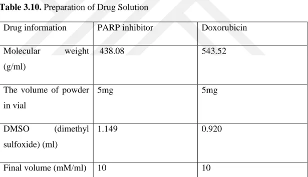
PC3, DU145 wild type, and DU145 knock-out were cultured in 35-mm plates with 5 ml serum-contained EMEM medium and incubated for 48 hours until reaching to appropriate confluence. 2 sets of plates were prepared for each cell line to test drug resistance against Doxorubicin as well as Doxorubicin+PARP inhibitor. The drugs were obtained from a pharmacy in the form of 5mg-powder in a vial and they were made in the form of solution using DMSO according to the following calculation below;
Table 3.10. Preparation of Drug Solution
Drug information PARP inhibitor Doxorubicin Molecular weight
(g/ml)
438.08 543.52
The volume of powder in vial 5mg 5mg DMSO (dimethyl sulfoxide) (ml) 1.149 0.920 Final volume (mM/ml) 10 10
After the drugs were made into a solution, there were kept at -80οC to preserve them. For the desired concentration, a serum-contained EMEM medium was used to dilute the drugs. After 48 hours of incubation, the plate for the drug combination test (Doxorubicin+PARP inhibitor) was removed from the incubator, and 10µM of PARP inhibitor was added to each well. Next, the plate was incubated at 37οC for 1 hour. The plates were removed and the following doxorubicin concentrations were added to each
32 well and incubated for 48 hours. Then the cells underwent through trypsinization process to collect the cells. After centrifugation, cells were mixed with Methylene blue (1:4 volume) to calculate cell viability using a haemocytometer. Methylene Blue can pass dead cells’ cell walls and turn blue under a microscope, while live cells remain intact without a change in their colour.
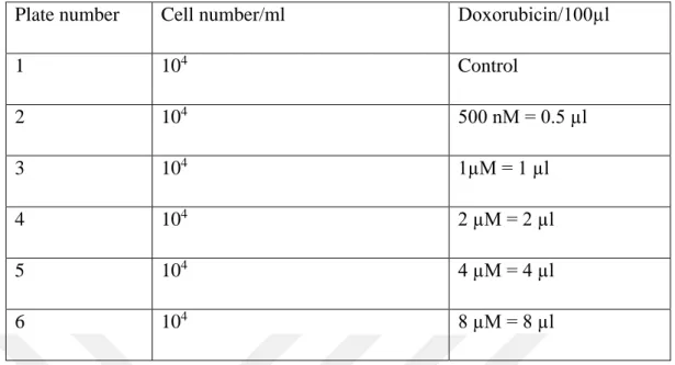
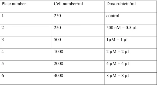
Table 3.11. Cell Numbers and Single Drug Concentration
Plate number Cell number/ml Doxorubicin/ml
1 104 Control 2 104 500 nM = 0.5 µl 3 104 1µM = 1 µl 4 104 2 µM = 2 µl 5 104 4 µM = 4 µl 6 104 8 µM = 8 µl
Table 3.12. Cell Numbers and Single Drug Concentration Plate
number
Cell number/ml PARP inhibitor Doxorubicin/ml
1 104 control control 2 104 10 µM= 10 µl none 3 104 10 µM= 10 µl 500 nM = 0.5 µl 4 104 10 µM= 10 µl 1µM = 1 µl 5 104 10 µM= 10 µl 2 µM = 2 µl 6 104 10 µM= 10 µl 4 µM = 4 µl 7 104 10 µM= 10 µl 8 µM = 8 µl
33 3.27. MTT Assay
3.27.1. Material
1. PC3, DU145 wild type, and DU145 knockout cell lines 2. 96-well tissue culture plates
3. EMEM medium 4. CO2 incubator 5. Doxorubicin 6. PARP inhibitor 7. MTT reagent 8. DMSO 9. Micropipette
10. FLUOstar Omega microplate reader
3.27.2. Method
3-[4,5-dimethylthiazol-2-yl]-2,5 diphenyl tetrazolium bromide assay was performed to measure the cell viability under certain drug concentrations.
104 cells/well of PC3, DU145 wild type, and DU145 knockout were plated into 96-well tissue culture plates with a serum-contained EMEM medium. The plate was incubated for 48 hours at 37οC. For each cell line, two sets of treatments were prepared for doxorubicin and doxorubicin+ PARP inhibitor. And the concentration of the following drug was added to the wells;