642
http://journals.tubitak.gov.tr/medical/
Turkish Journal of Medical Sciences Turk J Med Sci
(2013) 43: 642-644 © TÜBİTAK
doi:10.3906/sag-1206-40
Double-J stent placement with grasping forceps through ureteroscope
working channel: a novel technique*
Akif KOÇ1,**, Ömer BAYRAK2
1Department of Urology, Faculty of Medicine, Balıkesir University, Balıkesir, Turkey 2Department of Urology, Faculty of Medicine, Gazi University, Ankara, Turkey
1. Introduction
The use of a double-J stent was first described by Finney in 1978 (1). Subsequently, indwelling ureteral stents have been used in a variety of urological diseases and procedures for many years. Retrograde ureteral stenting is usually performed to bypass ureteric obstruction such as tumors or stones. Traditional retrograde replacement of double-J ureteral stents can result in complications such as hematuria, catheter migration, encrustation, stone formation, fracture of the catheter, or renal, parenchymal, and ureteral perforation (2–4). We report a new technique for retrograde placement of double-J stents with grasping forceps via the ureteroscope working channel.
2. Materials and methods 2.1. Patients
Between November 2008 and March 2010, ureteroscopy was performed in 34 patients and a 4.8-Fr, 28-cm double-J stent was inserted using the aforementioned new
technique in 17 consecutive patients (9 males, 8 females; age range: 19–57 years, mean age: 38). Informed consent was obtained from all patients.
All of the 17 patients underwent retrograde ureteroscopy, 16 for stone extraction and 1 for ureteropelvic junction obstruction. In 14 of the 17 cases, the ureter was dilated with a 10-Fr double-lumen dilatation catheter (equivalent to 3.3 mm) to gain access prior to ureteroscopy. Following dilation, ureteroscopy procedures were performed and 4.8-Fr, 28-cm double-pigtail ureteral stents were inserted using our technique. On the first postoperative day, kidney–ureter–bladder radiographic images were taken from all patients.
2.2. Technique
The technique is performed with the patient in the lithotomy position. The ureter was dilated with a 10-Fr double-lumen catheter (balloon dilatation catheter) to gain access prior to ureteroscopy, as required. After introducing Aim: To report a new technique for the retrograde placement of a double-J stent with grasping forceps through the ureteroscope working channel.
Materials and methods: In our technique, the ureteroscope is advanced to the renal pelvis or proximal location of the obstruction in the ureteral lumen; a double-J stent is then introduced into the ureteroscope working channel. The double-J stent is pushed forward until its proximal tip is seen; then the clamp over the distal tip of the pusher is opened and the guide-wire and pusher are removed from the channel, respectively. The grasping forceps is inserted into the working channel until it is touching the stent; the ureteroscope is removed from the ureter by pulling it over the stent and grasping forceps. Thus, the stent is placed in the ureteral lumen.
Results: This technique was successful in 17 patients: 16 retrograde ureteroscopies for stone extraction and 1 retrograde ureteroscopy with ureteropelvic junction obstruction. No complications occurred during follow-up.
Conclusion: Our simple technique may be safely used to place double-J stent in cases of ureteral obstruction. It may reduce procedure time and it avoids the potential complications of blind catheter placement.
Key words: Urinary catheterization, ureteroscopy, stents, methods, ureter
Received: 12.06.2012 Accepted: 14.11.2012 Published Online: 29.07.2013 Printed: 19.08.2013 Research Article
* This technique was previously presented at the 1st National Congress of Minimally Invasive Urological Surgery (Ankara, 2010) and the 30th World Congress of Endourology and SWL (İstanbul, 2012).
643 KOÇ and BAYRAK / Turk J Med Sci
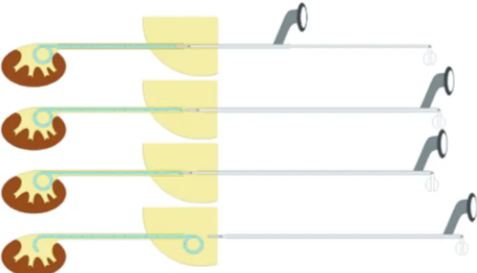
the ureteroscope to the renal pelvis or proximal location of the obstruction in the ureteral lumen, an appropriately sized double-J stent with guide-wire and pusher is introduced into the ureteroscope working channel (Figure 1a). A double-J stent is advanced until the proximal tip is visible. The clamp over the distal tip of the pusher is then opened, and the guide-wire and pusher are withdrawn from the channel, respectively (Figure 1b). A grasping forceps is inserted into the channel until it touches the stent (Figure 1c). The forceps is kept immobile, and then the ureteroscope is removed from the ureter by pulling it over the stent and grasping forceps (Figure 1d). Thus, the stent is placed into the ureteral lumen (Figure 1e). If the curl at the distal tip of the stent remains in the distal ureteral lumen, it can easily be grasped by the forceps and taken into the bladder lumen (Figure 2).
3. Results
Double-J ureteral stents were placed with a 100% success rate (17/17) in cases of ureteral obstruction including ureteropelvic junction obstruction (n = 1) and ureteral stone (n = 16). Double-J stents in 11 patients were
removed by cystoscopy at an average of 57 postoperative days (range: 29–168). Six patients could not be contacted. No complications occurred in any of the remaining 11 patients. Kidney–ureter–bladder radiographic images were normal in all patients.
4. Discussion
Ureteroscopy was first described by Young and McKay in 1929 (5). It has since been used for therapeutic and diagnostic aims in ureteral and renal pathologies in both adults and children (6,7). After ureteroscopy, replacement of ureteral stents might be necessary according to ureteral pathology. Ureteral stent replacement is traditionally performed using a cystoscope. If possible, this process should be performed under fluoroscopy. However, fluoroscopy is not universally available in endoscopy rooms (8), and so the procedure is often performed blind.
Following the definition by Finney in 1978, double-J stents have been used extensively within urology (1). Minor and major complications have been reported since then. Minor complications include hematuria, dysuria, frequency, and flank and suprapubic pain. Major complications include vesicoureteral reflux, migration, encrustation, urinary infection, stent fracture, necrosis, and ureteroarterial fistulas (9). Potentially life-threatening complications can also occur, such as acute postoperative pulmonary thromboembolism, hemoperitoneum, and renal vein perforation (3,4,10).
The technique described here may be particularly beneficial when it is planned to place a double-J stent to the ureter by means of a ureteroscope. Our method is especially useful for insertion of double-J stents to the ureteral lumen after passing the region of obstruction for any reason (e.g., stone, tumor, extrinsic compression) by ureteroscopy.
Occasionally, knots may form in a stent (11). Several techniques have been described for removal of knotted stents (12–14). At the end of ureteroscopy, if the ureteroscope has reached the renal pelvis, the risks of stent knotting and perforation of the urinary tract appear to Figure 2. Withdrawal of knotted stent from ureteral lumen to bladder.
Figure 1. Retrograde placement of double-J stent with grasping forceps via ureteroscope working channel. a: Ureteroscope is introduced to renal pelvis or proximity of obstruction in ureteral lumen. Double-J stent (appropriate diameter for ureteroscope working channel) with guide-wire and pusher is inserted into the ureteroscope working channel. b: Double-J stent is advanced until the proximal end is seen. The clamp over the distal tip of the pusher is opened, and the guide-wire and pusher are withdrawn from the channel, respectively. c: Grasping forceps is advanced along the channel until it is touching the stent. d: Grasping forceps is kept immobile and the ureteroscope is withdrawn from the ureter by pulling it over the stent and grasping forceps. e: The stent is thus placed in the ureteral lumen.
a
b
c
d
e
Bladder644
KOÇ and BAYRAK / Turk J Med Sci be eliminated in our technique. Moreover, the technique
appears to be safer, even though the ureteroscope cannot reach the renal pelvis, because the double-J stent is placed under endoscopic supervision in our technique. Additionally, this method can be safely applied in clinics without fluoroscopy guidance, as in the present study.
Sometimes, a double-pigtail stent cannot be inserted into the ureter for reasons such as tuberculosis of the ureter, retroperitoneal fibrosis, malignant strictures, or postoperative urinary fistula (15). Although none of our patients had such disorders, our technique may be useful in those situations.
The double-J stent can also be placed with a pusher via the ureteroscope working channel. However, this method requires a second pusher, as one pusher is not long enough to place the stent. Occasionally, another double-J stent set is needed for the second pusher, thereby increasing the cost of the procedure. In such cases with double-J stent knots
in the ureteral lumen: 1) the ureteroscope is removed from the body; 2) pushers are removed from the ureteroscope working channel; 3) the ureteroscope is advanced to the region of stent knotting; 4) grasping forceps are advanced through the working channel; 5) and the double-J stent must be caught by grasping forceps, then withdrawn into the bladder. In contrast, in our technique, a second pusher is not needed; in addition, if the distal tip of the stent curls within the distal ureteral lumen, the stent is caught by the grasping forceps, which is already present in the working channel; the stent can then be withdrawn into the bladder. Nevertheless, our technique may reduce procedure time and prevent complications due to additional processes.
In conclusion, our simple technique may be used to safely place double-J stents in cases of ureteral obstruction. It reduces procedure time and avoids the potential complications of blind catheter placement.
References
1. Finney RP. Experience with new double J ureteral catheter stent. J Urol 1978; 120: 678–81.
2. Damiano R, Oliva A, Esposito C, De Sio M, Autorino R, D’Armiento M. Early and late complications of double pigtail ureteral stent. Urol Int 2002; 69: 136–40.
3. Kidd RV 3rd, Confer DJ, Ball TP Jr. Ureteral and renal vein perforation with placement into the renal vein as a complication of the pigtail ureteral stent. J Urol 1980; 124: 424–6.
4. Chitale SV, Ho ET. Haemoperitoneum secondary to ureteric stenting. Int Urol Nephrol 2002; 34: 197–8.
5. Young HH, McKay, RW. Congenital valvular obstruction of the prostatic urethra. Surg Gynecol Obstet 1929; 48: 509.
6. Geavlete P, Jecu M, Geavlete B, Multescu R, Nita G, Georgescu D. Ureteroscopy--an essential modern approach in upper urinary tract diagnosis and treatment. J Med Life 2010; 3: 193–9. 7. Yapanoğlu T, Aydın HR, Aksoy Y, Özbey İ. Ureteroscopic
management of distal ureteral stones in children: holmium: YAG laser vs. pneumatic lithotripsy. Turk J Med Sci 2009; 39: 623–8.
8. Ioannis C, Ioannis Z, Evangelos I, Konstantinos G. Retrovesical migration of malpositioned double-j ureteral stent. Int Urol Nephrol 2003; 35: 325–6.
9. el Khader K. [Complications of double J ureteral stents]. J Urol (Paris) 1996; 102: 173–5.
10. Michalopoulos AS, Tzoufi MJ, Theodorakis G, Mentzelopoulos SD. Acute postoperative pulmonary thromboembolism as a result of intravascular migration of a pigtail ureteral stent. Anesth Analg 2002; 95: 1185–8.
11. Eisner B, Kim H, Sacco D. Repeat knot formation in a patient with an indwelling ureteral stent. Int Braz J Urol 2006; 32: 308–9. 12. Baldwin DD, Juriansz GJ, Stewart S, Hadley R. Knotted ureteral stent: a minimally invasive technique for removal. J Urol 1998; 159: 2065–6.
13. Kundargi P, Bansal M, Pattnaik PK. Knotted upper end: a new complication in the use of an indwelling ureteral stent. J Urol 1994; 151: 995–6.
14. Quek ML, Dunn MD. Knot formation at the mid portion of an indwelling ureteral stent. J Urol 2002; 168: 1497.
15. Smedley FH, Rimmer J, Taube M, Edwards L. 168 double J (pigtail) ureteric catheter insertions: a retrospective review. Ann R Coll Surg Engl 1988; 70: 377–9.