SELF-NANOPARTICLE FORMING IMMUNOSTIMULATORY
DNA: STRUCTURE-FUNCTION RELATIONSHIP STUDIES
A THESIS SUBMITTED TO
THE MATERIAL SCIENCE AND NANOTECHNOLOGY PROGRAMME OF THE INSTITUTE OF ENGINEERING AND SCIENCES
BILKENT UNIVERSITY
IN PARTIAL FULFILLMENT OF THE REQUIREMENTS FOR THE DEGREE OF MASTER OF SCIENCE
BY
RASHAD MAMMADOV JANUARY 2009
ii
I certify that I have read this thesis and that in my opinion it is fully adequate, in scope and in quality, as a thesis for the degree of Master of Science.
Assoc. Prof. Dr. Ihsan Gürsel
I certify that I have read this thesis and that in my opinion it is fully adequate, in scope and in quality, as a thesis for the degree of Master of Science.
Assist. Prof. Dr. Can Akçalı
I certify that I have read this thesis and that in my opinion it is fully adequate, in scope and in quality, as a thesis for the degree of Master of Science.
Assist. Prof. Dr. A. Elif Erson
Approved for the Institute of Engineering and Science
Director of Institute of Engineering and Science Prof. Dr. Mehmet Baray
iii ABSTRACT
SELF-NANOPARTICLE FORMING IMMUNOSTIMULATORY DNA: STRUCTURE-FUNCTION RELATIONSHIP STUDIES
Rashad Mammadov
M.Sc. in Material Science and Nanotechnology Supervisor: Assoc. Prof. Dr. Ihsan Gürsel
January 2009, 107 pages
Toll-like receptors (TLRs) are one the most critical and widely studied members of the family of pattern recognition receptors expressed on innate immune cells. They recognize microbial signatures, such as bacterial/viral DNA, LPS, from gram negative bacteria, peptidoglycan from gram positive bacteria, zymosan from yeast, lipopeptides or profilin protein from parasites, and even single or double stranded RNA of viruses. Among several members of TLR family, TLR9, that recognizes microbial unmethylated dinucleotide motifs on DNA initiate a robust Th1-biased inflammatory response. Synthetic oligodeoxynucleotides expressing unmethylated CpG motifs, mimic bacterial DNA effect and can be harnessed for the treatment of health problems ranging from infectious diseases to cancer, or to allergy/asthma as well as stand alone immunoprotective agents and also as a vaccine adjuvants that improve protection against pathogens. To date, various classes of CpG ODNs have been identified and were shown to induce differential immune activation in mice and man. Distinct structure-function relationship analyses revealed that these single-stranded linear ODNs alter the immune milieu as they are formulated to form complex multimeric DNA aggregates. Recently, Guanosine-rich D type CpG ODNs has been reported to form complex aggregates, that are differentially regulating immune cells to mount an anti-viral immunity. However, the clinical trials of this type are hampered mainly due to batch to batch variation during large-scale synthesis. To the best of our knowledge, there is no report on self-nanoparticle forming DNA except G-rich sequences. This thesis project was designed to generate stable, self-nanoparticle forming, G-run free, CpG expressing ODNs.
In this thesis, we designed a new generation CpG ODN, then characterized their structural and immunological properties. Our results suggest that dendrimeric structure confers higher immunostimulatory potential unparallel to conventional ODNs. Following four hours of in vivo ODN administration into mice indicated that nanoparticle-forming CpG ODNs initiated substantially high spleen and peritoneal exudate cell activation as evidenced by IFNγ and IL-12 production from culture medium. In order to shed light on the uptake and binding mechanisms, blocking experiments revealed that at least one type of scavenger receptor is critical for nanoparticle ODN internalization. Collectively, these data suggested that the improved stability to nucleases along with significantly higher binding to immune cells (no additional ODN formulation is required) seem to be the critical factors contributing to the nanoparticle CpG ODN mediated immune activation. The in vitro and in vivo performances implicated that these next generation immune stimulatory DNA molecules are promising candidates for various clinical applications.
Keywords: Innate immunity, TLR9, scavenger receptors, nanoparticulate and
iv ÖZET
KENDĐ KENDĐNE NANOPARÇAÇIK OLUŞTURAN BAĞIŞIKLIK UYARICI DNA: YAPI-FONKSĐYON ĐLĐŞKĐSĐ ÇALIŞMALARI
Rashad Mammadov
Malzeme Bilimi ve Nanoteknoloji Danışman: Doç. Dr. Đhsan Gürsel
Ocak 2009, 107 sayfa
Toll-benzeri reseptörler (TLR) doğal bağışıklık hücrelerinde ifade edilen patern tanıyıcı reseptörler ailesinin en çok çalışılan ve en kritik üyesidir. Bu reseptörler bakteri/virüs DNA’sı, gram negatif bakterilerdeki lipopolisakkaritler (LPS), gram pozitif bakterilerdeki peptidoglikan, maya mantarlarındaki zimosan, parazitlerdeki lipopeptidler veya profilin proteinleri, hatta virüslerdeki tek veya çift sarmallı RNA’lar gibi mikrobiyal işaretleri tanırlar. TLR ailesinin birçok üyesi arasından, mikrobiyal DNA üzerindeki metillenmemiş dinükleotid motifleri tanıyan TLR9 reseptörleri güçlü bir Th1 etkili enflamatuar yanıt başlatırlar. Metillenmemiş CpG motifleri taşıyan sentetik oligodeoksinükleotidler, bakteri DNA’sını taklit ettiklerinden bulaşıcı hastalıklardan kansere, alerji/astıma kadar birçok sağlık problemlerinin tedavisinde kullanılabilir olmalarının yanında bağışıklık koruyucu ajan ve patojenlere karşı korunmayı geliştirmek için aşı adjuvanı olarak da kullanılabilirler.
Şimdiye kadar birçok CpG ODN sınıfı tanımlanmış, bunların fare ve insanlarda
bağışıklık sistemini farklı şekillerde aktive ettiği gösterilmiştir. Yapı-fonksiyon ilişkisi analizleri göstermiştir ki tek zincirli lineer ODN’ler kompleks multimerik DNA kümeleri şeklinde formüle edildiklerinde farklı yönde bağışıklığa yol açmaktadırlar. Son zamanlarda Guanozilerce zengin CpG ODN’lerin kompleks kümeler oluşturup bağışıklık hücrelerini farklı şekilde yönlendirerek anti-viral bağışıklık oluşturduğu bildirilmiştir. Ancak bu çeşit ODN’ lerin klinikte kulanılması büyük ölçekli üretim esnasında üretimden üretime farklılıklar olması nedeniyle engellenmiştir. Bizim bilgimiz dahilinde, Guanozin zengini diziler haricinde kendi kendine nanoparçacık oluşturan DNA’lar bulunmamaktadır. Bu tez projesi dayanıklı, kendi kendine nanoparçacık oluşturan, G-yoksunu, CpG taşıyan ODN’ler ortaya çıkarmak için tasarlanmıştır.
Bu tez çalışmasında yeni jenerasyon CpG ODN tasarlanmış olup yapısal ve bağışıklık sistemi üzerinde etkileri karakterize edilmiştir. Sonuçlarımız dendrimerik yapıların bilinen diğer ODN’lere kıyasla çok daha yüksek oranda bağışıklık uyarıcı potansiyelinin olduğunu önermektedir. Farelere in vivo ODN uygulamasından dört saat sonra, nanoparçacık oluşturan CpG ODN’lerin, kültür ortamındaki IFNγ ve IL-12 üretiminden, dalak ve periton immün hücrelerini oldukça yüksek düzeyde aktive ettiği anlaşılmıştır. Hücreye alım ve bağlanma mekanizmalarını aydınlatmak için yapılan bloklama deneyleri en az bir çeşit skavenger (çöpçü) reseptörünün nanopartikül ODN’lerin hücreye alımında kritik rolü olduğunu ortaya çıkarmıştır. Sonuç olarak, bu verilerin ışığında nükleazlara karşı dirençli ve bağışıklık hücrelerine daha fazla bağlanmaları (ek bir ODN formülasyonu gerektirmeden) nanopartikül oluşturan CpG ODN kaynaklı bağışıklık aktivasyonuna katkıda bulunan faktörler olduğu söylenebilir. In vitro ve in vivo performansları bu yeni jenerasyon bağışıklık uyarıcı DNA’ların birçok klinik uygulama için güçlü adaylar olduğunu vurgulamıştır.
Anahtar kelimeler: Doğal bağışıklık, TLR9, scavenger reseptörleri, nanoparçacıklı ve
v
Dedicated to all dear members of my family who have
supported me throughout my life and showed heart to heart
affection and love which helped me not to feel alone...
vi
ACKNOWLEDGEMENTS
First and foremost I would like to thank my supervisor, Dr. Ihsan Gursel who had given me a chance to study in his lab and work on this very interesting project. His guidance helped me to focus on subject and not get destructed. I learned many from his skeptical approach to a subject which was very beneficial to make progress on my project. His patience will be a reference for me throughout my life.
I have special thanks to my labmates, Gizem, Fuat, Kutay, Erdem, Tamer and ex-labmate Hande, who were aware of their responsibilities and contributed to this thesis by minimizing routine technical problems in the lab. They were there whenever I needed them.
I would like to acknowledge Institute of Material Science and Nanotechnology (UNAM) administration, as they provided me opportunity to be an MSc student and supplied part of my stipend and health insurance. Dr. Aykutlu Dana and members of his group from UNAM, Burkan Kaplan and Musa Kurtuluş Abak, provided great assistance in AFM imaging studies, so I thank to them.
I appreciate Dr. Mayda Gürsel for her useful comments about my research which were very helpful.
Thanks to Dr. Ali O. Gure who boosted my interest to immunology in his Molecular Immunology course.
I would like to thank to administration of Molecular Biology and Genetics department, as they opened their labs to my use and let me to perform my experiments there. Moreover, I appreciate graduate studens and technicians of Molecular Biology and Genetics department for the assistance they provided in experiments.
Life wouldn’t be so interesting without my friends. They were with me in my hard times. Thanks to all of them.
My parents Irshad, Ezize and my sister Arzu deserves one of the bigger parts of acknowledgements as they supported me everytime and were very respectful to my decisions, especially one about being a molecular biologist. I have very special thanks to my grandparents who had great contributions to me, especially my grandfather who stimulated my interest to science subjects.
I have very sincere thanks to my dear lady Büşra. Most intense part of the experiments were coincided with first months of our marriage. Besides her patience, she supported me morally and made me to feel better in stressful conditions.
vii
TABLE OF CONTENTS
APPROVAL PAGE ...ii
ABSTRACT ...iii
ÖZET ...iv
DEDICATION PAGE ...v
ACKNOWLEDGMENTS ...vi
TABLEOF CONTENTS ...vii
LIST OF TABLES ...x
LIST OF FIGURES ...xi
ABBREVIATIONS ...xii
1. INTRODUCTION ...1
1.1.Innate Immune System ...1
1.1.1. Pathogen Recognition Receptors (PRRs) ...4
1.1.1.1. Scavenger receptors ...5
1.1.1.2. Toll-like receptors (TLRs) ...9
1.1.1.2.1. Extracellular TLRs ...9
1.1.1.2.2. Endosomal/Intracellular TLRs (nucleic acid-sensing TLRs) ...10
1.1.1.3. Toll-like receptor 9 (TLR9) ...10
1.1.1.3.1. Patterns recognized by TLR9 as stimulus ...10
1.1.1.3.2. Mechanism of TLR9 activation...11
1.1.1.3.2.1. Accessory molecules involved in TLR9 induction ...12
1.1.1.3.2.2. Signaling through TLR9 ...14
1.1.1.3.3. Cross-talk and cooperation of TLR9 with other TLRs ...15
1.2. Therapeutic potential of TLR ligands with emphasis on TLR9 ...16
1.2.1. CpG ODNs ...16
1.2.1.1. A/D-type CpG ODNs ...17
viii
1.2.1.3.C-type CpG ODN ...21
1.2.1.4. Other types of CpG ODNs ...22
1.2.2. Differential immune response mediated by particulate and linear CpG ODN ...23
1.2.3. CpG ODNs as therapeutics ...25
2. AIM AND HYPOTHESIS ...28
3. MATERIALS AND METHODS ...30
3.1. MATERIALS ...30
3.1.1. Reagents ...30
3.1.2. Buffers, Solutions, Culture Media ...33
3.2. METHODS ...33
3.2.1. Atomic Force Microscopy (AFM) ...33
3.2.2. Polyacrylamide Gel Electrophoresis (PAGE) ...34
3.2.3. Animals ...34
3.2.4. Cell culture ...34
3.2.4.1. Preparation of single cell splenocyte suspension culture...34
3.2.4.2. Cell counting & distribution ...35
3.2.4.3. Stimulation assay protocols ...36
3.2.4.4. Blocking assays in the presence of scavenger receptor ligands ...36
3.2.4.5. Experiments with ODN pre-incubation ...36
3.2.5. Enzyme-Linked Immunosorbent Assay (ELISA) ...37
3.2.6. Quantification of gene expression by PCR analyses ...38
3.2.6.1. Total RNA isolation ...38
3.2.6.2. Semi-Quantitative RT-PCR ...39
3.2.6.2.1. cDNA synthesis ...39
3.2.6.2.2. Polymerase Chain Reaction (PCR) ...39
3.2.6.2.2.1. Primers for PCR ...39
3.2.6.2.2.2. PCR protocol ...42
3.2.6.2.3. Agarose Gel Electrophoresis and band intensity measurement...43
3.2.7. DNAse treatment of the ODNs. ...44
3.2.8. ODN treatment of animals and detection of ex-vivo responses...44
3.2.9. Statistical Analysis ...45
ix
4.1. Synthesis of dendrimeric ODNs...47
4.2. Atomic Force Microscopy imaging of dendrimeric ODNs ...48
4.3. PAGE analysis of dendrimeric and conventional ODNs ...50
4.4. In vitro Stimulation assays using mouse spleen cells ...52
4.4.1. IL-6 production mediated by different CpG ODNs: Dose-dependent treatment at equimolar concentration ...52
4.4.2. IFNγ and IL-6 production mediated by different CpG ODNs: Treatment at equimolar concentrations ...54
4.5. Spleen cells and ODN mixing to study efficiency of binding ...56
4.6. Stability assays of the Dendrimeric ODNs: effect of DNAse pre-treatment on CpG mediated immune activation...62
4.7. Blocking of scavenger receptor interferes with dendrimeric CpG ODN- mediated immune activation ...65
4.8. Gene expression profile of murine splenocytes stimulated with dendrimeric CpG ODNs...67
4.8.1. Effects of dendrimeric CpG ODNs on different Toll-like receptor gene expression levels...68
4.8.2. Effects of dendrimeric CpG ODNs on cytokine & chemokine mRNA expression profile...69
4.9. In vivo stimulatory potential of dendrimeric nanoparticle forming ODN...72
5. DISCUSSION...75
6. FUTURE PERSPECTIVES...82
7. REFERENCES...84
x
LIST OF TABLES
Table 1.1. Cytokines affect behaviour of target cells ...3
Table 1.2. Chemokines induce chemotaxis of target cells to infection sites ...3
Table 1.3. Variations in TLR9 Expression among mice and human ...11
Table 3.1. The primer names, sequences, and expected product sizes of the primer pairs used throughout PCRassays...40 Table 3.2. PCR reactants...42
Table 3.3. PCR running conditions...43
Table 4.1. Mixing experiment: Murine splenocytes were pre-incubated with indicated ODNs for 2, 4 and 8 hours...59
Table 4.2. Retained immunostimulatory potential of ODNs after various treatment times with DNAseI ...64
Table 4.3. CpG ODN-mediated expression levels of TLR genes normalized with housekeeping gene (β-actin) ...70
Table 4.4. CpG ODN-mediated expression levels of cytokine/chemokine genes normalized with housekeeping gene (β-actin) ...71
xi
LIST OF FIGURES
Figure 1.1. Classification of scavenger receptors ...6
Figure 1.2. Signaling pathway activated through TLR7 and TLR9 ...15
Figure 1.3. Model for higher-order structure formation ...19
Figure 1.4. Duplex formation through palindromic sequences by 2 monomeric C- ODNs (ODN2395) ...21
Figure 1.5. Y-shaped CpG ODN . ...23
Figure 1.6. Dichotomy of different CpG ODN types delivered to early or late endosomes induce innate or adaptive immune responses, respectively, in PDCs ...25
Figure 1.7. Hypothetical effect of CpG ODN on tumorigenic tissue...27
Figure 2.1. Neubaer cell counting chamber...35
Figure 4.1. Bifunctional dendrimeric ODNs spontaneously form uniform-sized nanoparticles ...49
Figure 4.2. Trifunctional dendrimeric ODNs fold into heteregenous aggregates ...50
Figure 4.3. Polyacrylamide gel electrophoresis of different types of ODNs ...51
Figure 4.4. IL-6 production by murine splenocytes in response to dose-dependent CpG ODN stimulation ...54
Figure 4.5. Cytokine response of murine splenocytes stimulated with equal amount of CpG/Control ODNs ...56
Figure 4.6. Mixing experiment ...60
Figure 4.7. Maximal ODN activity assay (no mixing and washing is done) using 8µg/ml CpG ODNs and control ODNs ...62
Figure 4.8. Dendrimeric nanoparticle forming ODNs (420,421) were resistant to DNase I treatment ...64
Figure 4.9. Effects of scavenger receptor ligand on CpG mediated immune activation...67
Figure 4.10. Agarose gel image of PCR products for TLRs ...69
Figure 4.11. Agarose gel image of PCR products for cytokines/chemokines...70
Figure 4.12. Ex-vivo cytokine secretion from murine PECs ...73
xii
ABBREVIATIONS
AFM Atomic Force Microscopy
APC Antigen presenting cell
AVA Anthrax vaccine adsorbed
BP Base pairs
CD Cluster of differentiation
cDNA Complementary Deoxyribonucleic Acid
CMV Cytomegalovirus
CS Chondroitin sulfate
CpG Unmethylated cytosine-phosphate-guaniosine motifs
CXCL CXC-chemokine ligand
DC Dendritic cell
DNA Deoxyribonucleic acid
DOTAP 1,2- dioleoyl-3-trimethylammonium-propane
DS Dextran sulfate
dsRNA Double-stranded RNA
ELISA Enzyme Linked-Immunosorbent Assay
FBS Fetal Bovine Serum
G-run Guanosine run
HBV Hepatitis-B Virus
HEK Human embryonic kidney
HIV Human Immunodeficiency Virus
Ig Immunoglobulin
IκK Inhibitor kappa B kinase
IL Interleukin
iNOS Inducible Nitric Oxide Synthase
IFN Interferon
IRAK IL-1 receptor-associated kinase
xiii
LBP LPS-binding protein
LPS Lipopolysaccharide
LRR Leucine-rich repeats
LTA Lipotheicoic Acid
MALP-2 Macrophage-Activating Lipopeptide-2
MCP Monocyte Chemoattractant Protein
MDP Muramyl dipeptide
Mf Macrophage
MHC Major Histocompatibility Complex
MIP Macrophage Inflammatory Protein
MyD-88 Myeloid Differentiation Primary Response gene 88
NF-κB Nuclear factor-kappa B
NK Natural killer
NLR Nucleotide-binding oligomerization domain like
receptors
NO Nitric oxide
NOD Nucleotide-binding oligomerization domain
ODN Oligodeoxynucleotide
OVA Ovalbumin
PAMP Pathogen associated molecular patterns
PBS Phosphate buffered saline
PCR Polymerase chain reaction
pDC Plasmacytoid dendritic cells
PGN Peptidoglycan
pI:C Polyriboinosinic polyribocytidylic acid
PLG Polylactide-co-glycolide
PNPP Para-nitrophenyl pyro phosphate
PRR Pattern recognition receptors
xiv
RPMI Roswell Park Memorial Institute
RSV Respiratory Syncytial Virus
RT Reverse transcriptase
SA-AKP Streptavidin Alkaline-phosphatase
SLE Systemic Lupus Erythematosus
ssRNA Single-stranded RNA
SR Scavenger Receptor
STF Soluble tuberculosis factor
TCR T-cell receptor
Th T-helper
TIR Toll/IL-1 receptor
TLR Toll-like Receptor
1
1.
INTRODUCTION
Infectious diseases are one of the main causes of death worldwide. Although there is a significant decrease in mortality rate due to diseases caused by infectious agents, millions of people still die every year due to AIDS, respiratory infections, malaria, tuberculosis and diarrheal diseases (World Health Organization, 2008). More effective vaccines and biodrugs are needed to provide effective protection to these inflictions and decrease current mortality rate. In achieving this, a continued research effort is required in order to better understand how mammalian immune system recognize and discriminate non self “pathogens” and mount a response that leads to effective pathogen eradication before it causes any harm to the host. Developing novel strategies in order to accelerate this early immune response as well as to increase its magnitude and duration will help us to pave the way for better curative treatments during combat with these and newly arising deadly pathogenic diseases.
Immune system while discriminating self from non-self, relies on a very simple rule. This dogma is known as the “Danger Signal” and is based on the recognition of specific signature molecules expressed on pathogens, and are missing on our cells. With this, innate arm of the immune system mount a robust orchestral activation in order to contain and furthermore eradicate the invading infectious organisms. This first line of defence not only contains the invading intruders but instructs the formation of a strong adaptive immune response first by migrating to the local lymph nodes and there attains the function of an antigen presenting cell that interacts with the T helper and B cells. This interaction controlled and initiated by the “Innate Immunity” leads to “Adaptive Immunity”.
1.1. Innate Immune System
Innate immune system has many arms which are so organized that they form several layers of barriers (that varies with their sophistication and complexity) in order to prevent the access of infecting organisms into the body (i.e. skin, mucous, cilia, tears, soluble proteins etc are some examples of this complex network). Infectious microrganisms, although rarely, find access to reach internal parts of our body, and due to rich nutrient sources, and availability of an optimum growth environment, they start multiplying inside our body at an unprecedent rate. At this stage, several ‘danger’ signals are wandering around and are recognized by one or many members of the innate immune system cells. Cells of the innate immune system
2
includes mucosal epithelia, dendritic cells (DC), macrophages (Mf), B-cells, natural killer (NK) cells, eosinophils, basophils and mast cells. Soluble components of the innate immune system includes, acute-phase proteins, complement system proteins, opsonins, polyreactive immunoglobulins (Ig) and mediators of immune response such as cytokines and chemokines. Mucosal epithelia, which is found in all metazoans, is the main barrier between host and microorganisms. Epithelial cells and keratinocytes secrete antimicrobial peptides to decrease colonization of microorganisms (Medzhitov R, 2007). Microorganisms that pass epithelial barrier meet with phagocytes (macrophages, dendritic cells and neutrophils) in local tissues. Resident macrophages recognize acute-phase and complement proteins, host plasma proteins that are already bound to bacterial surfaces (opsonization), and engulf microorganism with further initiation of bactericidal mechanisms - such as acidification, degrading enzymes, nitric oxide and antimicrobial peptide production which destroy microbes. Mf and DC induce a number of early acting cytokines and chemokines (please see Table 1.1. and Table 1.2.) which recruit neutrophils and monocytes to site of infection. Monocytes generally mature further into DC and Mf at infection site. NK cells are specialized cells to sense especially viral infections (already infecting a host cell) and induce death of these host cells (Janeway CA, 2005). Eosinophils, basophils and mast cells are mainly involved in protection from multicellular parasites, while mast cells, also can sense microbial byproducts (Medzhitov R, 2007). Resident immature dendritic cells, which express numerous pathogen-recognition receptors (PRR) including Toll-Like Receptors (TLR) and Scavenger Receptors (SR) (these will be discussed in more detail in the subsequent sections), recognize certain microbial signatures and furthermore phagocytose them. Immature DC becomes mature and can no longer continue phagocytosis. They become Antigen Presenting Cells (APC) and migrate to draining lymph nodes. They are involved in further degradation and processing of microbial proteins for ‘antigen presentation’. This process is the sentinel stage that shapes adaptive immunity. The T and B cells are instructed and converted to antibody producing B-Cells, and effector T cells (Janeway CA, 2005).
3
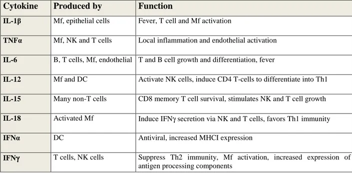
Table 1.1. Cytokines affect behaviour of target cells (Adopted from Janeway CA, 2005).
Mf: Macrophage, T: T-cells, DC: Dendritic cells, NK: Natural Killer cells
Table 1.2. Chemokines induce chemotaxis of target cells to infection sites (Janeway CA, 2005)
Chemokines Produced by Attracted cells Major effect
MIP-1α Monocyte, T, Mast cell , fibroblast
Monocyte,NK, T, basophil, DC
promotes Th1, antiviral defense and competes with HIV-1
MIP-1β Monocyte, Mf, Neutrophil, endothelial
Monocyte, NK, T, DC
competes with HIV-1
MCP-1 Monocyte, Mf, fibroblast, keratinocyte
Monocyte, NK, T, basophil, DC
promotes Th2, activate Mf and histamine release from basophils
IP10 T, fibroblast, endothelial, monocyte, keratinocyte
Resting T, NK, monocyte
promotes Th1, antiangiogenic, immunostimulant
Cytokine Produced by Function
IL-1β Mf, epithelial cells Fever, T cell and Mf activation
TNFα Mf, NK and T cells Local inflammation and endothelial activation IL-6 B, T cells, Mf, endothelial T and B cell growth and differentiation, fever
IL-12 Mf and DC Activate NK cells, induce CD4 T-cells to differentiate into Th1 IL-15 Many non-T cells CD8 memory T cell survival, stimulates NK and T cell growth IL-18 Activated Mf Induce IFNγ secretion via NK and T cells, favors Th1 immunity IFNα DC Antiviral, increased MHCI expression
IFNγγγγ T cells, NK cells Suppress Th2 immunity, Mf activation, increased expression of antigen processing components
4 1.1.1. Pathogen Recognition Receptors (PRRs)
Microorganisms have shared structures, which are necessary for their survival like LPS (lipopolysaccharide), PGN (peptidoglycan) including single-stranded and double stranded genome of viruses or bacteria. Most of these molecules are recognized by host immune system and named as pathogen-associated molecular patterns (PAMPs). Certain properties of PAMPs fit very well with innate immune recognition of host (Medzhitov R, 2007):
1) PAMPs are similar among different classes of microorganisms.
2) PAMPs are molecules unique to microorganism world, thus immune system cells use to discriminate self and non-self.
3) PAMPs have critical roles in microbial physiology, thus, these molecules can not be altered throughout the course of adaptive evolution.
Specific innate immune receptors sensing these diverse structures is known as the pathogen-recognition receptors (PRRs) (Medzhitov R, 1997). Similar to PAMPS, PRRs also share common properties (Akira S, 2006):
1) Each PRR recognizes a specific PAMP, activates signaling pathway and distinct immune response against pathogen
2) PRRs are expressed constitutively by host immune system and detect the pathogens regardless of their life-cycle stage.
3) PRRs are germline encoded, nonclonal, expressed on all cells of a given type, and independent of immunologic memory.
4) They are highly conserved structures from plants and fruit flies to mammals
PRRs can be classified as cytosolic PRRs which include nucleotide binding oligomerization domain (NOD)-like receptors (NLRs) and RIG-I-like helicases (RLHs) and membrane-bound PRRs which include Toll-like receptors (TLRs) and Scavenger Receptors (SR) (Kumagai Y, 2008, Mukhopadyay S, 2004).
PRRs collectively are divided into two broad groups: i) non-signaling PRRs, such as Scavenger Receptors and ii) signaling PRRs, such as the famous family of Toll like receptors. This section will summarize the features of these major players of immunity.
5 1.1.1.1. Scavenger Receptors
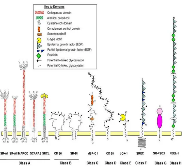
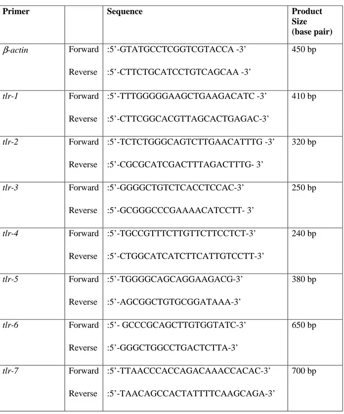
Scavenger receptors are large family of receptors expressed primarily by myeloid cells, but also expressed on selected endothelial cells (Mukhopadhyay S, 2004). They recognize various endogenous ligands such as phosphatidyl serine (apoptotic cells), low density lipoproteins (LDL) and its modified forms (acetylated LDL, oxidized LDL), modified proteins and also molecular patterns from foreign organisms. Scavenger receptors are divided into 8 major classes, mainly due to their molecular structure (Figure 1.1.). Among their diverse physiological roles, in accordance with the aim of this thesis, only the roles of scavenger receptors in recognition of microbial molecular signatures and their contribution to shape innate immunity will be covered .
Class A : Scavenger Receptor class-A (SRA)- I/II, Macrophage Receptor with Collagenous structure (MARCO), Scavenger Receptor class A member 5 (SCARA5) and SR with C type Lectin-I /Collectin from Placenta receptor-I (SRCL-I/CL-PI) are members of this class which have roles in pathogen recognition. These receptors recognize different species of Gram-positive and Gram-negative bacteria (Greaves DR, 2008) and modified LDL. Some of them also specialized in yeast (Saccharomyces cerevisiae) recognition such as SRCL-I (Mukhopadhyay S, 2004). SRCL-I was also shown to be involved in zymosan recognition and phagocytosis of fungi by human vascular endothelial cells (Jang S, 2008). Two well studied and known members i) SRA and ii) MARCO will be covered briefly.
SR-A I/II : SR-A, which is a trimeric type II transmembrane glycoprotein, has 3 isoforms - SR-AI, SR-AII and SR-AIII – that are alternative splice variants of same gene. SR-A was found to be expressed in myeloid cells (except monocytes), mast cells, specific subpopulations of bone marrow-derived and splenic dendritic cells (Plüddemann A, 2007). It binds to LPS (Hampton RY, 1991) and to LTA (Dunne DW, 1994) of Gram-negative and Gram-positive bacteria, respectively. Recently it is also reported that SR-A plays a role in recognition and internalization of dsRNA (Limmon GV, 2008). However, SR-A was found to be not essential for uptake and immunostimulatory activity of K-type CpG ODN and E.coli DNA (Zhu FG, 2001, please note that “D-type” was not used in this study). SR-A-/- mice are more susceptible to Listeria monocytogenes (Suzuki H, 1997), Staphylococcus aureus
6
(Thomas CA, 2000) and Streptococcus pneumoniae (Arredouani MS, 2006) which shows critical role of SR-A in recognition of these pathogens.
Figure 1.1. Classification of scavenger receptors. Each domain is indicated in the key above. (adopted from Plüddemann A, 2007)
MARCO: MARCO is constitutively expressed by peritoneal and spleen marginal zone macrophages and also by medullary cord macrophages of lymph node (Kraal G, 2000). However, low level MARCO expression can be upregulated via TLR-mediated induction in macrophages derived from other sources (Doyle SE, 2004). CpG ODN (K-type) binding to peritoneal macrophages from MARCO-/- mice was unchanged, while CpG-induced IL-12 production was severely impaired (Jozefowski S, 2006). MARCO recognizes LPS, LTA (Greaves DR, 2008), Gram-positive bacteria such as Streptococcus pneumoniae (Arredouani M, 2004) and Gram-negative bacteria such as Neiserria meningitides (Mukhopadhyay S,
7
2006). MARCO-/- mice infected with murine model of pneumococcal pneumonia, were unable to clear bacteria from lungs and showed decreased survival, while it induced increased cytokine release (Arredouani M, 2004).
Class B : CD36 and Scavenger Receptors class B I (SR-BI) are the members of this family involved in innate immunity.
CD36: CD36, type III transmembrane receptor, is expressed in microvascular (but not large vessel) endothelium, adipocytes, skeletal muscle, dendritic cells, epithelia of the retina, breast, and intestine, smooth muscle cells, and hematopoietic cells, including erythroid precursors, platelets, monocytes/macrophages, and megakaryocytes (Febbraio M, 2001). In addition to TLR2/6 heterodimer, studies showed that CD36 is indispensible for recognition of microbial diacylglycerides such as R-enantiomer of MALP-2 (a diacylated bacterial lipopeptide) as well as lipoteichoic acid (Hoebe K, 2005). CD36 was found to be critical for recognition and uptake of Staphylococcus aureus (Stuart LM, 2005). Finally, CD36 recognizes parasitic infection-induced membrane changes and mediates nonopsonic phagocytosis of P.
falciparum–infected erythrocytes which is critical for malaria infected erythrocyte clearance
(Febbraio M, 2001). CD36 has also been shown to have critical role in phagocytosis of apoptotic cells by macrophages (Fadok VA, 1998).
SR-BI: SR-BI, which has 2 alternative splice variants - SRBI & SRBII, is expressed on monocytes, macrophages and dendritic cells (only in humans), and is also found on hepatocytes, adipocytes and adrenal glands (Plüddemann A, 2007). SR-BI and SR-BII mediate uptake of Mycobacterium fortuitum by non-phagocytic cells such as HEK-293 and might have a role in non-opsonized uptake of several bacterial species (Philips JA, 2005). Human SR-BI binds LPS (Vishnyakova TG, 2003) and E2 glycoprotein of Hepatitis C virus (HCV) (Scarselli E, 2002) and mediates their internalization (Barth H, 2008).
Class C: Drosophila Scavenger Receptor-CI (dSR-CI), type I transmembrane, plays a role in phagocytosis of gram-positive and gram-negative bacteria and the recognition of microbial β -glucans in Drosophila (Mukhopadhyay S, 2004; Plüddemann A, 2007)
8
Class D: CD68 is not reported for pathogen recognition (Greaves DR, 2008).
Class E: Lectin-like oxidised LDL receptor-1 (LOX-1), a type II membrane glycoprotein, is the only member of the class E (Plüddemann A, 2007). CHO-K1 cells expressing LOX-1 stably have been shown to bind Gram positive and negative bacteria, which is Lox-1-dependent (Shimaoka T, 2001). Moreover, it has a contribution to antigen cross-presentation by recognizing Hsp70 (Delneste Y, 2002). Recently, a link between TLR9 and LOX-1 has been reported. Activation of TLR9 via CpG motifs increased LOX-1 expression (Lee JG, 2008). CpG type used in this study wasn’t mentioned, while it was ROS-inducer from macrophages, so probably it was K-type CpG ODN.
Class F: Scavenger Receptor(SR) expressed by Endothelial Cells-I (SREC-I) and SREC-II, type I transmembrane receptors, were not yet reported for any pathogen recognition (Plüddemann A, 2007; Greaves DR, 2008).
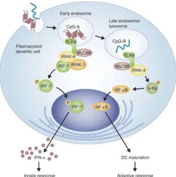
Class G: Scavenger Receptor that binds Phosphatidylserine and Oxidized lipoprotein (SR-PSOX), which is also known as CXC chemokine ligand 16 (CXCL16), is type I transmembrane molecule. SR-PSOX is expressed on macrophages and dendritic cells and in many organs (Plüddemann A, 2007). It can also perform function as chemokine ligand for an orphan G protein-coupled receptor Bonzo/CXCR6, when it is cleaved with ADAM10 – metalloproteinase (Mukhopadhyay S, 2004; Abel S, 2004). SR-PSOX/CXCL16 can recognize and phagocytose Gram-positive and negative bacteria, and maintain adherence of cells to CXCR6 expressing T cells (Plüddemann A, 2007). Chemokine form might have critical role in inflammatory valvular heart disease by recruiting CD8+ T cells to inflammed tissues and enhance IFNγ production (Yamauchi R, 2004). CXCL16 on plasmacytoid dendritic cells has been shown to recognize D-type CpG ODN and play critical role in its activity (Gursel M, 2006) (see section 1.2.2. for more details).
Class H: Fasciclin, EGF-like, laminin-type EGF-like and Link domain-containing scavenger receptor-1 and 2 (FEEL-1 and FEEL-2) are members of type I transmembrane proteins and are expressed on endothelial cells of the spleen, liver and lymph nodes. However, only FEEL-1 is expressed on monocytes and macrophages (Plüddemann A, 2007). These receptors also
9
increase uptake of Gram positive and negative bacteria when expressed in non-phagocytic cells such CHO (Mukhopadhyay S, 2004).
1.1.1.2. Toll Like Receptors (TLRs)
Among PRRs, probably the most widely studied and famous member is the TLR family that includes 12 murine (TLR1-9, TLR11-13) and 10 human (TLR1-10) receptors identified so far (Takeda K, 2007). While some TLRs (TLR1, 2, 4, 5, 6 and 10) are located on the plasma membrane of the innate immune cells, others such as TLR 3, 7, 8 and 9 are located in the intracellular endosomal and/or ER compartments. TLR 10-13 are not well characterized. Only TLR11 among them is known to recognize protozoan profilin (toxoplasma gondii) and uropathogenic bacteria in mice. In humans they are non-functional due to stop codon inside the gene (Takeda K, 2007). Because of special focus of this thesis, TLR9 receptor for CpG motifs will be discussed in details as a seperate section (please see 1.1.1.3.).
1.1.1.2.1. Extracellular TLRs
TLR1, 2 and 6 : TLR2 differs from other TLRs. It can be activated by a heterotypic interactions either with TLR1 or TLR6. TLR2/TLR6 and TLR2/TLR1 were known to recognize diacylated and triacylated lipopeptides, respectively (components of bacterial PGN layer). CD14 and CD36 are necessary accessory molecules required for TLR2/TLR6 activation (Miyake K, 2007). TLR2 is also triggered by zymosan from yeast and several glycolipids expressed by other specific bacteria (Takeda K, 2007).
TLR4: It is the first identified TLR member (Poltorak A, 1998). TLR4, senses endotoxin (lipopolysaccharide LPS) found in outer membrane of Gram-negative bacteria. Lipid A portion of LPS is responsible for its toxicity. LPS binding protein (LBP) and CD14 are accessory molecules that have a role in recruiting LPS and transfer and load onto TLR4/MD-2 complex (Miyake K, 2007). MD-2 is an accesory molecule, and its absence abolishes TLR4 signaling.
TLR5: Flagellin, a monomeric protein of bacterial flagella, is recognized by TLR5 receptor.
10
However, these mice were highly resistant to infections by this pathogen, where TLR5-mediated inflammation seems to have role in microbial invasion of host (Takeda K, 2007). 1.1.1.2.2. Endosomal/Intracellular TLRs (nucleic acid-sensing TLRs)
TLR3: Double-stranded RNA (dsRNA) appears during viral replication and induces type I IFN production. TLR3-/- mice have defective immune response against dsRNA (Alexopoulou L, 2001). TLR3 knockout mice do not show high susceptibility to infection with most of viruses (Takeda K, 2007).
TLR7/8: These two members are highly homologous and both of them recognize synthetic compounds, imidazoquinolines, which are clinically used for treatment of genital warts. That is associated with viral infection in humans and mice (mouse TLR8 is non-functional) (Takeda K, 2005; Takeda K, 2007). Moreover, TLR7 and human TLR8 recognize viral guanosine or uridine-rich single-stranded RNA (ssRNA) which is found in human immunodeficiency virus, vesicular stomatitis virus and influenza virus (Takeda K, 2005).
1.1.1.3. Toll Like Receptor 9 (TLR9)
1.1.1.3.1. Patterns Recognized by TLR9 as Stimulus
TLR9 is the only known member of TLR family that can recognize specific DNA motifs. Hemmi et al., five years after the identification of CpG motifs, reported that TLR9 is responsible for recognizing the CpG DNA in mice. They showed that splenocytes, lymph node cells, dendritic cells, and macrophages from TLR9 null mice do not respond to CpG motif expressing ODNs (CpG ODN), as evidenced by the loss of pro-inflammatory cytokine production or cell surface maturation marker downregulation (please see table 1.2. for TLR9 expression profile). Moreover, D-galactosamine (D-GalN) sensitized-mice are resistant to lethal effects of CpG ODN (Hemmi H, 2000). Then, it was shown that human TLR9 also is prerequsite for bacterial DNA/CpG DNA-dependent immunostimulation in both primary cells (B-cells and CD123+ DC) and TLR9 transfected cell lines (Bauer S, 2001; Takeshita F, 2001). Later findings challenged the idea that recognition of foreign DNA is restricted to CpG motifs. Non-CpG phosphodiester ODNs (PO-ODNs), which were delivered into endosomes after complex formation with DOTAP (Yasuda K, 2006), and self chromatin DNA/IgG autoantibody complexes (Boule MW, 2004) have been shown to be recognized via TLR9 and
11
stimulate innate immunity. Recently, TLR9 has been shown to recognize 2’-deoxyribose sugar backbone (base-free) of phophodiester DNA and activate cytokine secretion, but not phosphorothioate (PS) modified 2’-deoxyribose, PS or PO modified 2’-ribose backbones (Haas T, 2008a). Immunostimulatory activity and TLR9 affinity is increased further, when bases and CpG motifs were added to this backbone. PolyG addition (24 extra guanosines at 3’end) or DOTAP complexations were used here to target sugar backbone or PD-ODN into endosomes. PS modified sugar backbones were highly affine to TLR9 and TLR7 (significantly higher than PO-modified ligands) by PD-ODNs in vitro and in vivo (Haas T, 2008a).
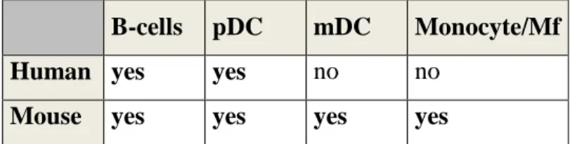
Table 1.3. Variations in TLR9 expression among mice and human (adopted from Krieg AM, 2006).
B-cells pDC mDC Monocyte/Mf
Human yes yes no no
Mouse yes yes yes yes
pDC: plasmacytoid DC, mDC: myeloid DC. Mf: macrophage
1.1.1.3.2. Mechanism of TLR9 Activation
In non-stimulated cells TLR9 is localized to endoplasmic reticulum, which is transported to early endosomes and then lysosomes upon CpG stimulation to interact with CpG (Latz E, 2004). Kim et. al, suggested that the transfer from ER to endosomes is mediated by UNC93B1 protein (Kim Y, 2008). Two independent studies, revealed that the ER to endosome docking was mediated by endosomal localization motifs present on TLR9 which are conflicting on region of these motifs at TLR9 protein (CA Leifer, 2006; GM Barton, 2006). There is also other contradicting ideas on movement of TLR9 from ER to endosomes: Latz et al. reported that this is maintained by non-secretory pathway (Latz E, 2004), while Chockalingam et al. reported recently that secretory pathway is involved and TLR9 passes through Golgi on the way to endosomes (Chockalingam A, 2008). In endosomes, TLR9 exists
12
as homodimers, ligand unbound form is the inactive conformation of the receptor. When ligand (CpG DNA) binds, seperate TIR domains gets closer and recruit MyD88 for inititation of signaling cascade (Latz E, 2007). Fitting with this idea, it was shown that only aggregated, multimeric forms of ODNs are stimulatory. As concentration of multimeric forms increase in ODN solution, it becomes more immunostimulatory (Wu CC, 2004). Multimeric ligand seems to have more advantage to stimulate homodimer receptor, otherwise two monomeric receptor should be activated seperately with 2 different monomeric form of ODN at the same time in endosomes/lysosomes.
Interaction of TLR9 with bacterial/viral DNA in endosomes of wild-type cells seems to prevent recognition of self-DNA, since artificially expressing TLR9 as chimeric protein (TLR9/TLR4) on the cell surface makes mammalian DNA stimulatory. Viral DNA becomes nonstimulatory in this case possibly due to existing protecting coat around the DNA (GM Barton, 2006). Mammalian DNA with phosphodiester backbone showed parallelism with PO-ODNs, which become more stimulatory against surface expressed TLR9, in sensitivity to DNase. (GM Barton, 2006). This finding challenged previous notion accepting CpG motifs as a determinant of self/non-self discrimination. Lastly, TLR9 has been shown to get activated after proteolytic cleavage of carboxy terminal fragment at endosomes via cathepsin proteases (Ewald SE, 2008; Park B, 2008), which is concordant with previous data that TLR9 signaling is blocked by inhibition of endosomal acidification (Ahmad-Nejad P, 2002). This acidification is required for optimal activity of TLR9-cleaving proteases. These recent findings strengthened the hypothesis that TLR9 functionality at endosomes is mainly for self/non-self discrimination. Since to get activated, it should have to go to endosomes where self DNA does not have access.
1.1.1.3.2.1. Accessory Molecules Involved in TLR9 Induction
LL37: LL37 is an endogenous antimicrobial peptide which is expressed highly in psoriatic skin, which is an autoimmune disease of skin. During physical injury, disease condition worsens, self-DNA release and pDC activation occurs. LL37 form complexes with self-DNA to be delivered to early endosomes of pDC and stimulate IFNα production. Otherwise,
self-13
DNA is inactive because it can’t colocalize with TLR9 in endosomes to stimulate it (Lande R, 2007).
HMBG1 and RAGE: HMBG1 (High-mobility group box 1) protein is a nuclear protein which binds DNA and is involved in bending of double-helix to increase affinity of DNA-transcription factor interactions. In inflammatory cytokine stimulation, it is released from dendritic cells and regulates IFNα stimulation in an autocrine manner. Recent work shows that HMBG1 complexation with A/D-type ODN (but not B/K-type ODN) increases its B cell activating and IFNα-inducing potential from pDC. Moreover, this additional stimulation depends on interaction of RAGE (receptor for advanced glycation end-products), which is multi-ligand receptor of immunoglobulin superfamily, with HMBG1-TLR9 complex. They also showed that HMBG1 and RAGE have critical role in IFNα induction with DNA-containing immune complexes in lupus patients (Tian J, 2007).
Cathepsin-K: Cathepsin-K is an osteoclast-specific protease which is involved in degradation of bone matrices. It is seen as a therapeutic target to treat diseases, such as osteoporosis and autoimmune arthritis, in which osteoclast activity abnormally increases. Recently its role in CpG-induced TLR9 activation has been delineated (Asagiri M, 2008). Specific inhibition of Cathepsin-K by pharmacological inhibitor in dendritic cells, dose-dependently reduced TLR9-mediated, but not TLR2 or TLR4-TLR9-mediated, production of IL-12, IL-23 and upregulation of maturation markers such as CD40, CD80 and CD86 (Asagiri M, 2008). Moreover, cathepsin K-/- mice were refractive to stimulation with CpG ODNs but not with LPS, regarding IL-6 and IL-12 production from dendritic cells.
UNC93B1: Missense mutation in Unc93b1 gene causes impaired signalling via TLR3 ,7, 9 (called as ‘3d mutation’ due to defects in 3 different signal pathway) and exogenous presentation of MHCII. Mice with these mutations show increased susceptibility to murine cytomegalovirus (MCMV) infection (Tabeta K, 2006) . This impaired signaling is caused by defects in delivering these TLRs from endoplasmic reticulum to endosomes, which is mediated by UNC93B1 protein. TLR9 artificially expressed on plasma membrane is not affected from ‘3d’ mutation and responds to CpG motifs (Kim Y, 2008).
14 1.1.1.3.2.2. Signaling Through TLR9
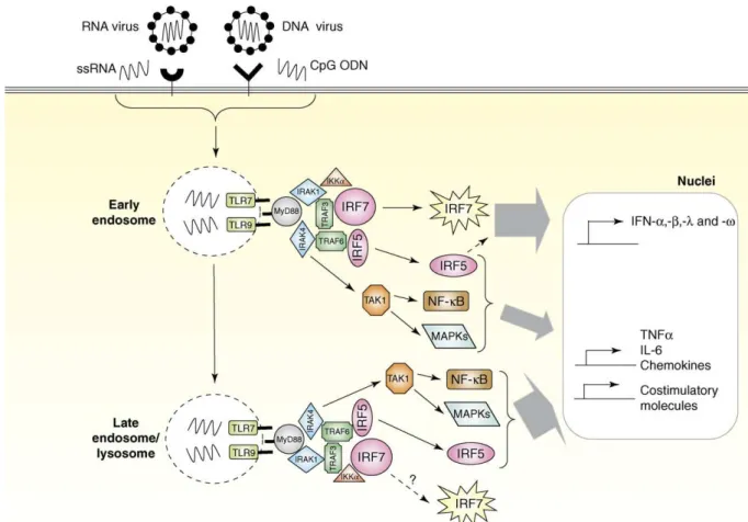
After 2h stimulation of TLR9-expressing cells with CpG ODN, number and size of CpG-containing vesicles increases significantly, some of which co-stain with TLR9. Colocalization of TLR9 with CpG in same endosomes have been shown to depend on presence of intracellular ‘Toll/IL-1R homologous region’ (TIR) domain of TLR9. (Takeshita F, 2004) CpG-TLR9 interaction recruits MyD88, which is an adaptor protein involved in signalling through most of TLRs (for clear depiction please look Figure 1.2.). MyD88 deficiency completely impairs TLR9 signaling, which indicates that it does not use other adaptor molecules like several TLRs. MyD88 has two domains: TIR domain to interact with TIR of TLR9 and death domain (DD) to interact with interleukin-1 receptor-associated kinase-1 (IRAK-1) and IRAK-4. IRAK-4 phosphorylates IRAK-1, which in turn upregulates its kinase activity, which leads to recruitment of tumor necrosis factor associated factor 6 (TRAF6)- E3 ubiquitin ligase (Klinman DM, 2004b; Ishii KJ, 2006, Cao W, 2007; Kumagai Y, 2008b). TRAF6 stimulates the protein kinase TAK1, which in turn phosphorylates IκB to induce nuclear translocation of NF-κB and initiates signaling by the mitogen-activated protein kinase (MAPK) kinase family. Interferon production in pDC, is initiated at the early endosomes, IRF7 transclocates to nucleus, which is mediated by phosphorylation of IRAK1 and TRAF6, and starts type I interferon (IFN) expression (Cao W, 2007; Kumagai Y, 2008b). Recently it has been shown that when rapamycin sensitive PI(3)K-mTOR-p70S6K pathway is blocked through mTOR or downstream mediators, IRF7 activation and type I IFN production is also impaired due to inhibited interaction between TLR9 and MyD88 (Cao W, 2008). IRF5 is also an important transcription factor activated by TRAF6 and together with transcription factors NFκB and AP1 (activated by MAPK) it induces the expression of pro-inflammatory cytokines, chemokines and costimulatory molecules (Cao W, 2007).
15
Figure 1.2. Signaling pathway activated through TLR7 and TLR9 (Cao W, 2007)
1.1.1.3.3. Cross-talk and Cooperation of TLR9 with other TLRs
When a microbial infection takes place, cells of the immune system recognize the pathogen through interacting with multiple TLR ligands present on the pathogen surface. While it achieves this recognition and mounts a respond necessary for pathogen clearence, the response it generates must not harm self tissues. To achieve this, co-operation and cross-talk is necessary between different TLRs and other members of immune system. Recent evidence strengthen this hypothesis that sometimes TLRs synergize and induce an immune response which is quantitatively more than additive of individual TLR response. In other cases, one TLR member antagonizes the other and brings down the response generated.
Several in vitro and in vivo studies have been performed and demonstrated that this cooperation does exist. Synergistic cooperation of TLR9 and TLR3 led to a more pronounced activation of TLR genes in, and proinflammatory cytokine production from mouse
16
macrophages (Tincer G, 2007 and Trinchieri G, 2007). During the course of Trypanazoma
cruzi infection, TLR9 and TLR2 cooperates to resist infection. TLR2, TLR3, and TLR9
contributes to control murine cytomegalovirus (MCMV) infection (Trinchieri G, 2007). During the Mycobacterium tuberculosis infection TLR9 and TLR2 cooperates to yield much stronger IL-12 (Th1 cytokine) induction. The double-knockout mice for TLR2 and TLR9 revealed that IL-12 production is completely impaired, and none of the animals could survive after bacterial aerosol challenge (Bafica A, 2005).
TLR9 along with other TLRs were also studied to establish whether there is a cross-talk between them. It was shown that TLR9 along with either TLR5 or TLR7, showed antagonistic character to certain TLR9-mediated immune effects. Substimulatory concentrations of TLR7 ligands such as R848, loxoribine or ssRNA significantly antagonized IFNα production by A/D-ODN and C-ODN from pDC, when both ligands (TLR9 and TLR7) given together (Berghöfer B, 2007). Moreover, when TLR5 and TLR9 were used together for stimulation, CpG-induced IFNα production and subsequent NK cell-mediated lysis were suppressed (Merlo A, 2007).
1.2. Therapeutic Potential of TLR Ligands with Emphasis on TLR9 1.2.1. CpG ODNs
Oligodeoxynucleotide (ODN) sequences designed for antisense therapy were first shown to have immunostimulatory effect in 1992 by Yamamato et al. They observed that some of antisense ODNs containing palindromic sequences induced strong type I and/or type II IFN production and led to profound interferon mediated NK cell killing activity. Later, it was understood that unmethylated CpG motifs, which are nearly 20 times more frequently expressed in microbial DNA than mammalian DNA, are responsible for this immune activation (Krieg AM, 1995). CpG motif expressing ODNs stimulated murine B-cell proliferation and IgM production significantly, whereas control ODNs in which cytosines are methylated or CpG is inverted to GpC didn’t have any stimulatory effect. Minimum ODN length for stimulatory effect was 8 bp. Location of CpG was also important that methylation of 5’ CpG completely abolished while methylation of CpG at 3’ end didn’t reduce
17
immunogenicity of ODN (Krieg AM,1995). CpG ODNs stimulate BALB/c mouse spleen cells to produce IL-6, IL-12 and IFNγ. If a sequence contains multiple CpG motifs over its single strand, it increases its stimulatory effect. Number of cytokine secreting cells peaked after 12h stimulation which then declined gradually (Klinman DM, 1996). CpG ODN stimulates human PBMC also to release TNFα, IL12, IL-6 and upregulate monocyte and B-cell activation markers. (Bauer M, 1999). They induce polyclonal activation and differentiation into plasma cells of memory B cells without BCR (B-cell receptor) signaling or T cell help which makes them powerful adjuvant for induction of humoral immunity. However, additional BCR signaling is needed for activation of naive B cells (Bernasconi NL, 2002). Overall response to CpG ODNs by innate immune system is Th1-polarized. The immunization of mice with CpG ODN plus an antigen, induces IFNγ, IL-12, antigen specific IgG2a (all are Th-1 associated), and suppress IL-5 (Th-2 associated) production in response to specific antigen (Chu RS, 1997). So far, specificity of sequences flanking CpG motifs were studied by many scientists. Central hexameric sequence should be PuPuCpGPyPy for mouse and PuPyCpGPuPy for humans to get optimal immune activation that substituting a Purine (Pu) for a Pyramidine (Py), or vice versa, significantly reduces or eliminates ODN activity (Krieg A, 1995; Verthelyi D, 2001). However, further sequences can be modified so that resulting ODNs elicit distinct immune profile. CpG ODNs designed up to now can be grouped into i) A/D-type CpG, ii) B/K-type CpG, and iii) C-type CpG which are discussed below. Moreover, additional less well established sequences, which do not fit to any of these three groups will be also covered.
1.2.1.1. A/D type CpG ODNs
This group of CpG ODNs is named as A-ODN by some groups and D-ODN by others. It will be referred to as D-ODN hereafter. D-ODNs have a mixed backbone of phosphodiester and phosphorothiote linkages. Generally, a single CpG motif is present within the middle of a palindromic region with phosphodiester (PO) backbone. This palindrome is capped by G (guanosine)-runs at both ends where the linkages between the guanosine pairs at 3` and 5` ends are phosphorothioate (PS) which confers resistance to exonuclease digestion. Minimum length of an active D-ODN is 18 bp. Representative sequences are given below:
18 1. D19: 5`-GGtgcatcgatgcagGGGGG-3` 2. D29: 5`-GGtgcacggtgcagGGGG-3` 3. D35: 5`-GGtgcatcgatgcaggggGG-3` 4. D no poly(G): 5`-GGtgcttcgatgcaaaaaAA-3` 5. D3CG: 5`-GGtcgatcgatcgaggggGG-3` 6. ODN 2216 : 5’-GGgggacgatcgtcggggGG-3’
Please note that lowercase letters indicate phophodiester linkages between bases, whereas uppercase letters indicate phosphorothioate linkages between bases. Bold bases are CpG motifs, and underlined bases represent unmethylated CpG dinucleotides.
Inversion, replacement, or methylation of the CpG abolishes the stimulatory activity completely. The polyG ends (minimum 4 G at 3’ end is required for optimal activity) and central palindromic region of the ODN also contribute to D-type activity significantly (Verthelyi D, 2001). D-ODN has a high tendency to spontaneously assemble into higher-order structures, heterogenous particles mainly composed of globular (~50nm size) and linear structures (~100nm size), under physiological concentration and this structure formation depends on the presence of poly(G) motifs and on the palindromic region (Costa LT, 2004). The model for this multimerization was suggested by Kerkmann et.al., and according to this, two monomeric D-ODN form a duplex via Watson-Crick base pairing at their palindromic regions. Moreover, four poly(G) ends of the two duplexes are linked together via a non Watson-Crick base pairing. This is an interaction established between Guanosines and is known as “Hoogsteen Base Pairing”. The interaction between the planar four Gs is called “G-tetrads” and multiple forms of these G-tetrads are named as G-Quadruplexes (Figure 1.3.). Exposing to high temperatures generally breaks down G-tetrads to their monomeric molecules (Kerkmann M, 2005).
19
Figure 1.3. Model for higher-order structure formation. G-tetrad links 4 D-ODN strands. (Kerkmann M, 2005)
D-ODN induces high amounts of IFN-α/β directly from plasmacytoid dendritic cells (pDCs) and IFN-γ from PBMC (peripheral blood mononuclear cells). Maximum activity is reached at 3 µM. They also indirectly induce IFN-γ, IP-10 from PBMC and NK cell activation strongly. NK cell-mediated IFN-γ production by D-ODN is IFNa/b-dependent, and IL12-independent (Krug A, 2001; Verthelyi D, 2001). They induce moderate levels of costimulatory molecules such as CD80, CD86 and HLA DR on pDC and B cells which are lower than K/B type CpG ODNs (Krug A, 2001). D-ODN stimulates monocytes to mature into CD83+/CD86+ dendritic cells which is IFN-α-dependent (Gursel M, 2002b). NK cell-mediated cytolytic activity, which is a very critical part of innate immunity against intracellular pathogens, was also increased considerably when PBMC (not pure NK cells themselves) were incubated with this type of CpG ODN (Krug A, 2001). D-type ODN stimulates mouse splenocytes to produce moderate IL-6, IL-12 and high levels of IFN-γ. Interestingly, IFN-γ induction by D-type ODN reaches maximal activity at 1µg/ml and declined as dose increased further (Vollmer J, 2004). Totally, these ODNs drive CD4 T cells to Th-1 direction. The clinical future of this class of ODN is hampered by the fact that G-rich containing sequences are problemmatic during syntesis. Poly dG-based oligonucleotides lack pharmaceutical attributes due to unpredictable secondary structure formation depending on the experimental conditions, nonsequence-specific protein binding, including immune stimulation (Wang D, 2008).
1.2.1.2. K/B-type CpG ODNs
This CpG ODN class is also called differently by different groups, B or K-type ODN. It will be referred to as K-ODN hereafter. These are full phosphorothioate backbone ODNs with at least one CpG motif, which is highly active as it is placed closer to 5’ terminus of the ODN, and at least one more base upstream of the 5`-CpG motif is required. Thymidine in the
20
immediate 5’ position is most favorable for humans (ODN2006). For mouse active CpG ODNs, CpG motif doesn’t have to be at the very 5’ end (ODN1555, ODN1826). Minimum length of ODN should be 12 bases for sustained immune activation, where ODNs with base number less than this have reduced activity on immune cells (Verthelyi D, 2001). This type doesn’t contain any polyG sequences and form any particulate complex structure under physiological conditions, it is believed to remain single stranded linear sequences (Costa LT, 2004). Optimal K-ODN sequences for human and mouse are given below:
1. ODN1826: 5`-TCCATGACGTTCCTGACGTT-3`
2. ODN1555: 5`-GCTAGACGTTAGCGT-3`
3. ODN2006: 5`-TCGTCGTTTTGTCGTTTTGTCGTT-3`
4. K23: 5`-TCGAGCGTTCTC-3`
K-ODNs trigger IL-6 and IgM secretion from B-cells and their proliferation. They also activate CD19+ B cells by upregulating CD69 (early activation marker) and CD25 (late activation marker) (Gursel M, 2002a). These ODNs are superior for maturation of pDCs and induction of proinflammatory cytokines from pDCs (Krug A, 2001). These ODNs can synergize with GM-CSF and activate the maturation markers of dendritic cells; MHC II, CD40, and CD83 (Hartmann G, 1999). Poor IFNα induction from pDCs is also characteristic of this type of ODNs, whereas it is reported that even this trace amount of IFNα is enough to induce MHC-I cross presentation in dendritic cells (Gray RC, 2007). K-ODN induced IFNγ, IP10 secretion from PBMC is little also if compared to other ODN types. However, splenocytes are stimulated better, regarding IFNγ response if mouse-active sequence is used. Spleen cells also secrete very high amounts of IL-6 and IL-12 in response to K-type ODNs (Krug A, 2001; Verthelyi D, 2001; Vollmer J, 2004). Mouse-active K-type ODN induces very high levels of nitric oxide (NO) from mouse-macrophage cell line (RAW264.7), whereas D-ODN induces significantly lower NO amount from these cells (Utaisincharoen P, 2002).
21 1.2.1.3. C-type CpG ODNs
C-ODNs have fully phosphorothioate backbone with 5’ CpG sequences (‘TCGTCG motif’) and 12-16 bp palindromic sequence at 3’ end for most optimal activity (Vollmer J, 2004). TCG at 5’ end is necessary for ODN activity as shifting it to 3’ end reduces IFNα inducing capacity. Nevertheless, 2-O-methyl (RNA-like) modification within the 5’-CpG motif led to complete loss of IFNα secretion. Immunostimulatory activity of C-ODN depends on the ODN length, the base content and a 5’-TCG. Further nucleotides from the palindromic region at 3’ end do not have significant additive effect on stimulatory capacity, unless length of sequence is shorter for optimal stimulatory activity. Minimum length for optimal activity is 22 bp, where adding polyT to 3’ end of shorter sequences significantly increases their activity (Jurk M, 2004). Palindromic sequence, which is believed to allow duplex formation in an endosomal environment (Fig 1.4.), is vital for immunostimulatory activity of C-ODN (Krieg AM, 2006). Since similar sequences without palindromic property have severely reduced immunostimulatory effect, especially in the context of IFNα induction.
Figure 1.4. Duplex formation through palindromic sequences by 2 monomeric C-ODNs (ODN2395). (Krieg A., 2006)
These ODNs were reported to combine the immunostimulatory activity of D-type and K-type ODNs. They directly activate human B cells with similar strength as K-ODNs (IL-6 secretion, proliferation and upregulation of activation markers such CD86) and induce IFN α production from pDCs weaker than D-ODN but stronger than K-ODN. They induce also higher levels of IFNγ, IP-10 from PBMC and upregulate activation markers on NK cells better than K-ODN. B-cell activation with this type of ODN is pronounced in whole human PBMC stimulation which is not the case for K type CpG ODN. IFNα production from pDC seems to have an additive effect to B-cell activation in whole PBMC stimulation. Depleting pDC from PBMC
22
lowers cell activation by C-type while it does not effect output of K-type stimulation of B-cells, significantly (Hartmann G, 2003).
Same sequences active in humans induce strong IL-6 and moderate IL12 and IFNγ from BALB/c splenocytes. Moreoever, mice immunized with mixture of recombinant HbsAg (surface antigen of hepatitis-B virus) and C-ODN induced several times higher HbsAg specific IgG2a isotype response, which is a clear demonstration of developing a Th1-biased immune response (Vollmer J, 2004).
To sum up, C-ODNs are somewhere between D-ODNs and K-ODNs both structurally and immunologically. They are not linear as K-ODN but have tendency to form duplexes in body fluid, inspite of not forming highly multimeric structures as D-ODN. Immune profile is also somewhat mixture of ‘D’ and ‘K’ that they induce lower levels of interferon than D-type, whereas induce B cells directly as K-ODNs.
1.2.1.4. Other Types of CpG ODNs
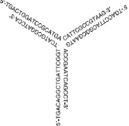
In order to better modulate immune system, several different CpG ODN were designed other than ‘K’, ‘D’ and ‘C’ types. One of them is Y-shaped ODN which is formed by 3 linear CpG ODNs which have complementary regions with each other (Figure 1.5 ). This ODN have fully phophodiester backbone and induces higher levels of IL-6 and TNFα from RAW264.7 cells, from mouse macrophage cell line, than single- and double-strands of same ODNs when used with same amounts. It is claimed in that report than this potency in cytokine release is related with efficient uptake into macrophages (M. Nishikawa, 2008).
Another work reported that 3’ extension of phophodiester linear (CpG or non-CpG ) ODNs with 24 guanine nucleotides turns these sequences into an effective IFNα inducer. Further extension doesn’t improve activity, whereas shorter polyG addition reduces. CpG motif has an additive effect to this stimulation. They showed that polyG addition confers complexation and efficient uptake into immune cells, like same sequences were mixed with DOTAP (complexation agent) (Haas T, 2008b).
23
Figure 1.5. Y-shaped CpG ODN (Nishikawa M, 2008).
1.2.2. Differential Immune Response Mediated by Particulate and Linear CpG ODN. Although both D-type and K-type ODNs contain CpG motifs and initiate their effect by triggering through TLR9 receptor, and even if they stimulate same cell types (B-cell and pDCs in human) they end up inducing quite distinct immune activation from each other. Understanding mechanism behind this dichotomy is very critical, since it will help to modulate immune response by administering K- or D- type according to the desired therapy. Firstly, D-ODNs have been reported to localize to different vesicles than K-ODNs which is claimed to be due to poly-G motifs of D-ODNs. Moreover, D-ODN was taken up four fold more efficiently than K-ODN by PBMC, where excess concentrations of each ODN did not inhibit binding or uptake of the other (Gursel M, 2002a). Collectively these data suggest that these ODNs use different mechanisms of uptake. Poly-G tail, which confers particulate nature to D-ODNs, is possibly responsible in efficient uptake of D-ODNs. Higher efficiency of uptake of particulate ODNs into immune cells such as mouse macrophages and DCs (De Jong S, 2007; Anderson RB, 2008) and human pDC (Kerkmann M, 2006) has been confirmed by other studies. Furthermore, ‘Interferon Regulatory Factor (IRF)-7’ was found to be critical for IFNα induction from pDC by D-ODN, as pDC from IRF7-/- mice is unresponsive to D-ODN, while cDC (conventional dendritic cells) release cytokines such IL-12 and IL-6 upon D-ODN stimulation. K-ODN-mediated stimulation of pDC or cDC from this IRF7-null mice is not affected (Honda K, 2005a). Two recent works helped to explain this unexpected feature of D