321
Pakistan Veterinary Journal
ISSN: 0253-8318 (PRINT), 2074-7764 (ONLINE) DOI: 10.29261/pakvetj/2018.031
Antibacterial Effect of Different Concentrations of Silver Nanoparticles
A Ebru Borum1* and Ertan Güneş2
1Department of Microbiology, Faculty of Veterinary Medicine, Balıkesir University, Balıkesir, Turkey 2Vocational School of Technical Sciences, Uludag University, Bursa, Turkey
*Corresponding author: [email protected]
ARTICLE HISTORY (17-327) A B S T R A C T Received: Revised: Accepted: Published online: September 27, 2017 December 19, 2017 December 20, 2017 February 12, 2018
Silver has been in use since time immemorial in the form of metallic silver, silver nitrate, silver sulfadiazine for the treatment of burns, wounds and several bacterial infections. Silver has long been known to show a strong antimicrobial effect to microorganisms. The antimicrobial effect of 30 and 100 ppm silver nanoparticles were investigated against Escherichia coli, Staphylococcus aureus, Salmonella
typhimurium, Enterococcus faecalis, Bacillus cereus, Bacillus subtilis,
Paenibacillus larvae, Candida albicans and Aspergillus niger. The microorganisms
were diluted with sterile distilled water and prepared dilutions of 106 of test
microorganisms. Dilutions of microorganisms cultured to blood agar base and incubated at 37°C for 24 hours. One mL dilution of 106 of all of microorganisms
was centrifuged at 3500 rpm for 20 minutes then 30 and 100 ppm of silver nanoparticle solutions were added. Samples were inoculated in blood agar for different time intervals i.e., 0, 2, 5, 10, 30, 60 minutes and 24 hour. Antibacterial activity of silver nanoparticles against various microorganisms was detected at 0, 2, 5, 10, 30, 60 minutes and 24 hours. As results, yeast, fungi and bacteria were inhibited at 30 and 100 ppm. But, P. larvae were not inhibited, while B. subtilis also could not be inhibited at 30 ppm. The antibacterial activity of 100 ppm was stronger than the antibacterial activity of 30 ppm of nanoparticles. Nanosilver is very effective to important pathogens.
©2017 PVJ. All rights reserved
Key words:
Antibacterial effect Microorganisms Silver nanoparticles
To Cite This Article: Borum AE and Güneş E, 2018. Antibacterial effect of different concentrations of silver
nanoparticles. Pak Vet J, 38(3): 321-324. http://dx.doi.org/10.29261/pakvetj/2018.031
INTRODUCTION
Silver is used in medical and surgical equipment such as endotracheal tubes, surgical meshes, catheters, dental filling materials, bandages, medical dressings and a topical cream to prevent burn-associated infections (Silver, 2003). Silver has long been known to show a strong antimicrobial effect to microorganisms (Liau et al., 1997). The nanosilver is effective against bacteria resistant to antibiotics, fungi and virus (Feng et al., 2000; Radzig et al., 2013). Silver nanoparticles are reported to be effective against fungi and bacteria as well as multidrug-resistant bacteria i.e., Escherichia coli (E. coli),
Staphylococcus aureus (S. aureus), Pseudomonas aeruginosa (P. aeruginosa), Proteus vulgaris (P. vulgaris), Bacillus subtilis (B. subtilis), Aspergillus niger
(A. niger), Candida albicans (C. albicans), Penicillium
citrinum (Pen. citrinum) (Kim et al., 2007; Lara et al.,
2010; Marambio-Jones and Hoek 2010; Lalueza et al., 2011).
Silver nanoparticles show great antibacterial effectiveness on important foodborne pathogens (include:
Escherichia coli O157:H7, Listeria monocytogenes, Salmonella typhimurium and Vibrio parahaemolyticus)
(Zhang et al., 2016). Also, nanosilver has antifungal effect on Candida albicans, Candida glabrata, Candida crusei,
Candida parapsilosis and Trichophyton mentagrophytes
(Kim et al., 2008). Silver nanoparticles have antiviral effect on human immunodeficiency virus-I (Lara et al., 2010) and herpes simplex (Barm-pinto et al., 2009).
Silver has been used as in creams, wound dressing, different medical devices, food containers, and water disinfection for antimicrobial agent. The new strains of bacteria were resistant to antibiotics. Therefore, new bactericides were development. The nanosilver is very effective for multidrug-resistant bacteria (Morones et al., 2005).
Silver in ionized form or in nanoparticles have got excellent antimicrobial, antifungal activities and was used for coating medical devices for preventing biofilm
Pak Vet J, 2018, 38(3): 321-324.
322 formation by pathogenic bacteria, water purification, (Bandyopadhyay et al., 2008; Chang et al., 2008; El-Naggar et al., 2016) and wound dressing for the promotion of healing (Feng et al., 2000; Abboud et al., 2014).
In this study, antimicrobial effect of 30 and 100 ppm silver nanoparticles were determined against E. coli, S.
aureus, S. typhimurium, Ent. faecalis, B. cereus, B. subtilis, P. larvae, C. albicans, A. niger. We detected
antibacterial effect of nanosilver on P. Larvae, which causes American foulbrood for honeybees the first time. We compared the efficacy of 30 and 100 ppm concentrations of silver.
MATERIALS AND METHODS
Escherichia coli (ATCC 25922), Staphylococcus aureus (ATCC 25923), Salmonella typhimurium (CCM
5445), Enterococcus faecalis (ATCC 29212), Bacillus
cereus (ATCC 6633), Bacillus subtilis (ATCC 6051), Paenibacillus larvae (ATCC 25747), Candida albicans
(ATCC 90028), Aspergillus niger (Clinical isolate, Uludag University, Medicine Faculty) were used for of antibacterial and antifungal activity of nanosilver. Bacteria and C. albicans were purchased from American Type Culture Collection (ATCC). The activity of nanosilver on P. larvae was determined for the first time in this study. E. coli, S. aureus, S. typhimurium, Ent.
faecalis, B. cereus, B. subtilis, P. larvae and C. albicans
were incubated in Fluid Thioglycollate Medium at 37°C for 24 hours and A. niger at 37°C for 7 days. The cells were washed twice and centrifuged at 3500 rpm for 20 minutes then suspended in distilled water, obtaining a final concentration of 106 cells/100 ml (Sondi and
Salopek-Sondi, 2004). Each culture was divided into two replicates. The cultures were then supplemented with 100 µl of 30 ppm and 100 ppm nanosilver solutions, respectively. Samples with different concentration of nanosilver were inoculated on blood agar at the exposure time of 0, 2, 5, 10, 30, 60 minutes and 24 hours. The cultures were incubated at 37°C for 24 hours. The number of bacteria was determined by counting the colonies.
Antifungal effect of nanosilver for A. niger was determined according to The National Committee for Clinical Laboratory Standards (NCCLS) M29 method. A.
niger was incubated Potato dextose agar at 37°C for 7
days. After, A. niger was inoculated to 1 ml of 0.85% sterile saline and, added 0.01 ml of Tween 20. Suspension of A. niger was homogenized with vortex for 15 seconds Spacciapoli et al. (2001). 1 ml dilution of 106 of A. niger
was centrifuged at 3500 rpm for 20 minutes. A 100 µl of 30 and 100 ppm silver solutions were added to two different cultures. Samples with nanosilver were inoculated on Potato Dextrose Agar (Bragg and Rannie, 1974) at 2, 5, 10, 30, and 60 minutes. The cultures were incubated at 37°C for 7 days. Plates without silver nanoparticles were used as negative control. We counted microorganisms growing on plates as colonies following Ki-Young et al. (2007) and Samarajeewa et al. (2017).
RESULTS AND DISCUSSION
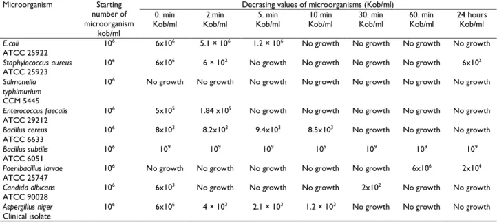
Thirty ppm of nanosilver solution inhibited the growth of S. typhimurium in 2 minutes. E. coli, S. aureus,
A. niger, C. albicans, Ent. faecalis and B. cereus were
inhibited on different time intervals of the exposure. But
S. aureus was not inhibited in 24 hours time interval of
exposure. P. larvae were not inhibited even in 60 min and 24 hours exposure. Bacillus subtilis was not inhibited by 30 ppm of nanosilver solution. Micro-organisms and inhibition periods are shown in Table 1.
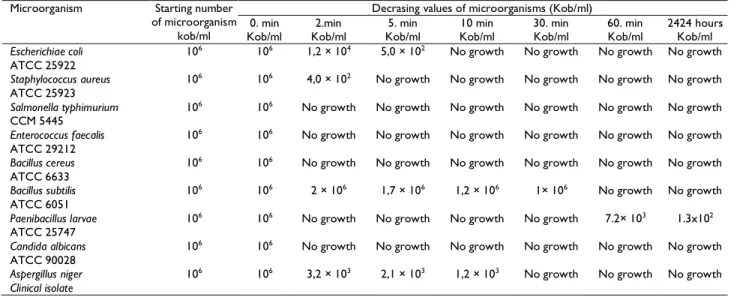
Ent. faecalis, S. typhimurium, B. cereus and, C. albicans were inhibited by 100 ppm of nanosilver within 2
minutes. E. coli did not grow at 10 minutes. S. aureus were inhibited at 5 minutes. A. niger and B. subtilis were not inhibited in the first 10 minutes but lost activity completely after 30 minutes. P. larvae were inhibited in the first 30 minutes but it grown 60 minutes and 24 hours. Microorganisms and inhibition periods are shown in Table 2.
Susceptibility to nanosilver is depended on the concentration. P. larvae inhibited in the first 30 minutes but it was not inhibited by 30 and 100 ppm of nanosilver solutions.
Table 1: Microorganisms and inhibition periods (30 ppm of nanosilver)
Microorganism Starting number of microorganism
kob/ml
Decrasing values of microorganisms (Kob/ml) 0. min
Kob/ml Kob/ml 2.min Kob/ml 5. min Kob/ml 10 min 30. min Kob/ml 60. min Kob/ml 24 hours Kob/ml
E.coli
ATCC 25922 10
6 6x10⁶ 5.1 × 10⁶ 1.2 × 10⁶ No growth No growth No growth No growth
Staphylococcus aureus
ATCC 25923 10
6 6x10⁶ 6 × 10² No growth No growth No growth No growth 6x10²
Salmonella typhimurium
CCM 5445
106 No growth No growth No growth No growth No growth No growth No growth
Enterococcus faecalis
ATCC 29212 10
6 5x10⁵ 1.84 x10⁵ No growth No growth No growth No growth No growth
Bacillus cereus
ATCC 6633 10
6 8x10³ 8.2x10³ 9.4x10³ 8.5x10³ No growth No growth No growth
Bacillus subtilis
ATCC 6051 10
6 109 109 109 109 109 109 109
Paenibacillus larvae
ATCC 25747 10
6 No growth No growth No growth No growth No growth 6x10⁶ 2x10⁴
Candida albicans
ATCC 90028 10
6 6x10³ No growth No growth No growth 2x10² No growth No growth
Aspergillus niger
Clinical isolate 10
Pak Vet J, 2018, 38(3): 321-324.
323 Table 2: Microorganisms and inhibition periods (100 ppm of nanosilver)
Microorganism Starting number of microorganism
kob/ml
Decrasing values of microorganisms (Kob/ml) 0. min
Kob/ml Kob/ml 2.min Kob/ml 5. min Kob/ml 10 min 30. min Kob/ml 60. min Kob/ml 2424 hours Kob/ml
Escherichiae coli
ATCC 25922 10
6 106 1,2 × 10⁴ 5,0 × 10² No growth No growth No growth No growth
Staphylococcus aureus
ATCC 25923 10
6 106 4,0 × 10² No growth No growth No growth No growth No growth
Salmonella typhimurium
CCM 5445 10
6 106 No growth No growth No growth No growth No growth No growth
Enterococcus faecalis
ATCC 29212
106 106 No growth No growth No growth No growth No growth No growth
Bacillus cereus
ATCC 6633 10
6 106 No growth No growth No growth No growth No growth No growth
Bacillus subtilis
ATCC 6051 10
6 106 2 × 10⁶ 1,7 × 10⁶ 1,2 × 10⁶ 1× 10⁶ No growth No growth
Paenibacillus larvae
ATCC 25747 10
6 106 No growth No growth No growth No growth 7.2× 10³ 1.3x10²
Candida albicans
ATCC 90028 10
6 106 No growth No growth No growth No growth No growth No growth
Aspergillus niger Clinical isolate
106 106 3,2 × 10³ 2,1 × 10³ 1,2 × 10³ No growth No growth No growth
Esherichia coli, Staphlococcus aureus, Aspergillus niger, Candida albicans, Enterococcus faecalis and Bacillus cereus were inhibited in different time intervals
(minutes).
P. larvae inhibited the first 30 minutes but after it
was not inhibited by 30 and 100 ppm of nanosilver solutions. Nanosilver was not effective for P. larvae. E.
faecalis, S. typhimurium, B. cereus and, C. albicans were
inhibited by 100 ppm of nanosilver within 2 minutes. We did not detect growth on plates. E. coli did not grow at 10 minutes. S. aureus were inhibited at 5 minutes. A. niger and, B. subtilis were not inhibited in first 10 minutes but it lost activity completely after 30 minutes.
The antibacterial activity of 100 ppm of nanoparticles was stronger than the antibacterial activity of 30 ppm of nanoparticles. But P. larvae were not inhibited after 60 minutes and 24 hours by both nanoparticle solutions. B.
subtilis was not inhibited by 30 ppm nanosilver. S. aureus
was inhibited at 5, 10, 30, 60 minutes but it was grown in 24 hours.
In one study, different concentrations of silver on E.
coli were investigated in vitro. According to the research,
it was determined that nano silver at 10 μg/cm3
concentration was effective at 70% on 105 CFU of E. coli.
50-60 μg/cm3 nanosilver concentration was effective for
100%. In the same study, 20 μg/cm3 of nano silver completely inhibited 104 CFU of E. coli. As the number of
bacteria decreased, nano silver was effective at lower concentrations (Yoon et al., 2007). 70 μg/mL concentration of silver nanoparticles were found to be effective on B. subtilis and E. coli. B. subtilis was found to be more susceptible to silver than E. coli (Ki-Young et al., 2007).
In another study, antimicrobial effect of a commercial nanosilver product, NanoCidR L2000, against some foodborne pathogens was evaluated. The MIC values of Ag NPs against tested pathogens were in the range of 3.12-6.25 μg/mL. While Listeria monocytogenes showed the MIC value of 6.25 μg/mL, Escherichia coli O157:H7,
Salmonella typhimurium and Vibrio parahaemolyticus all
showed the MIC values of 3.12 μg/mL. However, all the pathogens showed the same MBC value of 6.25 μg/mL (Zarei et al., 2014). Sixty-five bacterial isolates were isolated from 40 diabetic patients, S. aureus (37%) and P.
aeruginosa (18.5%) were the predominant isolates in the
ulcer samples. Squilla chitosan silver nanoparticles (Sq. Cs-Ag(0)) showed the maximum activity against the resistant bacteria (El-Naggar et al., 2016).
Thirty ppm of nanosilver solution inhibited
Salmonella typhimurium in 2 minutes. These results are
similar to our research findings.
Conclusions: The antibacterial activity of 100 ppm of
nanoparticles was stronger than the antibacterial activity of 30 ppm of nanoparticles. Nanosilver is very effective to important pathogens. Antimicrobial activity of nanosilver can be used for pathogens.
Author contribution: AEB helped in study design,
conduction of the laboratory investigations, collection of data and manuscript preparation. MEG helped in conduction of laboratory investigations and collection of data.
REFERENCES
Abboud EC, Settle JC, Legare TB, et al., 2014. Silver-based dressing for the reduction of surgical site infection: review of current experience and recommendation for future studies. Burns 40:30-9. Barm-pinto D, Shukla S, Perkas N, et al., 2009. Inhibitionof herpes simplex virus type 1 infection by silver nanoparticles capped with mercaptoethane sulfonate. Bioconjugate Chem 20:1497-502. Bandyopadhyay R, Sivaiah MV and Shankar PA, 2008. Silver-embedded
granular activated carbon as an antibacterial medium for water purification. J Chem Technol Biot 83:1177-80.
Bragg PD and Rannie DJ, 1974. The effect of silver ions on the respiratory chain of E. coli. Can J Microbiol 20:883-9.
Chang Q, He H and Ma Z, 2008. Efficient disinfection of Escherichia
coli in water by silver loaded alumina. J Inorganic Biochem102:
1736-42.
El-Naggar MY, Gohar YM, Sorour MA, et al., 2016. Hydrogel dressing with a nano-formula against methicillin-resistant Staphylococcus
aureus and Pseudomonas aeruginosa. Diabetic Foot Bacteria. J
Microbiol Biotechnol 26:408-20.
Feng QL, Wu J, Chen GQ, et al., 2000. A mechanistic study of the antibacterial effect of silver ions on Escherichia coli and
Staphylococcus aureus. J Biomed Mater Res 52:662-8.
Ki-Young Y, Jeong HB, Jae-Hong P, et al., 2007. Susceptibility constants of Escherichia coli and Bacillus subtilis to silver and copper nanoparticles. Sci Total Environ 373:572-5.
Kim JS, Kuk E, Yu KN, et al., 2007. Antimicrobial effects of silver nanoparticles. Nanomedicine 3:95-101.
Kim KJ, Sung WS, Moon SK, et al., 2008. Antifungal effect of silver nanoparticles on dermatophytes. J Microbiol Biotechnol 18:1482-4.
Pak Vet J, 2018, 38(3): 321-324.
324
Lalueza P, Monzon M, Arruebo M, et al., 2011. Bactericidal effects of different silver-containing materials. Mater Res Bull 46:2070-6. Lara HH, Ayala-Nunez NV, Ixtepan-Turrent L, et al., 2010. Bactericidal
effect of silver nanoparticles against multidrug-resistant bacteria. World J Microbiol Biotechnol 26:615-21.
Lara HH, Ayala-Nunez NV, Ixtepan-Turrent L, et al., 2010. Mode of antiviral action of silver nanoparticles against HIV-I. J Nanobiotechnol 8:1-10.
Liau SY, Read DC, Pugh WJ, et al., 1997. Interaction of silver nitrate with readily identifiable groups: relationship to the antibacterialaction of silver ions. Lett Appl Microbiol 25:279-83. Marambio-Jones C and Hoek EMV, 2010.A review of the antibacterial
effects of silver nanomaterials and potential implications for human health and the environment. J Nanopart Res 12:1531-51. Morones JR, Elechiguerra JL, Camacho A, et al., 2005. The bactericidal
effect of silver nanoparticles. Nanotechnology 16:2346-53. Radzig MA, Nadtochenko VA, Koksharova OA, et al., 2013.
Antibacterial effects of silver nanoparticles on gram-negative bacteria: Influence on the growth and biofilms formation, mechanisms of action. Colloids Surf B Biointerfaces 102:300-6.
Samarajeewa AD, Velicogna JR, Princz JI, et al., 2017. Effect on silver nano-particles on soil microbial growth, activity and community diversity in a sandy loam soil. Environ Pollut 220:504-13.
Silver S, 2003. Bacterial silver resistance: molecular biology and uses and misuses of silver compounds. FEMS Microbiol Rev 27:341-53. Sondi I and Salopek-Sondi B, 2004. Silver nanoparticles as antimicrobial
agent: a case study on E. coli as a model for gram-negative bacteria. J Colloid Interface Sci 275:177-82.
Spacciapoli P, Buxton D, Rothstein D, et al., 2001. Antimicrobial activity of silver nitrate against periodontal pathogens. J Periodontal Res 36:108-13.
Yoon KY, Byeon JH, Park JH, et al., 2007. Susceptibility constants of
Escherichia coli and Bacillus subtilis to silver and copper
nanoparticles. Sci Total Environ 373:572-5.
Zarei M, Jamnejad A and Khajehali E, 2014. Antibacterial Effect of silver nanoparticles against four foodborne pathogens. Jundishapur J Microbiol 7:1-4.
Zhang FH, Li M, Wei ZJ, et al., 2016. The effect of a combined nanoparticulate/calcium hydroxide medication on the biofilm of
Enterococcus faecalis in starvation phase. Shanghai Kou Qiang Yi