Association of neutrophil to lymphocyte ratio with presence
of isolated coronary artery ectasia
Nötrofilin lenfosite oranının izole koroner arter ektazisi varlığı ile ilişkisi
Department of Cardiology, Balikesir University, Faculty of Medicine, Balikesir; #Department of Cardiology, Bezmialem Vakif University, Faculty of Medicine, Istanbul;
*Department of Cardiology, Erzurum Training and Research Hospital, Erzurum;
†Department of Cardiology, Dr. Siyami Ersek Cardiovascular and Thoracic Surgery Training and Research Hospital, Istanbul Turgay Işık, M.D., Erkan Ayhan, M.D., Hüseyin Uyarel, M.D.,# İbrahim Halil Tanboğa, M.D.,*
Mustafa Kurt, M.D.,* Mahmut Uluganyan, M.D.,† Mehmet Ergelen, M.D.,# Abdurrahman Eksik, M.D.†
Objectives: Coronary artery ectasia (CAE) has been de-fined as a dilated artery luminal diameter that is at least 50% greater than the diameter of the normal portion of the artery. Isolated CAE is defined as CAE without significant coronary artery stenosis and isolated CAE has more pronounced in-flammatory symptoms. Neutrophil to lymphocyte ratio (NLR) is widely used as a marker of inflammation and an indicator of cardiovascular outcomes in patients with coronary artery disease. We examined a possible association between NLR and the presence of isolated CAE.
Study design: In this study, 2345 patients who underwent coronary angiography for suspected or known ischemic heart disease were evaluated retrospectively. Following the appli-cation of exclusion criteria, our study population consisted of 81 CAE patients and 85 age- and gender-matched subjects who proved to have normal coronary angiograms. Baseline neutrophil, lymphocyte and other hematologic indices were measured routinely prior to the coronary angiography.
Results: Patients with angiographic isolated CAE had sig-nificantly elevated NLR when compared to the patients with normal coronary artery pathology (3.39±1.36 vs. 2.25±0.58, p<0.001). A NLR level ≥2.37 measured on admission had a 77% sensitivity and 63% specificity in predicting isolated CAE at ROC curve analysis. In the multivariate analysis, hyper-cholesterolemia (OR=2.63, 95% CI 1.22-5.65, p=0.01), obe-sity (OR=3.76, 95% CI 1.43-9.87, p=0.007) and increased NLR (OR=6.03, 95% CI 2.61-13.94, p<0.001) were indepen-dent predictors for the presence of isolated CAE.
Conclusion: Neutrophil to lymphocyte ratio is a readily avail-able clinical laboratory value that is associated with the pres-ence of isolated CAE.
Amaç: Koroner arter ektazisi (KAE), koroner arterlerin nor-mal koroner arter bölgesine göre en az %50’den daha fazla genişlemesi olarak tanımlanır. İzole KAE ciddi koroner arter darlığı olmaksızın KAE’nin bulunması olarak tanımlanır ve belirgin enflamatuvar özelliklere sahiptir. Nötrofilin lenfosite oranı (NLO) enflamasyonun yaygın kullanılan bir belirtecidir ve koroner arter hastalığı olan kişilerde hastalığın seyrinin bir göstergesidir. Bu çalışmada, NLO ile izole KAE arasındaki olası ilişki araştırıldı.
Çalışma planı: Bilinen veya şüphenilen iskemik kalp hasta-lığı nedeniyle koroner anjiyografi yapılan 2345 hasta geriye dönük olarak değerlendirildi. Dışlanma kriterleri sonrası, ça-lışma popülasyonu KAE olan 81 hasta ve bu gurupla yaş ve cinsiyet olarak eşleşmiş anjiyografileri normal 85 hastayı kap-samaktaydı. Koroner anjiyografi öncesinde tüm hastalarda nötrofil, lenfosit ve diğer hematolojik göstergelerin ölçümleri rutin olarak yapılmıştı.
Bulgular: İzole KAE’si olan hastalarda NLO düzeyinin nor-mal koroner arterli olgulara kıyasla belirgin olarak artmış ol-duğu görüldü (3.39±1.36 ve 2.25±0.58, p<0.001). Receiver operating curve (ROC) eğrisi analizlerinde, başvuru anında ölçülen NLO ≥2.37 değerinin izole KAE’yi öngörmede du-yarlılığının %77 ve özgüllüğünün %63 olduğu saptandı. Çok değişkenli lojistik regresyon analizinde hiperkolesterolemi (OO=2.63, %95 GA 1.22-5.65, p=0.01), obezite (OO=3.76, %95 GA 1.43-9.87, p=0.007) ve artmış NLO (OO=6.03, %95 GA 2.61-13.94, p<0.001) izole KAE varlığı için bağımsız be-lirleyiciler olarak saptandı.
Sonuç: Nötrofilin lenfosite oranı izole KAE varlığı ile ilişkili olan, kolaylıkla ölçülebilen bir laboratuvar bulgusudur. Received: March 11, 2012 Accepted:October 17, 2012
Correspondence: Dr. Turgay Işık. Balıkesir Üniversitesi Tıp Fakültesi, Kardiyoloji Anabilim Dalı, Balıkesir. Tel: +90 266 - 612 14 55 e-mail: [email protected]
© 2013 Turkish Society of Cardiology
he role of inflammation in cardiovascular dis-eases is known.[1] White blood cell (WBC) count and related parameters are markers of inflammation in cardiovascular disease and neutrophil to lympho-cyte ratio (NLR) has been shown to have the greatest predictive power for poor outcomes in patients diag-nosed with or at high risk for coronary artery disease. [2,3] NLR is more predictive because the two inflam-matory markers, neutrophil and lymphocyte, indicate opposing clinical outcomes.[3]
Coronary artery ectasia (CAE), an abnormality of the coronary anatomy, has been defined as a dilated artery luminal diameter that is at least 50% greater than the diameter of the normal portion of the artery. [4] Isolated CAE is defined as CAE without significant coronary artery stenosis. It has been demonstrated that isolated CAE is associated with an increased cardio-vascular event rate.[5] The underlying mechanisms re-sponsible for ectasia formation are not well understood, however previous studies have demonstrated that CAE may be a form of atherosclerosis with more active in-flammatory properties than normal vessels.[6-10]
Given that the mechanism of CAE may be asso-ciated with increased inflammation and that NLR is a marker of inflammation, we hypothesized that in-creased NLR would be associated with the presence of isolated CAE. We evaluated this hypothesis in a popu-lation of patients who underwent coronary angiogra-phy for suspected or known ischemic heart disease.
PATIENTS AND METHODS Study population
In this study, 2345 patients who underwent coronary angiography for suspected or known ischemic heart disease were retrospectively evaluated between April 2009 and July 2011. Out of these, 102 (4.3%) patients had isolated CAE. 21 patients were excluded due to exclusion criteria (8 patients with previous history of myocardial infarction (MI) or percutaneous coronary intervention; 4 patients with heart failure; 2 patients with valvular heart disease; 4 patients with inflam-matory diseases; 1 patient with malignancy; 1 patient with hematological disorders and 1 patient with cur-rent use of corticosteroids). Finally, 81 patients with isolated CAE were included in the study. The control group consisted of 85 age- and gender-matched sub-jects who were selected in a consecutive manner from
among catheterized patients during the same study peri-od who had normal coronary angiograms. All patients re-cruited into the study under-went coronary angiography for presence of chest pain or had objective signs of isch-emia (treadmill exercise or myocardial SPECT).
The patients’ laboratory and clinical characteristics, such as age, sex, diabetes mellitus (DM), hyperten-sion, hypercholesterolemia,
smoking, family history of coronary artery disease (CAD), height and weight, were accessed through medical records. In cases of inconsistencies, the pa-tients were contacted by telephone. The body mass index (BMI) was calculated by dividing weight in ki-lograms by height in meters squared (kg/m2).
Laboratory measurements
In our hospital, blood samples are collected from the ante-cubital vein by an atraumatic puncture prior to the coronary angiography and are sent to the laborato-ry for analysis within 1 hour after collection. Venous blood is collected in a tube containing K3 EDTA for measurement of hematologic indices in all patients undergoing the coronary angiography. Hematologic indices are evaluated from CBC (complete blood count) analysis performed by a Coulter LH 780 He-matology Analyzer (Beckman Coulter Ireland Inc. Mervue, Galway, Ireland). The glomerular filtration rate (GFR) was estimated by the simplified Modifica-tion of Diet in Renal Disease EquaModifica-tion.
Coronary angiography and echocardiography Coronary angiographies were performed in our clinic using the standard Judkins technique without nitro-glycerin. The right anterior oblique view was used to evaluate ectasia in the left coronary system and left anterior oblique view was used for the evaluation of the right coronary artery. Evaluations were performed visually by two experienced angiographers. The ves-sel diameter was calculated quantitatively in case of the presence of conflict about CAE.
Transthoracic echocardiography was performed on patients before discharge using a system V
(Ving-T
Abbreviations:BMI Body mass index CAD Coronary artery disease CAE Coronary artery ectasia DM Diabetes mellitus GFR Glomerular filtration rate LVEF Left ventricular ejection fraction
MI Myocardial infarction MMP Matrix metalloprotease NGAL Neutrophil gelatinase-
associated lipocalin NLR Neutrophil to lymphocyte ratio
ROC Receiver operating characteristics WBC White blood cell
med, GE, Horten, Norway) with a 2.5 MHz phased-array transducer. Recordings were taken on patients positioned in the left lateral decubitus position. The left ventricular ejection fraction (LVEF) was mea-sured using the modified Simpson’s rule.[11]
Definitions
The diagnosis of DM was based on previous history of diabetes treated with or without drug therapies. Hypercholesterolemia was defined as total cholesterol of ≥200 mg/dl. A BMI of ≥30 kg/m2 was defined as obese. According to the results of coronary angiogra-phy, significant coronary artery stenosis was defined as ≥50% of major coronary arteries. Absence of any atherosclerotic plaques was regarded as normal coro-nary artery pathology.
Stable angina was defined as discomfort in the chest, jaw, shoulder, back, or arms, typically elicited by exertion or emotional stress and relieved by rest or nitroglycerin. Current smokers were defined as those who had smoked for some period during the past year. Patients were considered as having ESRD, if they were dependent on chronic dialysis. Renal insuffi-ciency was defined as a GFR of <60 ml/min/1.732 m2.
beta-blockers and increased NLR) were included in the model. Two-tailed p values <0.05 were considered to indicate statistical significance. Statistical analyses were performed using SPSS, version 15.0 for Win-dows.
RESULTS
Among 166 patients (mean age 58.25±9.6, 69.3% male), NLR ranged from 1.30 to7.40 (median 2.50, mean 2.81±1.18). The baseline characteristics of the normal vessels or vessels where isolated CAE was present angiographically are summarized in Table 1. Patients with isolated CAE were more frequently diagnosed with diabetes, hypercholesterolemia, and obesity relative to the control group and had signifi-cantly elevated neutrophil counts and NLR and de-creased lymphocyte values. There was no difference between groups for presence of hypertension, smok-ing, family history for CAD, renal insufficiency and medications. In addition, the baseline LVEF, WBC, hemoglobin, platelet and monocyte parameters were similar in both groups.
Moreover, we found that isolated CAE most com-monly affects the right coronary artery (63.4%), left
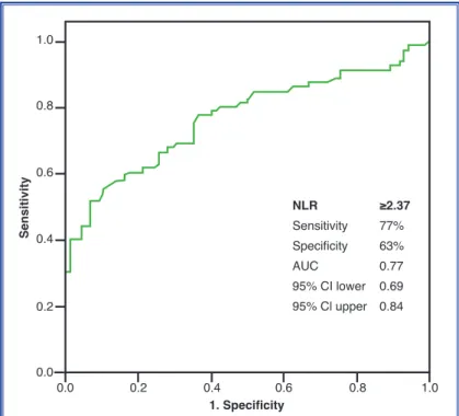
1.0 0.8 0.6 0.4 0.2 0.0 0.0 0.2 0.4 0.6 0.8 1.0 Sensitivity 1. Specificity NLR ≥2.37 Sensitivity 77% Specificity 63% AUC 0.77 95% CI lower 0.69 95% Cl upper 0.84
Figure 1. The receiver-operating characteristic curve of neutrophil to lym-phocyte ratio for predicting isolated coronary artery ectasia. NLR: Neutro-phil to lymphocyte ratio; AUC: Area under curve.
The local ethics committee approved this study.
Statistical analysis
Continuous variables are expressed as mean ± standard deviation. Categorical variables are expressed as percentages. Adequacy of all parameters to normal distribution, was tested by using the Kolmogorov-Smirnov test. To compare parametric continuous variables, the Student’s t test was used; to compare nonparametric continuous vari-ables, the Mann-Whitney U-test was used; to compare categorical variables, the chi-square test was used. The Receiver Operat-ing Characteristics (ROC) curve was used to demonstrate the sensitivity and specific-ity of NLR and its respective optimal cut-off value for predicting isolated CAE. Mul-tivariate logistic regression analysis was used to identify the independent predictors of the presence of isolated CAE. All vari-ables showing significance values <0.25 on univariate analysis (diabetes, hypercholes-terolemia, smoking, obesity, WBC, use of
the presence or absence of isolated CAE (Area Under curve [AUC]: 0.77, CI 95%, 0.69-0.84). A NLR value ≥2.37 yielded a sensitivity of 77%, specificity of 63%, positive predictive value of 67.0% and a negative pre-dictive value of 75.0% (Fig. 1). When we divided the study population into two groups according to the NLR anterior descending artery (57.3%), and left
circum-flex artery (46.3%). Isolated CAE most frequently af-fects one vessel (43.2%) or two vessels (43.2%) and less frequently three vessels (13.6%).
In a ROC curve analysis, a NLR value of 2.37 was identified as an effective cutpoint in the segregation of
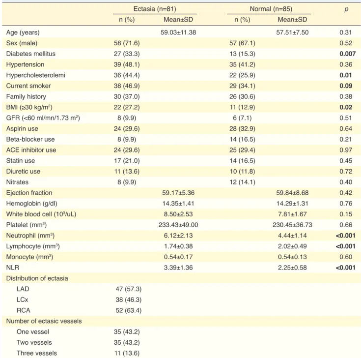
Table 1. Baseline characteristics of angiographicly normaly and ectatic coronary vessels
Ectasia (n=81) Normal (n=85) p n (%) Mean±SD n (%) Mean±SD Age (years) 59.03±11.38 57.51±7.50 0.31 Sex (male) 58 (71.6) 57 (67.1) 0.52 Diabetes mellitus 27 (33.3) 13 (15.3) 0.007 Hypertension 39 (48.1) 35 (41.2) 0.36 Hypercholesterolemi 36 (44.4) 22 (25.9) 0.01 Current smoker 38 (46.9) 29 (34.1) 0.09 Family history 30 (37.0) 26 (30.6) 0.38 BMI (≥30 kg/m2) 22 (27.2) 11 (12.9) 0.02 GFR (<60 ml/mn/1.73 m2) 8 (9.9) 6 (7.1) 0.51 Aspirin use 24 (29.6) 28 (32.9) 0.64 Beta-blocker use 8 (9.9) 14 (16.5) 0.21
ACE inhibitor use 24 (29.6) 25 (29.4) 0.97
Statin use 17 (21.0) 14 (16.5) 0.45
Diuretic use 11 (13.6) 10 (11.8) 0.72
Nitrates 8 (9.9) 12 (14.1) 0.40
Ejection fraction 59.17±5.36 59.84±8.68 0.42
Hemoglobin (g/dl) 14.35±1.41 14.29±1.31 0.76
White blood cell (103/uL) 8.50±2.53 7.81±1.67 0.15
Platelet (mm3) 233.43±49.00 230.45±36.73 0.66 Neutrophil (mm3) 6.12±2.13 4.44±1.14 <0.001 Lymphocyte (mm3) 1.74±0.38 2.02±0.49 <0.001 Monocyte (mm3) 0.54±0.17 0.54±0.13 0.60 NLR 3.39±1.36 2.25±0.58 <0.001 Distribution of ectasia LAD 47 (57.3) LCx 38 (46.3) RCA 52 (63.4)
Number of ectasic vessels
One vessel 35 (43.2)
Two vessels 35 (43.2)
Three vessels 11 (13.6)
Results are expressed as mean±SD and percentage. To compare parametric continuous variables (age, hemoglobine and platelet), the Student’s t-test was used; to compare nonparametric continuous variables (Ejection fraction, WBC, Neutrophil, Lymphocyte, Monocyte and NLR) the Mann-Whitney U-test was used. ACE: Angiotensin converting enzyme; BMI: Body mass index; GFR: Glomerular filtration rate; LAD: Left anterior descending artery; LCx: Left circumflex artery; NLR: Neutrophil to lymphocyte ratio; RCA: Right coronary artery.
level cut-off value or 2.37 used in the ROC analysis, diabetes, hypertension and the presence of isolated CAE were significantly in increased the NLR group (p<0.001, p=0.02, p<0.001, respectively, Table 2).
Variables found to be statistically significant in uni-variate analyses were included in multiuni-variate logistic regression analysis. Hypercholesterolemia (OR=2.63, 95% CI 1.22-5.65, p=0.01), obesity (OR=3.76, 95% CI 1.43-9.87, p=0.007), and increased NLR (OR=6.03, 95% CI 2.61-13.94, p<0.001) were independently as-sociated with the presence of isolated CAE (Table 3).
DISCUSSION
This is the first study that has been conducted evaluat-ing the relationship between NLR and the presence of isolated CAE. A greater baseline NLR value was
in-dependently associated with the presence of isolated CAE.
In previous publications, CAE exists in 1.5-5% of patients included in coronary angiographic studies. [12] In this study, we reported an incidence of isolated CAE of 4.3% among patients who underwent coro-nary angiography for the presence of chest pain or had objective signs of ischemia. CAE can cause angina pectoris and even MI with vasospasm, dissection or thrombus in patients without CAD.[5] In several stud-ies, patients with CAE have been shown to have in-creased risk of mortality equivalent to patients with CAD.[13]
Despite advances in cardiology, the pathogenesis of CAE remains unclear. The determination of fac-tors associated with the presence of CAE may have Table 2. Baseline characteristics of patients with normal and increased NLR
NLR <2.37 (n=72) NLR ≥2.37 (n=94) p n (%) Mean±SD n (%) Mean±SD Age (years) 57.47±8.11 58.86±10.60 0.35 Sex (male) 48 (66.7) 67 (71.3) 0.52 Diabetes mellitus 8 (11.1) 32 (34.0) <0.001 Hypertension 25 (34.7) 49 (52.1) 0.02 Hypercholesterolemi 24 (33.3) 34 (36.2) 0.70 Current smoker 25 (34.7) 42 (44.7) 0.19 Family history 24 (33.3) 32 (34.0) 0.92 BMI (≥30 kg/m2) 15 (20.8) 18 (19.1) 0.78 GFR (<60 ml/mn/1.73 m2) 4 (5.6) 10 (10.6) 0.24 Aspirin use 22 (30.6) 30 (31.9) 0.85 Beta-blocker use 11 (15.3) 11 (11.7) 0.50
ACE inhibitor use 25 (34.7) 24 (25.5) 0.19
Statin use 12 (16.7) 19 (20.2) 0.56 Diuretic use 6 (8.3) 15 (16.0) 0.14 Nitrates 12 (16.7) 8 (8.5) 0.11 Ejection fraction 59.38±8.82 59.61±5.79 0.67 Hemoglobine (g/dl) 14.31±1.38 14.32±1.34 0.96 WBC (103/uL) 7.20±1.68 8.87±2.21 <0.001 Platelet (mm3) 228.20±41.95 234.74±43.88 0.33 Monocyte (mm3) 0.52±0.14 0.56±0.16 0.09 Isolated CAE 18 (25.0) 63 (67.0) <0.001
Results are expressed as mean±SD and percentage. To compare parametric continuous variables (Age, Hemoglobine and Platelet), the Student’s t test was used; to compare nonparametric continuous variables (Ejection fraction, WBC and Monocyte) the Mann-Whitney U-test was used. ACE: Angiotensin converting enzyme; BMI: Body mass index; CEA: Coronary artery ectasia; GFR: Glomerular filtration rate; NLR: Neutrophil to lymphocyte ratio, WBC: white blood cell.
a salutary influence on the management of these pa-tients. Although CAE can be congenital, some factors associated with the presence of CAE include inflam-matory markers such as C-reactive protein, IL-6, tu-mor necrosis factor-alpha, matrix metalloprotease (MMP),[7,8] and atherosclerotic risk factors, including advanced age, DM and smoking.[9] In addition, Li et al. demonstrated that patients with isolated CAE had increased leukocyte, neutrophil and monocyte counts when compared to patients with normal coronary ar-teries.[10] Similarly, in our study, neutrophil, NLR and cardiovascular risk factors, such as diabetes, hyper-cholesterolemia and obesity were found to be signifi-cantly increased in patients with isolated CAE. In this study, we determined a cut-off point NLR ≥2.37 for predicting isolated CAE, and a much lower cut-off point in comparison to many acute vascular condi-tions.[3] This may be related to chronic, mild inflam-mation occurring in CAE.
Antoniadis et al.[6] reported that infiltration of the media layer by inflammatory cells can be seen in ec-tatic segments.Neutrophils cause damage to the tis-sue and may play an important role in pathogenesis of CAE by secreting elastase, MMP and oxygen free radicals.[14,15] Akyel et al.[16] reported that the protein neutrophil gelatinase-associated lipocalin (NGAL) is increased in patients with isolated CAE. NGAL is secreted from activated neutrophils. NGAL prevents degradation of MMP-9 by the formation of an hibitory complex. Therefore, MMP-9 expression in-creases in the presence of NGAL, and this leads to increased degradation of gelation and collagen. As a result of this destructive chronic inflammatory state, artery walls are weakened potentially resulting in ec-sasia.
At the same time, in this study we observed that
the lymphocyte level was decreased among the CAE group. The probable causes of lymphopenia include decreased production of lymphocyte as a result of increased steroid level due to CAE induced stressed condition,[17] and increased apoptosis of lymphocyte triggered by increased inflammation.[18] Lymphocytes may have a pivotal role in modulating the inflamma-tory response in this slow chronic inflammainflamma-tory state, similar to atherosclerosis.[19,20] In addition, several pre-vious studies have reported decreased lymphocytes in acute cardiovascular events, and there is a negative correlation between cardiovascular prognosis and the lymphocyte level.[3,21-23]
In this study, in addition to increased NLR in iso-lated CAE, there is an independent association of both obesity and hyperlipidemia with CAE. Sudhir et al.[24] demonstrated that CAE is more prevalent in patients with hypercholesterolemia. They have suggested that lipoproteins may be directly involved in this process, perhaps reducing the tensile strength of the artery wall.Waly et al.[25] reported an associated between CAE and obesity.Previous studies have indicated a positive and significant association between serum elastase activity and BMI. Increased elastase activi-ties may contribute to CAE.[26]
This study has several limitations. First, cytokines related to inflammation were not investigated as a re-sult of the retrospective design. Second, we did not perform an analysis of the prognostic value of NLR in CAE. The pathogenesis of CAE and CAD is similar and many studies of stable CAD have demonstrated the prognostic value of NLR.[27] Based on this point, we hypothesized that there may be a prognostic value of NLR in CAE patients. Prospective randomized controlled studies would aid in understanding this phenomena. Third, as the definition of normal coro-Table 3. Independent predictors of angiographic ectasia in multivariate logistic regression analysis
Univariate OR (95% CI) p Mulivariate OR (95% CI) p
Diabetes mellitus 2.76 (1.30-5.86) 0.008 1.85 (0.77-4.44) 0.16
Hypercholesterolemia 2.29 (1.19-4.40) 0.01 2.63 (1.22-5.65) 0.01
Current smoker 1.70 (0.91-3.19) 0.09 1.66 (0.80-3.45) 0.17
Body mass index (≥30 kg/m2) 2.50 (1.12-5.58) 0.02 3.76 (1.43-9.87) 0.007
Neutrophil to lymphocyte ratio (≥2.37) 6.09 (3.07-12.09) <0.001 6.03 (2.61-13.94) <0.001
Beta-blocker 0.55 (0.22-1.40) 0.21 0.43 (0.13-1.40) 0.16
nary arteries is based on angiographic views, athero-sclerotic plaques could not be excluded. However in clinical practice, patients with normal coronary anato-my do not undergo intravascular ultrasound routinely and the diagnosis of normal coronary artery is usu-ally by visual assessment at the coronary angiography procedure. Forth, although the formation of CAE is a slow, chronic condition, we only evaluated a single serum NLR in this study.
Conflict-of-interest issues regarding the authorship or article: None declared
REFERENCES
1. Ross R. Atherosclerosis-an inflammatory disease. N Engl J Med 1999;340:115-26. 2. Horne BD, Anderson JL, John JM, Weaver A, Bair TL, Jensen KR, et al. Which white blood cell subtypes predict increased cardiovascular risk? J Am Coll Cardiol 2005;45:1638-43. 3. Núñez J, Núñez E, Bodí V, Sanchis J, Miñana G, Mainar L, et al. Usefulness of the neutrophil to lymphocyte ratio in predict-ing long-term mortality in ST segment elevation myocardial infarction. Am J Cardiol 2008;101:747-52.
4. Swaye PS, Fisher LD, Litwin P, Vignola PA, Judkins MP, Kemp HG, et al. Aneurysmal coronary artery disease. Circu-lation 1983;67:134-8.
5. Krüger D, Stierle U, Herrmann G, Simon R, Sheikhzadeh A. Exercise-induced myocardial ischemia in isolated coronary artery ectasias and aneurysms (“dilated coronopathy”). J Am Coll Cardiol 1999;34:1461-70. 6. Antoniadis AP, Chatzizisis YS, Giannoglou GD. Pathogenetic mechanisms of coronary ectasia. Int J Cardiol 2008;130:335-43. 7. Aydin M, Tekin IO, Dogan SM, Yildirim N, Arasli M, Sayin MR, et al. The levels of tumor necrosis factor-alpha and in-terleukin-6 in patients with isolated coronary artery ectasia. Mediators Inflamm 2009;2009:106145. 8. Dogan A, Tuzun N, Turker Y, Akcay S, Kaya S, Ozaydin M. Matrix metalloproteinases and inflammatory markers in coro-nary artery ectasia: their relationship to severity of coronary artery ectasia. Coron Artery Dis 2008;19:559-63. 9. Finkelstein A, Michowitz Y, Abashidze A, Miller H, Keren G, George J. Temporal association between circulating pro-teolytic, inflammatory and neurohormonal markers in patients with coronary ectasia. Atherosclerosis 2005;179:353-9. 10. Li JJ, Nie SP, Qian XW, Zeng HS, Zhang CY. Chronic inflam- matory status in patients with coronary artery ectasia. Cyto-kine 2009;46:61-4. 11. Schiller NB, Shah PM, Crawford M, DeMaria A, Devereux R, Feigenbaum H, et al. Recommendations for quantitation of the left ventricle by two-dimensional echocardiography.
American Society of Echocardiography Committee on Stan-dards, Subcommittee on Quantitation of Two-Dimensional Echocardiograms. J Am Soc Echocardiogr 1989;2:358-67. 12. Boles U, Eriksson P, Zhao Y, Henein MY. Coronary
ar-tery ectasia: remains a clinical dilemma. Coron Arar-tery Dis 2010;21:318-20.
13. Harikrishnan S, Sunder KR, Tharakan J, Titus T, Bhat A, Sivasankaran S, et al. Coronary artery ectasia: angiographic, clinical profile and follow-up. Indian Heart J 2000;52:547-53.
14. Baldus S, Heeschen C, Meinertz T, Zeiher AM, Eiserich JP, Münzel T, et al. Myeloperoxidase serum levels predict risk in patients with acute coronary syndromes. Circulation 2003;108:1440-5. 15. Dollery CM, Owen CA, Sukhova GK, Krettek A, Shapiro SD, Libby P. Neutrophil elastase in human atherosclerotic plaques: production by macrophages. Circulation 2003;107:2829-36. 16. Akyel A, Sahinarslan A, Kiziltunc E, Yıldız U, Alsancak Y,
Akboga MK, et al. Neutrophil gelatinase-associated lipo-calin levels in isolated coronary artery ectasia. Can J Cardiol 2011;27:773-8. 17. Onsrud M, Thorsby E. Influence of in vivo hydrocortisone on some human blood lymphocyte subpopulations. I. Effect on natural killer cell activity. Scand J Immunol 1981;13:573-9. 18. Hotchkiss RS, Karl IE. The pathophysiology and treatment of sepsis. N Engl J Med 2003;348:138-50.
19. Ait-Oufella H, Salomon BL, Potteaux S, Robertson AK, Gourdy P, Zoll J, et al. Natural regulatory T cells con-trol the development of atherosclerosis in mice. Nat Med 2006;12:178-80. 20. Gupta S, Agrawal A, Agrawal S, Su H, Gollapudi S. A para-dox of immunodeficiency and inflammation in human aging: lessons learned from apoptosis. Immun Ageing 2006;3:5. 21. Dragu R, Khoury S, Zuckerman R, Suleiman M, Mutlak D, Agmon Y, et al. Predictive value of white blood cell subtypes for long-term outcome following myocardial infarction. Ath-erosclerosis 2008;196:405-12. 22. Ommen SR, Gibbons RJ, Hodge DO, Thomson SP. Useful-ness of the lymphocyte concentration as a prognostic marker in coronary artery disease. Am J Cardiol 1997;79:812-4. 23. Tamhane UU, Aneja S, Montgomery D, Rogers EK, Eagle
KA, Gurm HS. Association between admission neutrophil to lymphocyte ratio and outcomes in patients with acute coro-nary syndrome. Am J Cardiol 2008;102:653-7.
24. Sudhir K, Ports TA, Amidon TM, Goldberger JJ, Bhushan V, Kane JP, et al. Increased prevalence of coronary ectasia in heterozygous familial hypercholesterolemia. Circulation 1995;91:1375-80.
25. Waly HM, Elayda MA, Lee VV, el-Said G, Reul GJ, Hall RJ. Coronary artery ectasia in Egyptian patients with coronary ar-tery disease. Tex Heart Inst J 1997;24:349-52.
26. Bizbiz L, Bonithon-Kopp C, Ducimetierè P, Berr C, Alp-erovitch A, Robert L. Relation of serum elastase activity to ultrasonographically assessed carotid artery wall lesions and
cardiovascular risk factors. The EVA study. Atherosclerosis 1996;120:47-55.
27. Papa A, Emdin M, Passino C, Michelassi C, Battaglia D, Coc-ci F. Predictive value of elevated neutrophil-lymphocyte ratio on cardiac mortality in patients with stable coronary artery disease. Clin Chim Acta 2008;395:27-31.
Key words: Coronary angiography; coronary vessel anomalies/ complications; coronary vessels/pathology; neutrophils; lympho-cytes; dilatation, pathologic.
Anahtar sözcükler: Koroner anjiyografi; koroner damar anomalisi/ komplikasyon; koroner damarlar/patoloji; nötrofil; lemfosit; dilatas-yon, patolojik.