栽藻造押8629原愿圆圆源缘员苑圆 8629-82210956 耘皂葬蚤造押ijopress岳员远猿援糟燥皂
Comparison of pre-treatment and post-treatment use
of selenium in retinal ischemia reperfusion injury
窑Basic Research窑
1Department of Ophthalmology, Faculty of Medicine, Balikesir University, Balikesir 10010, Turkey
2Department of Biochemistry, Faculty of Veterinary, Balikesir University, Balikesir 10010, Turkey
3Department of Histology and Embryology, Faculty of Medicine, Erciyes University, Kayseri 38010, Turkey 4Department of Physiology, Faculty of Medicine, Balikesir University, Balikesir 10010, Turkey
5Department of Pharmacology, Faculty of Veterinary, Balikesir University, Balikesir 10010, Turkey
6Department of Ophthalmology, Faculty of Medicine, Adnan Menderes University, Aydin 09010, Turkey
7Department of Biochemistry, Faculty of Medicine, Balikesir University, Balikesir 10010, Turkey
Correspondence to: Alper Yazici. Department of Ophthalmology, Cagis Kampus, Faculty of Medicine, Balikesir University, Balikesir 10010, Turkey. [email protected] Received: 2014-05-14 Accepted: 2014-08-27
Abstract
·
AIM: To investigate the effects of selenium in rat retinal ischemia reperfusion (IR) model and compare pre-treatment and post-pre-treatment use.·
METHODS: Selenium pre-treatment group ( =8) was treated with intraperitoneal (i.p.) selenium 0.5 mg/kg for 7d and terminated 24h after the IR injury. Selenium post-treatment group ( =8) was treated with i.p. selenium 0.5 mg/kg for 7d after the IR injury with termination at the end of the 7d period. Sham group ( =8) received i.p. saline injections identical to the selenium volume for 7d with termination 24h after the IR injury. Control group ( =8) received no intervention. Main outcome measures were retina superoxide dismutase (SOD), glutathione (GSH), total antioxidant status (TAS), malondialdehyde (MDA), DNA fragmentation levels, and immunohistological apoptosis evaluation.·
RESULTS: Compared to the Sham group, selenium pre -treatment had a statistical difference in all parameters except SOD. Post -treatment selenium also resulted in statistical differences in all parameters except the MDA levels. When comparing selenium groups, the pre -treatment selenium group had a statistically higher success in reduction of markers of cell damage such as MDA and DNA fragmentation. In contrast, the post -selenium treatment group had resulted in statisticallyhigher levels of GSH. Histologically both selenium groups succeeded to limit retinal thickening and apoptosis. Pre -treatment use was statistically more successful in decreasing apoptosis in ganglion cell layer compared to post-treatment use.
·
CONCLUSION: Selenium was successful in retinal protection in IR injuries. Pre -treatment efficacy was superior in terms of prevention of tissue damage and apoptosis.·
KEYWORDS: retina; selenium; ischemia-reperfusion; apoptosis; pre-treatment; post-treatment; oxidative stress DOI:10.3980/j.issn.2222-3959.2015.02.09Yazici A, Aksit H, Sari ES, Yay A, Erken HA, Aksit D, Cakmak H, Seyrek K, Ermis SS Comparison of pre-treatment and post-treatment use of selenium in retinal ischemia reperfusion injury.
2015;8(2):263-268 INTRODUCTION
T
he retina is a sensory organ that converts photon energy into electrical impulses, which are then transmitted to the brain the optic nerve. Endless hit of light waves to the retina necessitates a perfect anti-oxidant system to buffer the photo-oxidative stress and a flawless blood supply to support this high metabolic function. For this reason, the retina is one of the most vital organs and has one of the highest blood flow rates in the body [1]. The high level of metabolism and continuous effort used to balance oxidant and antioxidant stress and the high lipid content of the retina, makes it more susceptible to ischemia and thus reperfusion injury. The critical time interval for retinal ischemia to create irreversible damage is between 60 to 90min [2]. Ischemia occurs when a tissue's blood supply is disrupted but the damage inflicted during is much greater [3]. Ischemia reperfusion (IR) injury results in overproduction of free radicals with depletion of antioxidants creating a vicious cycle with further production of free radicals through chain reactions [4]. Free radicals damage tissue morphology protein oxidation, lipid peroxidation and deoxyribonucleic acid (DNA) adducts and ultimately led to cell death[5]. Selenium is an essential dietary trace element that has neuroprotective as well as antioxidant properties. It is also an essential part of the antioxidant enzymes that regulate and buffer the oxidant/antioxidant system [6]. Selenium protects DNA, lipids and proteins the action of glutathioneperoxidase like activity with reduction of hydroperoxides and lipoperoxidases [7,8]. The neuroprotective effect of selenium is not solely dependent on antioxidative properties but also on denovo protein synthesis that is necessary for selenium-mediated neuroprotection [6]. The beneficial effects of selenium in IR injury in different organs such as the brain[8], liver[9], heart[10], ileum[11], kidney[12], spinal cord[13], and testis[14] has been demonstrated in previous studies. Various studies used different methodological approaches utilizing selenium pre- or post-treatment or applying it during IR [8,15,16]. We hypothesized that selenium having both antioxidant and neuroprotective properties might be an ideal element in preventing IR injury of the retina, a neurosensory organ that is highly dependent on oxygen supply and thus sensitive to ischemia. To the best of our knowledge, this is the first study to investigate the effects of selenium in retinal IR injury in rats and compare the efficacy of selenium use before or after the IR injury.
MATERIALS AND METHODS
Materials Institutional ethics committee approval for animal studies was obtained prior to the study. All animals used in the study received care in compliance with the guidelines established by the committee. All experiments were conducted in accordance with the Animal Care and Use Committee and The Association for Research in Vision and Ophthalmology (ARVO) guidelines.
Methods
Animals and study protocol The study included 32 male Wistar-Albino rats weighing approximately 200-250 g. The rats were kept in a stable environment at a constant room temperature and humidity they were placed on a constant 12h light/dark cycle and received ad libitum and tap water during the study. Rats were randomly divided into 4 groups. The first group was the selenium pre-treatment group ( =8) treated with intraperitoneal (i.p). selenium (sodium selenite 98% powder, Sigma S5261) 0.5 mg/kg for 7d. At the end of the treatment period IR injury was performed and eyes were enucleated 24h after the IR injury. The second group was the selenium post-treatment group ( =8). In this group IR injury was performed and the treatment [i.p. selenium (sodium selenite 98% powder, Sigma S5261) 0.5 mg/kg] was started at the same day and lasted 7d in total. The third group was the sham group ( =8) that received i.p. saline injections identical to the selenium volume for 7d with termination 24h after the IR injury. The fourth group was the control group ( =8) with no intervention.
Ischemia was induced by elevating intraocular pressure. After induction with 50 mg/kg of ketamine (Ketalar誖 , Eczacibasi, Turkey) and 5 mg/kg xyzaline (Rompun誖 , Bayer, Turkey), the anterior chamber of the rat's right eyes were cannulated with a 30 G needle which was then connected to a saline bottle. Intraocular pressure (IOP) was raised to 110 mm Hg for 60min by elevating the saline reservoir. Ischemia was confirmed by whitening of the anterior segment of the globe and blanching of the episcleral veins [17]. After 60min of
exposure to high IOP levels, the cannula was removed from the anterior chamber and reperfusion was confirmed with observation of episcleral veins. The retinas of the enucleated eyes were used for biochemical analysis [superoxide dismutase (SOD), malondialdehyde (MDA), glutathione (GSH), total antioksidan status (TAS) and DNA fragmentation] and histological evaluation.
Biochemical evaluation Tissue samples were immediately weighed and washed with 0.9% NaCl solution, homogenized (2000 rpm/min for 1min, 1:10 w/v) using a stirrer (Stuart SHM 1, UK) in 1.15% KCl solution in an ice bath. Then homogenate was centrifuged at 5000伊g for 60min at 4℃ . The resultant supernatant was used at the analyses. Protein analysis in homogenate and supernatant was performed according to the Lowry method[18]. SOD estimation was based on the generation of superoxide radicals produced by xanthine on xanthine oxidase, which reacts with 2-(4-iodophenyl)-3-(4- nitrophenol)-5-phenyltetrazolium chloride to form a red formazon dye. The SOD activity is then measured by the degree of inhibition of this reaction. SOD determined with commercially available kit (OxiSelect Superoxide Dismutase Activity Assay, Cell Biolabs, STA-340, USA). The results are shown as % inhibition/mg tissue protein.
GSH measurements were performed by the method of Fairbanks and Kiee [19]. Reduced GSH levels in supernatant were estimated with 5,5'-bis-dithionitrobenzoic acid reagent. The results were determined by aqueous standard solution of GSH (Sigma Chemical Co., St. Louis, Missouri, USA), and were expressed as mg/mg tissue protein.
MDA levels in the homogenate were determined by using the single heating method of Yoshioka [20] based on thiobarbituric acid (TBA) reactivity. For this purpose, 0.5 mL of tissue homogenate was mixed with 2.5 mL trichloroacetic acid solution (TCAA) (20%) and 1 mL thiobarbituric acid (0.67%), and then placed in a boiling water bath for 30min at 95℃ . After cooling in tap water, the reaction mixture was vortexed, 4 mL of n-butanol was added to it, and all vials were then centrifuged for 10min at 3000 rpm. Then, the organic layer was removed and its absorbance at 535 nm against n-butanol was measured. Finally, the concentration of MDA was calculated by the absorbance coefficient of the MDA-TBA comple (absorbance coefficient 着=1.56 伊 105/mol/cm) and was described as滋mol/mg tissue protein. TAS of the supernatant was determined using an automated measurement method with a commercially available kit developed by Rel (Total Antioxidant Status Assay kit, Rel Assay Diagnostics, Turkey). The antioxidative effect of the sample against the potent-free radical reactions initiated by the reduced hydroxyl radical is measured using this method. The results were expressed as millimoles of Trolox equivalent per mg tissue protein.
The extent of apoptosis was evaluated by the measurement of DNA fragmentation. This was assessed by quantification of cytosolic oligonucleosome-bound DNA by using the Cell
栽藻造押8629原愿圆圆源缘员苑圆 8629-82210956 耘皂葬蚤造押ijopress岳员远猿援糟燥皂 Death Detection ELISA plus kit (Roche, Mannheim,
Germany). Tissue of rats was treated with a homogenizer (Stuart SHM1, UK). The homogenate was made with the lysis buffer and then centrifuged at 20 000 g for 10min at 4℃ . The supernatant fraction was used as the antigen source for the immunoassay. This assay is based on the quantitative sandwich ELISA principle using mouse monoclonal antibodies directed against histones (coating antibody) and DNA (peroxidase-labelled antibody) respectively. The amount of peroxidase retained in the immunocomplex is determined photometrically with ABTS [2, 29-azino-di-(3-thylbenzthiazoline sulfonate)] as a substrate (Thermo Multiskan FC Microplate Photometer, USA). This allows the specific determination of mono-and oligonucleosomes in the cytoplasmic fraction of tissue lysates.
Histological analysis The specimens were fixed in 10% neutral buffered formalin, processed by routine histological methods and embedded in paraffin blocks. Sections were carefully cut to include the full length from superior to inferior along the vertical meridian through the optic nerve head. Sections (5滋m thick) were mounted on poly-l-lysine-coated microscope slides. All sections were stained by hematoxylin-eosin (H&E) staining. They were placed under the microscope with a digital camera connected to a computer system. Photographs were taken with an Olympus BX-51 photomicroscope (Olympus Optical Co. Ltd., Tokyo, Japan). Retinal thickness measurements were performed from microscopic images of each section within 0.5 to 1 mm superior and inferior to the optic disc. Three measurements from each section was taken and the average of superior and inferior sections were noted.
Terminal deoxynucleotidyl transferased UTP nick end labeling (TUNEL) staining It was performed by an observer blinded to the group assignments by a TUNEL assay kit according to the manufacturer's instructions (ApopTaq Peroxidase In Situ Apoptosis Detection Kit, S7101-KIT, Millipore). TUNEL positive cells in experimental groups were captured from the microscopic area of each experimental group at an original magnification 伊400. To evaluate percentage of TUNEL-positive cells, three visual fields per section were randomly chosen and the number of TUNEL-positive cell nuclei were counted in the inner nuclear layer (INL) and ganglion cell layer (GCL) with the same magnification (伊400). The apoptotic index (AI) was calculated using the following formula: AI=apoptotic nuclei/total nuclei伊100%.
Statistical Analysis The results were statistically analyzed with SPSS version 15.0 (SPSS Inc., Chi, IL, USA). Shapiro-Wilk test was used to evaluate the normality of distribution of the data and One-way ANOVA and Post-Hoc Tukey HSD tests were used for statistical comparison since the data was normally distributed. All data were given as the mean依SE (standard error) and <0.05 was considered as statistically significant.
RESULTS
Biochemical Parameters The SOD (% inhibition/mg protein), TAS (mmol trolox equivalent/mg protein) and GSH (mg/mg protein) levels decreased whereas DNA fragmentation (U/mg protein) and MDA (滋mol/ mg protein) levels increased in the sham group when compared with the control group. The differences in all parameters were statistically significant. Compared to the Sham group, selenium pre-treatment had a statistical difference in all parameters except SOD. Post-treatment selenium also resulted in statistical differences in all parameters except the MDA levels. When comparing selenium groups, selenium pre-treatment resulted in a higher reduction of cell damage markers such as MDA and DNA fragmentation. In contrast, the post- treatment selenium group had resulted in statistically higher levels of GSH. The results of the four groups are presented in Table 1.
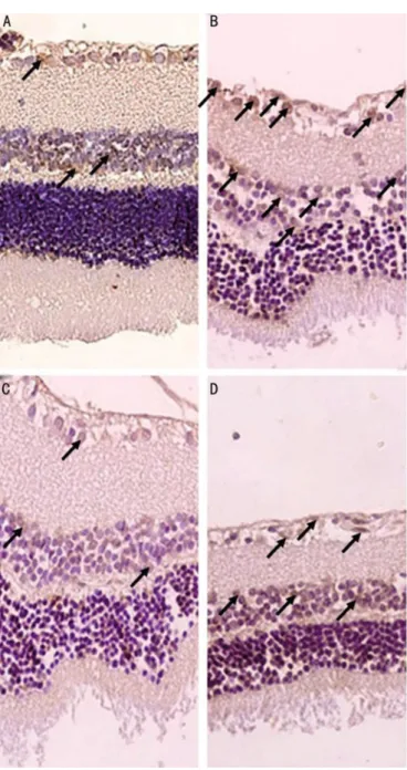
Histopathological Findings On histopathological examination of the retinal sections, there was a marked difference between the control and sham groups. Retinal sections from the control group revealed normal morphology. H&E staining of the sham retina showed that IR injury increased retinal thickness nearly 50% when compared with the normal retinal tissues of the control group. The thickness of the retinal layers in the pre-treatment and post-treatment selenium groups were statistically lower than the Sham group with insignificant difference from the control group ( <0.001). Administration of selenium after the IR ameliorated histopathological consequences of IR (Figure 1). The TUNEL staining in the Sham group revealed that TUNEL-positive cells were mainly distributed in the ganglion cell layer of the retina. Compared to the control group, AI was increased nearly to 2 fold in the Sham group. Furthermore, TUNEL staining showed that treatment with selenium before or after IR injury, significantly attenuated IR induced cell death in the retina of the experimental rats. Compared to the Sham group, AI was significantly reduced in pre-treatment and post-treatment selenium groups( <0.001). Between treatment groups, the AI was lower in the pre-treatment group in both GCL and INL layers but the difference was significant only in the GCL layer ( =0.03). The present results suggest that selenium is a potentially beneficial agent in IR induced retinal injury in rats (Figure 2). The retinal thickness, the AI in GCL and INL layers were summarized in Table 2 and Figure 3.
DISCUSSION
Due to its unique characteristics, the retina is vulnerable to damage induced by ischemia and following reperfusion. IR injury has a very important role in many blinding diseases like retinopathy of prematurity, diabetic retinopathy, and ischemic vascular occlusion. Therefore, understanding the IR pathophysiology and developing therapeutic options is crucial in order to avoid visual loss and related morbidities.
In physiological conditions, free radicals are scavenged by normal cellular metabolism. IR causes an imbalance in favor
of oxidative stress leading to depletion in antioxidants [1]. In the light of these findings, it is obvious that the IR insult needs antioxidant support. For this purpose, various agents have been studied in retinal IR models[21-26]. Selenium with its anti-inflammatory, anti-apoptotic and neuroprotective properties through selenoenzymes and selenoproteins has been studied in different organ IR models. Some of these studies used selenium before, some during and some after the induction of IR injury [8,15,16]. Literature search reveals particular studies that studied mixture of antioxidants including selenium for possible protective role in diabetic retinopathy which has IR based tissue damage[27-29]. However, selenium has not been studied in retinal IR models solely. For the first time, we tried to express the effect of selenium in
retinal IR injury and tried to establish if there is any superiority in preventive (pre-treatment) or therapeutic (post-treatment) approach.
The retinal IR injury we induced was successful and resulted Table 1 The levels of all studied parameters in study groups expressed as mean±SE n=8
Parameters Control Sham Selenium pre-treatment Selenium post-treatment P
SOD (% inhibition/mg protein) 35.31±1.18b 23.99±0.85d 27.14±1.40c,d 31.04±1.07b,c < 0.001
GSH (mg/mg protein) 3.39±0.10b 2.18±0.02e 2.66±0.07d 2.96±0.02c < 0.001
TAS (mmol trolox equiv./mg protein) 0.66±0.007a 0.28±0.009c 0.45±0.01b 0.41±0.016b < 0.001
MDA (μmol/ mg protein) 9.66±0.30c 14.43±0.35a 11.76±0.30b 13.17±0.12a < 0.001
DNA Fragmentation (U/mg protein) 1.93±0.009d 2.88±0.07a 2.35±0.02c 2.63±0.02b < 0.001 a. b, c, d, eStatistical significance exists in the same row in case of having different letters.
Figure 1 Representative photomicrographs of H&E -stained retinal sections obtained from experimental groups A: Normal histopathological image of the retinal thickness in the control group; B: The total retina thickness in the sham group was increased compared with the control group; C: Selenium pre-treatment ameliorated histopathological consequences of IR; D: Selenium post-treatment also apparently alleviated IR-induced histopathological damage in the retina. Original magnification, 400× .
Figure 2 Representative photomicrographs of retinal immunostaining for TUNEL in different groups A: In a normal control group; B: In Sham group TUNEL-positive cells (arrows) were increased in retinal layers; C: Pre- treatment selenium groups showed lower TUNEL-positive cells; D: Post- treatment selenium groups showed lower TUNEL-positive cells. TUNEL staining, 400伊.
栽藻造押8629原愿圆圆源缘员苑圆 8629-82210956 耘皂葬蚤造押ijopress岳员远猿援糟燥皂
in an increase in MDA, DNA fragmentation as well as a decrease in SOD, GSH and TAS. The approximate percentages of depletion of the tissue antioxidants (SOD, GSH, and TAS) and increment in cellular damage parameters ( MDA , DNA fragmentation ) after ischemic insult were 30%-40% and 50% respectively. Histologically the IR injury resulted in inflammation and edema leading to a nearly 50% increment in retinal thickness that again confirmed the reliability of the insult. The TUNEL staining of the retinas also revealed 100% increase in AI in the IR induced eyes. Pre-treatment use of selenium effectively protected the retina from IR insult. It created statistically higher tissue levels of GSH and TAS and higher but not significant levels of SOD. Pre-treatment selenium resulted in an enrichment of antioxidants in the retina with a subsequent increase in the free radical buffering capacity, and thus an increased resistance against the ischemic insult. The increase of reactive oxygen species in IR degrade the polyunsaturated fatty acids found abundantly in the membranes of cells and cellular organelles with consequent formation of MDA that
cause DNA adducts [30]. Thus, increased MDA and DNA fragmentation might be considered as markers of the severity of tissue damage and apoptotic cell death. Both markers were significantly reduced in the selenium pre-treatment group exhibiting a decrement in tissue damage and apoptosis of the retina. This protective effect was also seen in histologic evaluation of TUNEL stainings. In both GCL and INL layers the AI was lower when compared to post-selenium group but the statistical significance was present just for GCL. Yousuf [15]studied selenium pre-treatment in brain IR injury and found that selenium pre-treatment rats had lower neurotoxicity with diminished caspase staining of neural tissues, which is compatible with our results.
We also experienced excellent retinoprotection in the selenium post-treatment group. All parameters were favorably affected with the exception of MDA levels in which the difference did not reach statistical significance. Compared to the Sham group, histopathological examinations also revealed a decrement in inflammation, retinal thickness and TUNEL stained apoptotic cells. Ozbal [8]performed IR injury in brain tissue and investigated the possible neuroprotective effect of selenium use after the IR insult. They concluded that post-treatment selenium significantly decreased apoptosis in neural tissues, which was consistent with our results.
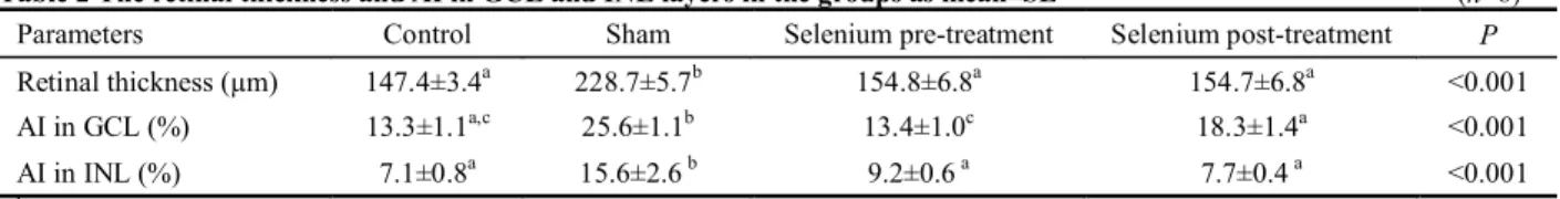
GCL is critically important to evaluate the viability of retina and is an important determining factor of the extent of visual disability due to ischemic retinal injury. The importance of this layer can easily be verified as technological instruments are being developed to assess this layer such as in determining the retinal damage of glaucoma patients. Similar to previous studies, we also experienced increased number of TUNEL positive cells in the retinal GCL in the Sham group with the IR insult [31]. Our experiments also revealed the effectiveness of pre and post-treatment selenium use in increasing both the antioxidant capacity (SOD, GSH and TAS) and decreasing apoptosis of the neural retina. In both groups, the number of apoptotic cells in the INL and GCL were markedly reduced again implying its success in neuroprotection. The statistically lower MDA and DNA fragmentation levels also confirmed the neuroprotective effect of selenium. We also found that the pre-treatment use is superior in GCL protection compared to post-treatment use. The main objective of this study was to assess for the first time the efficacy of selenium in retinoprotection in IR injury. Selenium succeeded in preserving retinal morphology, limiting retinal inflammation and thickness increment and ameliorating apoptosis (both histopathologically and Table 2 The retinal thickness and AI in GCL and INL layers in the groups as mean±SE (n=8)
Parameters Control Sham Selenium pre-treatment Selenium post-treatment P
Retinal thickness (µm) 147.4±3.4a 228.7±5.7b 154.8±6.8a 154.7±6.8a <0.001
AI in GCL (%) 13.3±1.1a,c 25.6±1.1b 13.4±1.0c 18.3±1.4a <0.001
AI in INL (%) 7.1±0.8a 15.6±2.6 b 9.2±0.6 a 7.7±0.4 a <0.001
a. b, c: Statistical significance exists in the same row in case of having different letters.
biochemically). The second objective was again for the first time, was to compare the efficacy of pre-treatment and post-treatment strategies. We have realized that the pre-treatment was superior to post-treatment in terms of tissue damage and apoptosis with statistically lower MDA levels (marker of tissue damage severity), DNA fragmentation (apoptosis) and lower levels of apoptotic TUNEL positive cells.
As a conclusion, selenium was very effective in ameliorating IR induced damage in the retina and the pre-treatment use was superior to post-treatment use in decreasing apoptosis especially in the GCL.
ACKNOWLEDGEMENTS
We special thanks to Assist Prof. Hasmet Yazici for the positive reinforcement in studying this issue.
Conflicts of Interest: Yazici A, None; Aksit H, None; Sari ES, None; Yay A, None; Erken HA, None; Aksit D, None;Cakmak H, None; Seyrek K, None; Ermis SS, None.
REFERENCES
1 Li SY, Fu ZJ, Lo AC. Hypoxia-induced oxidative stress in ischemic
retinopathy. 2012;2012:426769
2 Aydemir O, Celebi S, Yilmaz T, Yekeler H, Kukner AS. Protective effects of vitamin E forms (alpha-tocopherol, gamma-tocopherol and d-alpha-tocopherol polyethylene glycol 1000 succinate) on retinal edema during
ischemia-reperfusion injury in the guinea pig retina. 2004;
25(5-6):283-289
3 Tsujikawa A, Ogura Y, Hiroshiba N, Miyamoto K, Kiryu J, Tojo SJ, Miyasaka M, Honda Y. Retinal ischemia-reperfusion injury attenuated by blocking of adhesion molecules of vascular endothelium.
1999;40(6):1183-1190
4 Elahi MM, Kong YX, Matata BM. Oxidative stress as a mediator of
cardiovascular disease. 2009;2(5):259-269
5 Ozden S, Kildaci B, Muftuoglu S, Cakar N, Yildirim C. Effect of
trimetazidine on retinal ischemia/reperfusion injury in rats.
2001;215(4):309-317
6 Savaskan NE, Br覿uer AU, K俟hbacher M, Ey俟poglu IY, Kyriakopoulos A, Ninnemann O, Behne D, Nitsch R. Selenium deficiency increases
susceptibility to glutamate-induced excitotoxicity. 2003;17 (1):
112-114
7 Chen J, Berry MJ. Selenium and selenoproteins in the brain and brain
diseases 2003;86(1):1-12
8 Ozbal S, Erbil G, Ko觭dor H, Tugyan K, Pekgetin C, Ozogul C.The effects
of selenium against cerebral ischemia-reperfusion injury in rats. 2008;438(3):265-269
9 Zapletal C, Heyne S, Golling M, Kraus T, Gebhard MM, Herfarth C, Klar E. Influence of selenium therapy on liver microcirculation after warm ischemia/reperfusion: an intravital microscopy study.
2013;33(1-2):974-975
10 Ostadalova I, Vobecky M, Chvojkova Z, Mikova D, Hampl V, Wilhelm J, Ostadal B. Selenium protects the immature rat heart against
ischemia/reperfusion injury. 2007;300(1-2):259-267
11 Ozturk C, Avlan D, Cinel I, Cinel L, Unl俟 A, Camdeviren H, Atik U,
Oral U. Selenium pretreatment prevents bacterial translocation in rat
intestinal ischemia/reperfusion model. 2002;46(2):171-175
12 Karayaylali I, Emre M, Seyrek N, Yildiz SM, Erdogan S, Balal M, Paydas S, Tuncer I, Alparslan N. Nonsynergistic effects of trimetazidine and selenium combination therapy on renal ischemic-reperfusion injury in
rats. 2004;21(1):47-60
13 Anderson DK, Saunders RD, Demediuk P, Dugan LL, Braughler JM, Hall ED, Means ED, Horrocks LA. Lipid hydrolysis and peroxidation in injured spinal cord: partial protection with methylprednisolone or vitamin E
and selenium. 1985;2(4):257-267
14 Avlan D, Erdougan K, Cimen B, D俟鬤mez Apa D, Cinel I, Aks觟yek S. The protective effect of selenium on ipsilateral and contralateral testes in
testicular reperfusion injury. 2005;21(4):274-278
15 Yousuf S, Atif F, Ahmad M, Hoda MN, Khan MB, Ishrat T, Islam F. Selenium plays a modulatory role against cerebral ischemia-induced
neuronal damage in rat hippocampus. 2007;1147:218-225
16 Bozkurt S, Arikan DC, Kurutas EB, Sayar H, Okumus M, Coskun A, Bakan V. Selenium has a protective effect on ischemia/reperfusion injury in a rat ovary model: biochemical and histopathologic evaluation.
2012;47(9):1735-1741
17 Yokota H, Narayanan SP, Zhang W, Liu H, Rojas M, Xu Z, Lemtalsi T, Nagaoka T, Yoshida A, Brooks SE, Caldwell RW, Caldwell RB. Neuroprotection from retinal ischemia/reperfusion injury by NOX2 NADPH
oxidase deletion. 2011;52(11):8123-8131
18 Lowry OH, Rosebrough NJ, Farr AL, Randall RJ. Protein measurement
with the Folin phenol reagent. 1951;193(1):265-275
19 Fairbanks SW, Kiee G. . 3rd ed.
Philadelphia: WB Saunders;1999
20 Yoshioka T, Kawada K, Shimada T, Mori M. Lipid peroxidation in maternal and cord blood and protective mechanism against
activated-oxygen toxicity in the blood. 1979;135(3):372-376
21 Ji YS, Park JW, Heo H, Park JS, Park SW. The neuroprotective effect of carnosine (beta-Alanyl-l-Histidine) on retinal ganglion cell following
ischemia-reperfusion injury. 2013;39(6):634-641
22 Park SW, Lee HS, Sung MS, Kim SJ. The effect of melatonin on retinal
ganglion cell survival in ischemic retina. 2012;48 (2):
116-122
23 Chen FT, Yang CM, Yang CH. The protective effects of the proteasome inhibitor bortezomib (velcade) on ischemia-reperfusion injury in the rat
retina. 2013;8(5):e64262
24 Vin AP, Hu H, Zhai Y, Von Zee CL,Logeman A, Stubbs EB Jr, Perlman JI, Bu P. Neuroprotective effect of resveratrol prophylaxis on experimental
retinal ischemic injury. 2013;108:72-75
25 Zhang XY, Xiao YQ, Zhang Y, Ye W. Protective effect of pioglitazone on retinal ischemia/reperfusion injury in rats.
2013;54(6):3912-3921
26 Fukuda K, Hirooka K, Mizote M, Nakamura T, Itano T, Shiraga F. Neuroprotection against retinal ischemia-reperfusion injury by blocking the
angiotensin II type 1 receptor. 2010;51 (7):
3629-3638
27 Kowluru RA, Koppolu P, Chakrabarti S, Chen S. Diabetes-induced activation of nuclear transcriptional factor in the retina, and its inhibition by
antioxidants. 2003;37(11):1169-1180
28 Di Leo MA, Ghirlanda G, Gentiloni Silveri N, Giardina B, Franconi F, Santini SA. Potential therapeutic effect of antioxidants in experimental diabetic retina: a comparison between chronic taurine and vitamin E plus
selenium supplementations. 2003;37(3):323-330
29 Kowluru RA, Engerman RL, Case GL, Kern TS. Retinal glutamate in
diabetes and effect of antioxidants. 2001;38(5):385-390
30 Pryor WA, Stanley JP. Letter: A suggested mechanism for the production of malonaldehyde during the autoxidation of polyunsaturated fatty acids.
Nonenzymic production of prostaglandin endoperoxides during
autoxidation. 1975;40(24):3615-3617
31 Tong N, Zhang Z, Gong Y, Yin L, Wu X. Diosmin protects rat retina
from ischemia/reperfusion injury. 2012;28 (5):