See discussions, stats, and author profiles for this publication at: https://www.researchgate.net/publication/334143499
The effect of melatonin in rats with uterine torsion on uterus contractions,
and the levels of ADMA, SDMA, arginine, Hsp90, TLR4, and NF-κB
Article in Veteriner Fakültesi dergisi · July 2019
DOI: 10.33988/auvfd.467062 CITATIONS 0 READS 26 7 authors, including:
Some of the authors of this publication are also working on these related projects:
türkiye de alaca atlarView project
Radioactivity, heavy metal and oxidative stress measurements in the follicular fluids of cattle bred near a coal-fired power plantView project Ali Risvanli
Firat University
62PUBLICATIONS 180CITATIONS SEE PROFILE
Halef Doğan
Namık Kemal Üniversitesi 15PUBLICATIONS 6CITATIONS SEE PROFILE Ibrahim Şeker Firat University 105PUBLICATIONS 462CITATIONS SEE PROFILE
All content following this page was uploaded by Ali Risvanli on 05 August 2019. The user has requested enhancement of the downloaded file.
The effect of melatonin in rats with uterine torsion on uterus
contractions, and the levels of ADMA, SDMA, arginine, Hsp90, TLR4,
and NF-κB
Halef DOĞAN
1a, Ali RIŞVANLI
2b, Nevzat SAAT
3c, Hüseyin Fatih GÜL
4d, Necip İLHAN
4e,
İbrahim ŞEKER
5f, Engin ŞAHNA
6g1Namık Kemal University, Faculty of Veterinary Medicine, Department of Obstetrics and Gynecology, Tekirdağ; 2Fırat University,
Faculty of Veterinary Medicine, Department of Obstetrics and Gynecology, Elazığ; 3Balıkesir University, Faculty of Veterinary
Medicine, Department of Obstetrics and Gynecology, Balıkesir; 4Fırat University, Faculty of Medicine, Department of Biochemistry,
Elazığ; 5Fırat University, Faculty of Medicine, Department of Zootechny, Elazığ; 6Fırat University, Faculty of Medicine, Department
of Pharmacology, Elazığ, Turkey.
aORCID: 0000-0003-1365-1729; bORCID: 0000-0001-5653-0025; cORCID: 0000-0002-8135-6142; dORCID: 0000-0002-9828-1298; eORCID: 0000-0001-9997-0418; fORCID: 0000-0002-4791-2993;
gORCID: 0000-0001-8311-9055
Corresponding author: [email protected]
Received date: 03.10.2018- Accepted date: 04.05.2019
Abstract: In this study was aimed at reducing uterine damage and increasing fertility after uterine torsion in pregnant animals.
With this aim, uterine torsion was experimentally formed in 35 rats that were between 18-19 days pregnant. The animals were randomly divided into five groups, and melatonin was administered prior to torsion, at the time of torsion, and detorsion (10 mg/kg/gün IP). Ovario-hysterectomy operation was performed on all animals on the first day following parturition. Subsequently, from the obtained uterus samples, determination of the levels of asymmetrical dimethyl arginin (ADMA), symmetrical dimethyl arginin (SDMA), and arginine was made using the high-performance liquid chromatography (HPLC) and levels of Heat shock protein 90 (Hsp90), TLR4 (Toll Like Receptor 4) and NFκB (Nuclear factor kappa-light-chain-enhancer of activated B cells) were measured using the Western blot technique. The contraction-relaxation responses of the myometrium were also determined in the organ baths. According to the results of the western blot, higher protein expressions than those of the control group were determined in the second, third, fourth, and fifth groups in Hsp90, TLR4, NF-κB. The lowest values of arginine and ADMA were found in Group 3, whilst the lowest SDMA value was determined in Group 1. It was determined that melatonin reduces tissue damage secondary to torsio uteri and, furthermore, that administration of this hormone at the time of torsion formation was more effective than its administration at the time of detorsion.
Keywords: Melatonin, pregnancy, rats, torsion, uterine
Torsiyo uterili ratlarda melatoninin uterus kontraksiyonları, ADMA, SDMA, arjinin, Hsp90, TLR4 ve
NFκB düzeylerine etkisi
Özet: Bu çalışmada gebe hayvanlarda torsiyo uteri sonrası şekillenen uterus hasarını azaltmaya ve fertiliteyi artırmaya yönelik
bir çalışma planlandı. Bu amaçla 35 adet 18-19 günlük gebe ratda deneysel olarak torsiyo uteri oluşturuldu. Hayvanlar rastgele 5 gruba ayrılarak torsiyon öncesi, torsiyon anında ve detorsiyon sırasında melatonin (10 mg/kg/gün IP) uygulandı. Doğum sonrası ilk gün tüm hayvanlara ovariohisterektomi operasyonu yapılarak kornulardan numuneler alındı. Daha sonra elde edilen uterus dokularında asymmetrical dimethyl arginin (ADMA), symmetrical dimethyl arginin (SDMA) ve Arjinin miktar tayinleri yüksek performanslı sıvı kromatografisi (HPLC) ve Heat shock protein 90 (Hsp90), TLR4 (Toll Like Receptor 4) ve NFκB (Nuclear factor kappa-light-chain-enhancer of activated B cells) düzeyleri ise Western Blot ile belirlendi. Miyometriyumun in vitro kasılma gevşeme cevapları da organ banyosunda tespit edildi. Western blot sonuçlarına göre HSP90, TLR4, NFκB’da 2, 3, 4 ve 5. gruplarda kontrol grubuna göre daha fazla protein ekspresyonu olduğu belirlendi. Arjinin ve ADMA değerleri en düşük olarak 3. grupta bulundu. En düşük SDMA değeri ise 1. grupta tespit edildi. Sonuç olarak; elde edilen tüm bu veriler melatoninin torsiyo uteriye bağlı doku hasarını azalttığı ayrıca bu hormonun torsiyonun şekillendiği anda uygulanmasının detorsiyon anında uygulanmasına göre daha etkili olduğu kanaatine varıldı.
Halef Doğan - Ali Rışvanlı - Nevzat Saat - Hüseyin Fatih Gül - Necip İlhan - İbrahim Şeker - Engin Şahna 268
Introduction
Uterine torsion is one of the frequent causes of dystocia in all ruminants, although it is primarily found in cattle, and it constitutes 7% of all dystocias. Treatment applications are generally developed in order to correct the torsion and enable parturition (11), although no routine application exists for the maintenance of fertility in animals with uterine torsion. Nevertheless, more than half of the surviving animals with uterine torsion have been reported to become isolated from the herd in later stages due to infertility. The cause of infertility in these animals has been suggested to be the ischaemic/reperfusion damage as a result of torsion and detorsion (10).
It has been reported that the physiological and pharmacological concentrations of melatonin affect the free radicals that increase during ischaemic/reperfusion damage and reduce myocardial damage (mortality, arrhythmias, and infarction areas). This effect of melatonin may be related to its anti-adrenergic property, as well as its free-radical scavenging and anti-oxidant effects (7). The ischaemic/reperfusion preventive effect of melatonin is not limited to the heart, but has also been detected in hepatic (13)and cerebral (9) cells. This study set out to determine the effects of melatonin administrations on uterine tissue in pregnant rats in which uterine torsion was formed experimentally.
Materials and Methods
A total of 35 female Wistar rats aged 3-4 months, with a gestational age of 18-19 days and a weight of 200-250 g, were used. The animals were obtained from the Experimental Investigations Centre of Firat University, Turkey. They were kept in individual cages and were exposed to a rhythm of 12 hr dark and 12 hr light, and were fed ad libitum. Ethical approval was obtained from the Local Ethical Committee of Fırat University Laboratory Animals (17.12.2014 - 2014/128).
The animals were grouped as follows: Group 1: rats that were given only anaesthesia on their 18-19th
gestational day (n=7). Group 2: rats with an experimentally formed uterine torsion of 360 degrees on their 18-19th gestational day, corrected six hr later (n=7).
Group 3: rats with an experimentally formed uterine torsion of 360 degrees on their 18-19th gestational day,
corrected six hr later, with melatonin (10 mg/kg IP) applied at the same time (n=7). Group 4: rats with an experimentally formed uterine torsion of 360 degrees on their 18-19th gestational day, with melatonin (10 mg/kg IP)
administered six hr later along with torsion correction (n=7). Group 5: rats receiving melatonin on the 15-16th
gestational day (10 mg/kg/day IP), and then uterine torsion of 360 degrees was formed experimentally in the uterus of these rats on their 18-19th gestational day, corrected six hr
later (n=7).
Vaginal irrigations were performed as described by Risvanli et al., (12) and involved using elastic pipettes and tips with distilled water. Animals whose slides included spermatozoids were accepted as coitus positive. These dates were recorded as the zero day of pregnancy.
All operations were performed with the animals under ketamine/xylazine anesthesia, and animals with a gestational age of 18-19 days underwent laparotomy following routine procedures. The right cornu uteri of the animals were passed through the hole formed in the non-vascular region of lig. lata uteri of the left cornu uteri at the level of the urinary bladder. Then, the right cornu was passed through the same hole for the second time, and torsion was thereby formed. Following this procedure, the abdomens of the animals were closed with proper suturing material. Six hr after the operation, another laparotomy was performed on the same animals under anaesthesia and the torsion was corrected.
On the first day post-partum, all animals underwent ovariohysterectomy and the samples were obtained from uterine cornus and kept at -80°C. The contraction-relaxation responses of the myometrium were also determined in the organ baths. Within 24 hr after parturition/abortion a tissue section comprising all the layers of the uterus, with dimensions of 15x2 mm and of 23.4±6.67 mg weight, was obtained from the middle part of the uterine cornu parallel to the long axis (Figure 1).
Figure 1. Spontaneous contraction within the first postpartum 24 hr.
Şekil 1. Postpartum ilk 24 saat içindeki kendiliğinden kasılma
The tissues that had been kept at -80°C in the deep freeze were removed and weighed on sensitive scales as pieces of 100 mg. Homogenization was performed by diluting 0.01 M phosphate buffer saline (pH 7.4) at a 1:10 ratio, and within ice blocks of 4°C. Following the homogenization procedure, centrifugation was carried out at 50.000 rpm for 10 min, and the supernatant parts were removed (5). The centrifugation procedure was repeated on the remaining pellet portions until these portions brightened up, and the obtained homogenates were apportioned. The amount of protein inside the homogenates was determined using the Lowry method
(µg/ml). A 20 μg total protein sample was loaded onto a 10% polyacrylamide gel and electrophoresed in SDS running solution (Running buffer; 2.4 mM Tris, 19.2 mM glisin, SDS of 0.01%) at 90 V for one hour. The separated proteins were transferred from the SDS-PAGE at 100 V in a period of one hour inside a transfer solution (25 mM Tris, 192 mM glisin, 20% methanol, pH 8.3) to the PVDF membrane. Following the transfer, the membrane was also blocked at 4°C inside the prepared milk powder of 5% inside PBS-T (PBS+ 1% Tween-20) solution. Following the blocking procedure, the membrane was treated with primary antibodies for two hours at room temperature. Subsequently, the membrane was washed four times with PBS-T for duration of 30 minutes. After the washing procedure, the membrane was treated with horseradish peroxidase conjugated secondary antibodies at a dilution of 1:10.000 for one hour at room temperature. The membrane was washed again four times with PBS-T for duration of 30 minutes. The protein bands were then visualized using the DAB chemical solution (5).
The analyses of ADMA, arginine, and symmetrical dimethyl arginine (SDMA) in the samples were carried out using the high performance liquid chromatography (HPLC) apparatus at the University of Firat’s Department of Biochemistry (16).
In the organ bath experiments, the Kruskal-Wallis test, which is the equivalent of the one-way non-parametric analysis of variance, was performed in the statistical analyses of the data due to the presence of five groups, the use of different experimental subjects, and the lack of a normal distribution. The Kruskal-Wallis test was used in groups in which the medians were not equal, and the Bonferroni Corrected Mann-Whitney U test (P<0.01) was used as the post hoc multiple comparison method by lowering the level of significance in groups in which the significance was lower than 0.05. The Corrected Mann-Whitney U test was performed following the Kruskal-Wallis variance analysis in the evaluation of the distribution of arginine, SDMA, and ADMA values according to the groups. The SPSS for Windows version 22.0 (SPSS Inc. Chicago, Illinois, USA) program package was used for the statistical analyses.
Results
The parameters of the area under the contractile curve (AUC), amplitude, and frequency of spontaneously occurring contractions were determined in all five groups (Table 1). While the difference between the groups with regard to the parameters of amplitude and AUC was determined to be statistically significant (P<0.05), the difference regarding the frequency parameter was found to be insignificant (P>0.05).
When the difference between the groups with regard to AUC was addressed, the values belonging to Group 3
were determined to be higher than those of Groups 2, 4, and 5 (P<0.05). Still, the difference between Group 3 and Group 1 and 4 was found to be statistically insignificant (P>0.05) (Table 1).
When the difference between the groups with regard to amplitude was addressed, the values belonging to Group 3 were determined to be higher than those of Groups 2 and 5 (P<0.05). Still, the difference between Group 3 and Group 1 and 4 was found to be statistically insignificant (P>0.05) (Table 2).
Table 1. The 30-min analysis of spontaneous myometrial contractions.
Tablo 1. Spontan miyometriyal kasılmaların 30 dakikalık kasılma analizi Groups AUC (gXsec) Amplitude (g) Frequency 1 34.23±5.08a 0.14±0.05a,b 20.41±7.31 2 14.26±4.80b 0.07±0.02c 15.00±2.63 3 64.92±14.96a 0.29±0.08a 17.57±6.74 4 25.85±8.66c 0.12±0.03a,b 22.29±2.70 5 15.51±9.01b 0.08±0.02c 16.98±7.21 P * * **
*P<0.05, **P>0.05, a,b,c: The difference between the frequencies
demonstrated by different letters within the same column was significant (P<0.01).
Table 2. Distribution of the values of arginin, SDMA and ADMA according to the groups.
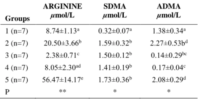
Tablo 2. Arjinin, SDMA, ADMA değerlerinin gruplara göre dağılımı ARGININE µmol/L SDMA µmol/L ADMA µmol/L Groups 1 (n=7) 8.74±1.13a 0.32±0.07a 1.38±0.34a 2 (n=7) 20.50±3.66b 1.59±0.32b 2.27±0.53bd 3 (n=7) 2.38±0.71c 1.50±0.12b 0.14±0.29bc 4 (n=7) 8.05±2.30ad 1.41±0.19b 0.17±0.04c 5 (n=7) 56.47±14.17e 1.73±0.36b 2.08±0.29d P ** * *
*P<0.01, **P<0.05 a, b, c, d, e: The difference between the frequencies demonstrated by different letters within the same column was significant (P<0.01).
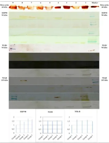
The bands appearing as a result of the western blot procedure were visualized using the image-j program and the relative protein amounts were calculated. Accordingly, the protein expression values for Hsp90, TLR4, and NF-κB in Group 1, which was accepted as the control group, were observed to be 100%. For Hsp90, the protein expression values in the second, third, fourth, and fifth groups were determined to be higher than those of the
Halef Doğan - Ali Rışvanlı - Nevzat Saat - Hüseyin Fatih Gül - Necip İlhan - İbrahim Şeker - Engin Şahna 270
control group, in the order of 56.3%, 38.0%, 77.6%, and 70.1%, respectively. For TLR4, higher protein expression values than those of the control group were determined in the second, third, fourth, and fifth groups, in the order of 55.4%, 50.5%, 61.1%, and 47.9%, respectively. For NF-κB, higher protein expression values than those of the control group were determined in the second, third, fourth, and fifth groups, in the order of 65.5%, 54.5%, 62.7%, and 45.5%, respectively (Figure 2).
Figure 2. The expression values of HSP90, TLR4 and NFκB proteins between the groups.
Şekil 2. HSP90, TLR4 ve NFκB proteinlerinin gruplararası ekspresyon değerleri
The distribution of the arginine, SDMA, and ADMA values according to the groups has been summarised in Table 2. Accordingly, the lowest values of arginine (2.38±0.71 µmol/L; P<0.05) and ADMA (0.14±0.29 µmol/L; P<0.01) were determined in Group 3. The lowest SDMA value, however, was determined in Group 1 (0.32±0.07 µmol/L; P<0.01).
Discussion and Conclusion
Melatonin plays an important role in the TLR4/NF-κB pathway, especially in ischaemic/reperfusion damage. When the secretion of NF-κB and TLR4 are blocked, inflammation has been reported to be suppressed as well. NF-κB is activated in response to oxidative stress and is a
redox sensible transcription factor, which is responsible for the production of inflammatory genes. In rats with TLR4 insufficiency, ischaemic/reperfusion damage-related inflammation has been suggested to decrease (6). The relationship between the Hsp90 and NF-κB molecules in various inflammatory disorders has been put forth in various studies. It has been reported that the transcription of many genes is up-regulated secondary to activation of κB following cerebral ischaemia. Inhibition of the NF-κB pathway has been reported to be protective against cerebral ischaemic damage, both genetically and pharmacologically (8). The inhibitor is found through the formation of complexes with NF-κB kinase and Hsp90, and impairment of these complexes by Hsp90 inhibitors blocks the inhibitor NF-κB kinase function; subsequently, the NF-κB activation may be blocked. Secondary to this, an increase in ischaemic/reperfusion damage has been reported (14). In the present study, according to the results of the western blot, higher protein expressions than those of the control group for Hsp90 are found in the second, third, fourth, and fifth groups, in the order of 56.3%, 38.0%, 77.6%, and 70.1%, respectively. For TLR4, higher protein expression values than those of the control group were determined in the second, third, fourth, and fifth groups, in the order of 55.4%, 50.5%, 61.1%, and 47.9%, respectively. For NF-κB, higher protein expression values than those of the control group were determined in the second, third, fourth, and fifth groups, in the order of 65.5%, 54.5%, 62.7%, and 45.5%, respectively.
In ischaemic/reperfusion damage, ADMA reduces the activity of dimethylarginine dimethylaminohydrolase (DDAH), and increases its concentration. Furthermore, it also exerts an effect on the production of nitric oxide (NO) by competing with arginine for binding to the active point of the nitric oxide synthetase (NOS) center. However, the molecular mechanisms in the ischaemic/reperfusion
damage have not been completely understood.
Nonetheless, the ADMA/DDAH pathway has the
potential ability to reduce the effects of
ischaemic/reperfusion damages (4, 8, 18). In a study carried out by Ferrigno et al. (5) on male rats with regard to hepatic ischaemic/reperfusion damage, it was reported that the serum ADMA levels had increased and that the intracellular ADMA levels had decreased after a 60-min ischaemic attack. In the same study, following reperfusion, the DDAH activity and the mRNA and protein expression were reported to have decreased. In the present study also, the lowest values of arginine (2.38±0.71 µmol/L; P<0.05) and ADMA (0.14±0.29 µmol/L; P<0.01) were determined at the time of detorsion in Group 3, in which melatonin had been administered. The lowest SDMA value, however, was determined in Group 1, in which torsion had not been formed (0.32±0.07 µmol/L; P<0.01).
In spite of the fact that the mechanism of melatonin cannot be precisely explained, it has been reported to
inhibit the spontaneous or oxytocin-stimulated
myometrial contractions in pregnant and non-pregnant rats (2). However, it has also been reported that melatonin at micromolar doses does not inhibit the uterine smooth muscles stimulated with prostaglandin f2-alpha in
ovariectomized rats (1).Moreover, in a study performed on pregnant sheep, melatonin administration was reported to have no effect on the myometrial contractility. Still, the administration of melatonin has been reported to inhibit the oxytocin-stimulated myometrial activity in rats (15). Despite all the research carried out in the literature, no publication regarding the effects of melatonin on uterine contractility in produced ischaemic/reperfusion damage or torsion/detorsion was encountered. In the present study, however, the contractility in the uterus of rats, in which uterine torsion was produced experimentally in advanced pregnancy, was observed to be decreased compared with the control group. Of the AUC, amplitude, and frequency parameters evaluated in the organ bath experiments, the difference between the melatonin-administered groups with regard to amplitude and AUC was found to be statistically significant. However, the difference between the groups with regard to frequency was determined to be statistically insignificant. When the inter-group differences were addressed with regard to AUC, the values belonging to Group 3 were higher than those of Groups 2, 4, and 5. When the intergroup differences were addressed with regard to amplitude, the values in Group 3 were determined to be higher than those of Groups 2 and 5. Administration of melatonin at the time of detorsion positively affected the uterine contractions, and not at the time of torsion formation.
In previous studies on ovarian torsion (Adnexal torsion), various materials such as vitamins E and C, mannitol, melatonin, caffeic acid, and erythropoietin have been used to reduce the reperfusion damage, and as a result these chemicals have been reported to reduce the damage caused by ischaemia/reperfusion to the ovary (3), to a certain extent. In a study carried out by Turkoz et al., (17) in which they produced ovarian torsion in rats, melatonin was reported to protect the ovary against oxidative damage resulting from ischaemia/reperfusion. In the present study also, according to the results of the organ bath and western blot experiments, administration of melatonin was observed to reduce the damage resulting from ischaemia/reperfusion caused by torsion/detorsion in rats, in which torsio uteri was experimentally produced.
According to these experimentally obtained results, administration of melatonin at the time of torsion formation and not at the time of torsion reduces uterine damage caused by uterine torsion in advanced pregnancy. However, in animals such as sheep and cattle, generally
due to the inability to determine the precise time of torsion, it was determined that administration of melatonin following the diagnosis of uterine torsion or just after correction of the torsion was beneficial.
Acknowledgement
This study was supported by the Scientific and Technological Research Council of Turkey (TUBITAK - 115O381).
References
1. Abd-Allah AR, El-Sayed ESM, Abdel-Wahab MH, et al. (2003): Effect of melatonin on estrogen and progesterone
receptors in relation to uterine contraction in rats.
Pharmacol Res, 47, 349-354.
2. Ayar A, Kutlu S, Yilmaz B, et al. (2001): Melatonin
inhibits spontaneous and oxytocin-induced contractions of rat myometrium in vitro. Neuroendocrinol Lett, 22,
199-207.
3. Bakan V, Çıralık H, Tolun F (2009): Protective effect of
erythropoietin on torsion/detorsion injury in rat model. J
Pediatr Surg, 44, 1988–1994.
4. Breen AP, Murphy JA (1995): Reactions of oxyl radicals
with DNA. Free Radic Biol Med, 18, 1033–1037.
5. Ferrigno A, Rizzo V, Bianchi A, et al. (2014): Changes in
ADMA/DDAH pathway after hepatic ischemia/reperfusion injury in rats: the role of bile. BioMed Res Int, Article ID
627434.
6. Ha T, Liu L, Kelley J (2011): Toll-like receptors: new
players in myocardial ischemia/reperfusion injury.
Antioxid Redox Signal, 15, 1875-1893.
7. Halici Z, Karaca M, Keles ON, et al. 2008. Protective
effects of amlodipine on ischemia–reperfusion injury of rat ovary: biochemical and histopathologic evaluation. Fertil
Steril, 90, 2408–2415.
8. Herrmann O, Baumann B, de Lorenzi R, et al. (2005):
IKK mediates ischemia-induced neuronal death. Nat Med,
11, 1322-1329.
9. Kilic E, Ozdemir YG, Bolay H, et al. (1999):
Pinealectomy aggravates and melatonin administration attenuates brain damage in focal ischemia. J Cereb Blood
Flow Metab, 19, 511-516.
10. Lyons NA, Knight JTJD, Aldridge BM, et al. (2013):
Incidence, management and outcomes of uterine torsion in dairy cows. Cattle Pract, 21:1-6.
11. Musal B, Köker A. (2015): Dystocia. 195-257. In: (Semacan A, Kaymaz M, Findik M, Risvanli A, Koker A. (eds), Obstetrics and Gynecology in Farm Animals, 1st ed.
Medipres, Malatya, Turkey.
12. Risvanli A, Aydin M, Kaygusuzoglu E, et al. (2003): The
effect of thyroidectomy on sexual cycle and pregnancy rates in rats. Turkish J Vet Anim Sci, 27, 873-877.
13. Sewerynek E, Reiter RJ, Melchorri D, et al. (1996):
Oxidative damage in the liver induced by ischemia-reperfusion: protection by melatonin. Hepatogastroenterol,
43, 898-905.
14. Shimizu H, Saito, S, Higashiyama Y, et al. (2013): CREB,
NF-kappaB, and NADPH oxidase coordinately upregulate indoxyl sulfate induced angiotensinogen expression in
Halef Doğan - Ali Rışvanlı - Nevzat Saat - Hüseyin Fatih Gül - Necip İlhan - İbrahim Şeker - Engin Şahna 272
proximal tubular cells. Am J Physiol Cell Physiol, 304,
685-692.
15. Taskin O, Birincioglu M, Aydin A, et al. (1998): The
effects of twisted ischemic adnexa managed by detorsion on ovarian viability and histology: an ischemia-reperfusion rodent model. Hum Reprod, 13, 2823-2827.
16. Teerlink T, Nijveldt RJ, De Jong S, et al. (2002):
Determination of arginine, asymmetric dimethylarginine, and symmetric dimethylarginine in human plasma and other biological samples by high-performance liquid chromatography. Anal Biochem, 303, 131-137.
17. Turkoz Y, Celik O, Hascalik S, et al. (2004): Melatonin
reduces torsion–detorsion injury in rat ovary: biochemical and histopathologic evaluation. J Pineal Res 37, 137–141.
18. Yagnik GP, Takahashi Y, Tsoulfas G, et al. (2002):
Blockade of the L-arginine/NO synthase pathway worsens hepatic apoptosis and liver transplant preservation injury.
Hepatol, 36, 573–581.
View publication stats View publication stats