[CANCER RESEARCH 59, 290 –293, January 15, 1999]
Advances in Brief
Human MLH1 Deficiency Predisposes to Hematological Malignancy and
Neurofibromatosis Type 1
1Marie D. Ricciardone,
2Tayfun O
¨ zc¸elik,
2Birsen Cevher, Hilal O
¨ zdagˇ, Murat Tuncer, Aytemiz Gu¨rgey,
O
¨ zden Uzunalimogˇlu, Hu¨lya C¸etinkaya, Attila Tanyeli, Eren Erken, and Mehmet O
¨ ztu¨rk
3Department of Molecular Biology and Genetics, Faculty of Science, Bilkent University, 06533 Ankara, Turkey [M. D. R., T. O¨ ., B. C., H. O¨., M. O¨.]; Department of Pediatrics, Hacettepe University School of Medicine, 06100 Ankara, Turkey [M. T., A. G.]; Department of Internal Medicine, Ankara University School of Medicine, 06100 Ankara, Turkey [O¨ . U., H. C¸.]; and Departments of Pediatrics and Internal Medicine, C¸ukurova University School of Medicine, 01330 Adana, Turkey [A. T., E. E.]
Abstract
Heterozygous germ-line mutations in the DNA mismatch repair genes lead to hereditary nonpolyposis colorectal cancer. The disease suscepti-bility of individuals who constitutionally lack both wild-type alleles is unknown. We have identified three offspring in a hereditary nonpolyposis colorectal cancer family who developed hematological malignancy at a very early age, and at least two of them displayed signs of neurofibro-matosis type 1 (NF1). DNA sequence analysis and allele-specific amplifi-cation in two siblings revealed a homozygous MLH1 mutation (C676T3Arg226Stop). Thus, a homozygous germ-line MLH1 mutation and consequent mismatch repair deficiency results in a mutator pheno-type characterized by leukemia and/or lymphoma associated with neuro-fibromatosis type 1.
Introduction
HNPCC4
is a common autosomal dominant disease characterized by an inherited predisposition to early onset of colorectal cancer and to an increased incidence of other cancers (1). Mutations in MLH1 and
MSH2 are seen in.90% of HNPCC kindreds, whereas mutations in PMS2, PMS1, and MSH6 have been seen in only a few families (2).
HNPCC individuals are heterozygous for the mutant allele. Thus, they retain a functional allele at the constitutional level, which is appar-ently sufficient for DNA MMR (3). However, a subsequent somatic mutation leading to loss of the wild-type allele results in a mutator cancer phenotype characterized by a high rate of base substitutions, as well as small insertions and deletions in mono- and dinucleotide repeats (3). Somatic mutations in HNPCC tumors have been identified in long mononucleotide tracts within the coding sequences of several genes involved in the control of cell growth, including TGFbRII,
IGFIIR, and BAX, as well as two additional MMR genes, MSH3 and MSH6 (Refs. 4 –7). These downstream gene mutations have been
implicated in the progression of colorectal cancer. Thus, the result of somatic loss of MMR gene function in individual cells has been well characterized. However, the phenotypic consequence of a constitu-tional loss of MMR gene function remains poorly understood. We describe here a germ-line homozygous mutation of the MLH1 gene that predisposes to NF1 and early childhood hematological malignan-cies.
Materials and Methods
Patient Samples. Informed consent was obtained from all participants in the study. Child IV-4 developed atypical chronic myeloid leukemia at 12 months, displayed more than 10 abdominal cafe´ au lait spots (.15 mm) and two fibromatous skin tumors (2 cm), and showed hepatosplenic enlargement (8 cm). A differential blood count done at the time blood was drawn for DNA analysis showed 28% polymorphonuclear cells, 9% lymphocytes, 48% mono-cytes, 14% blasts, and 1% eosinophils. Bone marrow smear revealed 10% promyelocytes, 8% myelocytes, 5% metamyelocytes, 22% polymorphonuclear cells, 12% eosinophils, 18% normoblasts, and 25% blasts. Child IV-3 devel-oped non-Hodgkin’s lymphoma at 39 months and displayed more than 10 cafe´ au lait spots (.15 mm). Bone marrow smear revealed 33% normoblasts, 6% promyelocytes, 12% myelocytes, 9% metamyelocytes, 6% polymorphnuclear cells, and 34% lymphoblasts. Megakaryocytes were seen, and dysplasia was not detected. Child IV-2 developed acute leukemia at 24 months; neurofibro-matosis could not be confirmed.
DNA Sequence Analysis. Genomic DNA was isolated from peripheral blood samples (except for child IV-3, for whom genomic DNA was isolated from a bone marrow smear) by proteinase K lysis, phenol/chloroform extrac-tion, and ethanol precipitation. MLH1 exon 8 primers, 5 9-CTCAGCCAT-GAGACAATAAATCC-39 (forward) and 59-GGTTCCCAAATAATGT-GATGG-39 (reverse), were used for both PCR amplification and cycle sequencing. PCR amplification reactions were performed using GeneAmp PCR Core Reagents (Perkin-Elmer, Foster City, CA). PCR parameters were denaturation (95°C for 30 s), annealing (55°C for 30 s), and extension (72°C for 60 s) for 30 cycles. Dye-terminator cycle sequencing reactions were performed using the ABI PRISM Dye-Terminator Cycle Sequencing Ready Reaction kit (Perkin-Elmer) according to the recommended protocol with the following modifications. DMSO (5%) was included in the forward primer reaction, and a denaturation temperature of 98°C was used for the reverse primer. Automated fluorescence sequence analysis was performed on the 377 DNA Sequencer (ABI; Perkin-Elmer).
Allele-specific Amplification. Forward primers were designed for allele specificity at the 39 terminal nucleotide: 59-GAAATGCTGTTAGTC-39 (wild-type) and 59-GAAATGCTGTTAGTT-39 (mutant). The reverse primer for both alleles was 59-AGTGTTGATTACGTG-39. Amplification reactions contained ;25 ng of genomic DNA and 10 pmol of primer. PCR parameters were 35 cycles of denaturation (94°C for 30 s), annealing (50°C for 60 s), and extension (72°C for 90 s). PCR products were analyzed by 3% Metaphor (FMC) gel electrophoresis. The competition experiment was performed using a constant amount of mutant DNA (50 ng) and dilutions (1:1, 2:1, 5:1, 50:1, and 500:1) of wild-type DNA.
Results and Discussion
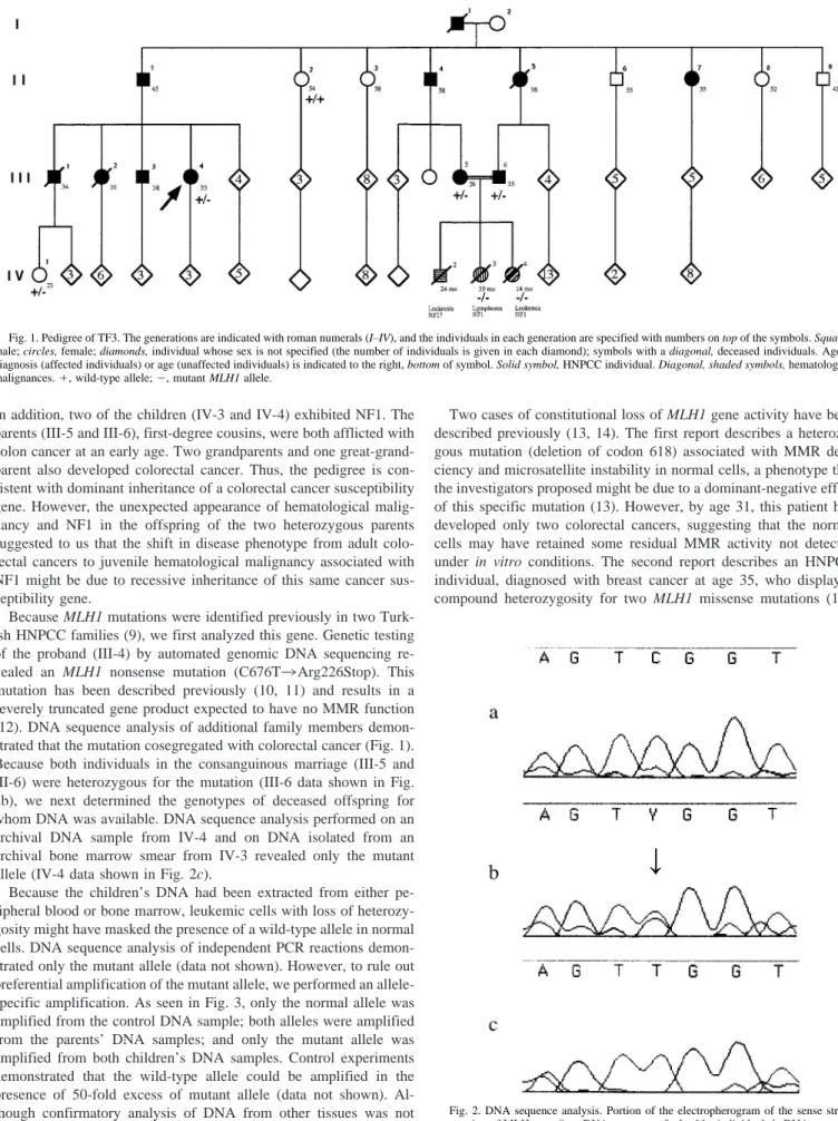
To identify individuals with a constitutional loss of MMR gene function, we inspected large HNPCC kindreds for putative homozy-gotes. TF3 is a large Turkish kindred that fulfills the Amsterdam criteria (8) for HNPCC: colorectal cancer diagnosed at an early age (26 –58 years) in multiple first-degree relatives over three consecutive generations (Fig. 1). Of particular genetic interest were three offspring (IV-2, IV-3, and IV-4) of a consanguinous marriage. All three chil-dren were diagnosed with hematological malignancy by the age of 3.
Received 9/16/98; accepted 11/25/98.
The costs of publication of this article were defrayed in part by the payment of page charges. This article must therefore be hereby marked advertisement in accordance with 18 U.S.C. Section 1734 solely to indicate this fact.
1This work was supported in part by a combined grant (Grant TTGV-172) from the
Technology Development Foundation of Turkey and Bilkent Holding. H. O¨ . received a Bu¨tu¨nles(tirilmis( Doktora Program fellowship from the Scientific and Technical Research Council (TU¨ BITAK), Turkey.
2These authors contributed equally to the work. 3To whom requests for reprints should be addressed.
4The abbreviations used are: HNPCC, hereditary nonpolypopsis colorectal cancer;
MMR, mismatch repair; NF1, neurofibromatosis type 1; TGFbRII, transforming growth factorb receptor II; IGFIIR, insulin-like growth factor II receptor.
In addition, two of the children (IV-3 and IV-4) exhibited NF1. The parents (III-5 and III-6), first-degree cousins, were both afflicted with colon cancer at an early age. Two grandparents and one great-grand-parent also developed colorectal cancer. Thus, the pedigree is con-sistent with dominant inheritance of a colorectal cancer susceptibility gene. However, the unexpected appearance of hematological malig-nancy and NF1 in the offspring of the two heterozygous parents suggested to us that the shift in disease phenotype from adult colo-rectal cancers to juvenile hematological malignancy associated with NF1 might be due to recessive inheritance of this same cancer sus-ceptibility gene.
Because MLH1 mutations were identified previously in two Turk-ish HNPCC families (9), we first analyzed this gene. Genetic testing of the proband (III-4) by automated genomic DNA sequencing re-vealed an MLH1 nonsense mutation (C676T3Arg226Stop). This mutation has been described previously (10, 11) and results in a severely truncated gene product expected to have no MMR function (12). DNA sequence analysis of additional family members demon-strated that the mutation cosegregated with colorectal cancer (Fig. 1). Because both individuals in the consanguinous marriage (III-5 and III-6) were heterozygous for the mutation (III-6 data shown in Fig. 2b), we next determined the genotypes of deceased offspring for whom DNA was available. DNA sequence analysis performed on an archival DNA sample from IV-4 and on DNA isolated from an archival bone marrow smear from IV-3 revealed only the mutant allele (IV-4 data shown in Fig. 2c).
Because the children’s DNA had been extracted from either pe-ripheral blood or bone marrow, leukemic cells with loss of heterozy-gosity might have masked the presence of a wild-type allele in normal cells. DNA sequence analysis of independent PCR reactions demon-strated only the mutant allele (data not shown). However, to rule out preferential amplification of the mutant allele, we performed an allele-specific amplification. As seen in Fig. 3, only the normal allele was amplified from the control DNA sample; both alleles were amplified from the parents’ DNA samples; and only the mutant allele was amplified from both children’s DNA samples. Control experiments demonstrated that the wild-type allele could be amplified in the presence of 50-fold excess of mutant allele (data not shown). Al-though confirmatory analysis of DNA from other tissues was not possible, these results strongly suggest that both children were truly homozygous for the mutant allele.
Two cases of constitutional loss of MLH1 gene activity have been described previously (13, 14). The first report describes a heterozy-gous mutation (deletion of codon 618) associated with MMR defi-ciency and microsatellite instability in normal cells, a phenotype that the investigators proposed might be due to a dominant-negative effect of this specific mutation (13). However, by age 31, this patient had developed only two colorectal cancers, suggesting that the normal cells may have retained some residual MMR activity not detected under in vitro conditions. The second report describes an HNPCC individual, diagnosed with breast cancer at age 35, who displayed compound heterozygosity for two MLH1 missense mutations (14).
Fig. 1. Pedigree of TF3. The generations are indicated with roman numerals (I–IV), and the individuals in each generation are specified with numbers on top of the symbols. Squares, male; circles, female; diamonds, individual whose sex is not specified (the number of individuals is given in each diamond); symbols with a diagonal, deceased individuals. Age of diagnosis (affected individuals) or age (unaffected individuals) is indicated to the right, bottom of symbol. Solid symbol, HNPCC individual. Diagonal, shaded symbols, hematological malignances.1, wild-type allele; 2, mutant MLH1 allele.
Fig. 2. DNA sequence analysis. Portion of the electropherogram of the sense strand sequencing of MLH exon 8. a, DNA sequence of a healthy individual; b, DNA sequence of father (III-6); c, DNA sequence of child (IV-4). Arrow, heterozygous C3T MLH1 mutation.
291
Although the investigators demonstrated microsatellite instability and increased in vitro mutation rate, one of the mutated gene products likely retained residual MMR activity because it was lost in the tumor cells. Thus, it is likely that this missense mutation was itself not deleterious and that, therefore, this case does not represent constitu-tional MLH1 deficiency. In contrast to these two studies, the mutation that we have identified is a nonsense mutation that results in a severely truncated, nonfunctional gene product. The children most likely inherited the same allele from each parent and thus represent true homozygotes. The resulting MLH1 deficiency appears to have induced a severe tumorigenic syndrome, involving both hematological malignancy and neurofibromatosis.
Our observations demonstrate that a homozygous MLH1 mutation is not lethal but predisposes to juvenile hematological malignancy associated with NF1, in contrast to HNPCC observed in heterozygous individuals. NF1, an autosomal dominant disorder, with a predispo-sition to neoplasms of neural crest origin and less frequently to juvenile hematological malignancy, develops as a result of NF1 gene mutations (15, 16). The observation of both NF1 and hematological malignancy in MLH1 homozygotes suggests that they also bear mu-tations in NF1 and probably other genes. The clinical signs and the inheritance mode of NF1 and hematological malignancy suggest that the putative secondary mutations were not inherited but occurred as early as the post-zygotic stage. Inheritance of de novo mutations from heterozygous parents is possible (16) but unlikely because offspring from the other apparent MLH1 heterozygotes displayed neither NF1 nor hematological malignancy (Fig. 1). Furthermore, these disorders are not frequently associated with HNPCC (8). Thus, homozygous
MLH1 mutation in humans appears to create a mutator phenotype of
severe NF1 syndrome associated with juvenile hematological malig-nancy. This contrasts with heterozygous MLH1 mutations that lead mainly to colorectal cancers at a later age with a mean of 44 years (8). These phenotypic differences provide important clues about MMR gene-mediated carcinogenesis.
On the basis of evidence provided here and in other models (17–20), we propose that tumorigenesis through an “MMR deficiency pathway” requires at least three steps in normal individuals: (a) heterozygous mutation creating a somatic “pro-mutator” phenotype; (b) wild-type allele loss producing a somatic mutator phenotype; and
(c) downstream gene mutation(s) generating a cancer phenotype. Tumors arising through this pathway would be extremely rare because of the multiple steps required. In an individual with a constitutional heterozygous MMR gene mutation, a “pro-mutator” phenotype would lead to a mutator phenotype with loss of the wild-type allele in preneoplastic cells, as observed in HNPCC (3). Clonal cells would then become genetically unstable, resulting in downstream gene mu-tations such as TGFbRII, IGFIIR, and BAX (4–6) that, in turn, lead to loss of growth control. The rate-limiting step in this case would be wild-type MMR gene loss preferentially occurring in colon epithelium because of high cell turnover. In this pathway, colon cancers will arise earlier and more frequently, as observed in HNPCC, because fewer steps are required. A homozygous MMR gene mutation would create a true mutator phenotype with constitutional genomic instability, as described for mice (21), leading directly to downstream gene muta-tions. The limiting factor in this case would be the susceptibility of downstream gene(s) to MMR deficiency. NF1 and other genes in-volved in hematopoiesis are good candidates as downstream genes. Indeed, the NF1 mutation rate is about 10-fold higher than most other genes (16). In this pathway, tumors would occur frequently and very early in life. The tissue specificity of the cancers (and other disorders) would be determined by the downstream genes affected, as illustrated by our results and the data on MMR-deficient mice (21).
Our description of a homozygous germ-line MLH1 mutation asso-ciated with hematological malignancy and neurofibromatosis appears not to be a unique and coincidental event. An independent study describes two individuals with childhood malignancies (including leukemia) and NF1 in a large HNPCC kindred from North Africa. At least one of the children was shown to be homozygous for a germ-line
MLH1 mutation inherited from heterozygous parents (22). The
strik-ing similarity of these observations strengthens our interpretation of a mutator phenotype of homozygous germ-line MMR gene mutations in humans.
Acknowledgments
We thank the TF3 family members for their cooperation, A. Puisieux and his colleagues for communicating their results to us prior to publication, and L. Mesci for technical assistance.
References
1. Kinzler, K. W., and Vogelstein, B. Lessons from hereditary colorectal cancer. Cell, 87: 159 –170, 1996.
2. Peltomaki, P., and Vasen, H. F. Mutations predisposing to hereditary nonpolyposis colorectal cancer: database and results of a collaborative study. The International Collaborative Group on Hereditary Nonpolyposis Colorectal Cancer. Gastroenterol-ogy, 113: 1146 –1158, 1997.
3. Parsons, R., Li, G. M., Longley, M. J., Fang, W. H., Papadopoulos, N., Jen, J., de la Chapelle, A., Kinzler, K. W., Vogelstein, B., and Modrich, P. Hypermutability and mismatch repair deficiency in RER1 tumor cells. Cell, 75: 1227–1236, 1993. 4. Markowitz, S., Wang, J., Myeroff, L., Parsons, R., Sun, L., Lutterbaugh, J., Fan, R. S.,
Zborowska, E., Kinzler, K. W., and Vogelstein, B. Inactivation of the type II TGF-b receptor in colon cancer cells with microsatellite instability. Science (Washington DC), 268: 1336 –1338, 1995.
5. Souza, R. F., Appel, R., Yin, J., Wang, S., Smolinski, K. N., Abraham, J. M., Zou, T. T., Shi, Y. Q., Lei, J., Cottrell, J., Cymes, K., Biden, K., Simms, L., Leggett, B., Lynch, P. M., Frazier, M., Powell, S. M., Harpaz, N., Sugimura, H., Young, J., and Meltzer, S. J. Microsatellite instability in the insulin-like growth factor II receptor gene in gastrointestinal tumors. Nat. Genet., 14: 255–257, 1996.
6. Rampino, N., Yamamoto, H., Ionov, Y., Li, Y., Sawai, H., Reed, J. C., and Perucho, M. Somatic frameshift mutations in the BAX gene in colon cancers of the microsat-ellite mutator phenotype. Science (Washington DC), 275: 967–969, 1997. 7. Yamamoto, H., Sawai, H., Weber, T. K., Rodriguez-Bigas, M. A., and Perucho, M.
Somatic frameshift mutations in DNA mismatch repair and proapoptosis genes in hereditary nonpolyposis colorectal cancer. Cancer Res., 58: 997–1003, 1998. 8. Lynch, H. T., Smyrk, T., and Lynch, J. An update of HNPCC (Lynch Syndrome).
Cancer Genet. Cytogenet., 93: 84 –99, 1997. Fig. 3. Allele-specific amplification analysis. a, PCR products resulting from
ampli-fication with wild type-specific primer; b, PCR products amplified with mutant-specific primer. Lane M, mother; Lane F, father; Lane C1, child III-4; Lane C2, child III-3; Lane WT, wild-type control (WT).
292
9. Wang, Q., Desseigne, F., Lasset, C., Saurin, J. C., Navarro, C., Yagci, T., Keser, I., Bagci, H., Luleci, G., Gelen, T., Chayvialle, J. A., Puisieux, A., and Ozturk, M. Germline hMSH2 and hMLH1 gene mutations in incomplete HNPCC families. Int. J. Cancer, 73: 831– 836, 1997.
10. Moslein, G., Tester, D. J., Lindor, N. M., Honchel, R., Cunningham, J. M., French, A. J., Halling, K. C., Schwab, M., Goretzki, P., and Thibodeau, S. N. Microsatellite instability and mutation analysis of hMSH2 and hMLH1 in patients with sporadic, familial and hereditary colorectal cancer. Hum. Mol. Genet., 5: 1245–1252, 1996. 11. Wehner, M., Buschhausen, L., Lamberti, C., Kruse, R., Caspari, R., Propping, P., and
Friedl, W. Hereditary nonpolyposis colorectal cancer (HNPCC): eight novel germline mutations in hMSH2 or hMLH1 genes. Hum. Mut., 10: 241–244, 1997. 12. Pang, Q., Prolla, T. A., and Liskay, R. M. Functional domains of the Saccharomyces
cerevisiae Mlh1p and Pms1p DNA mismatch repair proteins and their relevance to human hereditary nonpolyposis colorectal cancer-associated mutations. Mol. Cell. Biol., 17: 4465– 4473, 1997.
13. Parsons, R., Li, G-M., Longley, M., Modrich, P., Liu, B., Berk, T., Hamilton, S. R., Kinzler, K. W., and Vogelstein, B. Mismatch repair deficiency in phenotypically normal human cells. Science (Washington DC), 268: 738 –740, 1995.
14. Hackman, P., Tannergard, P., Osei-Mensa, S., Chen, J., Kane, M. F., Kolodner, R., Lambert, B., Hellgren, D., and Lindblom, A. A human compound heterozygote for two MLH1 missense mutations. Nat. Genet., 17: 135–136, 1997.
15. Hudson, S. M. Neurofibromatosis: historical perspective, classification and diagnostic criteria. In: S. M. Huson and R. A. C. Hughes (eds.), The Neurofibromatoses. A Pathogenetic and Clinical Overview, pp. 1–22. London: Chapman and Hall, 1994. 16. Colman, S. D., and Wallace, M. R. Molecular genetics of neurofibromatosis types 1
and 2. In: J. K. Cowell (ed.), Molecular Genetics of Cancer, pp. 43–70. Oxford: Bios Scientific Publishers, 1995.
17. Knudson, A. G. Hereditary cancer: two hits revisited. J. Cancer Res. Clin. Oncol., 122: 135–140, 1996.
18. Loeb, L. A. Mutator phenotype may be required for multistage carcinogenesis. Cancer Res., 51: 3075–3079, 1991.
19. Kinzler K. W., and Vogelstein B. Gatekeepers and caretakers. Nature (Lond.), 386: 761–763, 1997.
20. Horwitz, M. The genetics of familial leukemia. Leukemia (Baltimore), 11: 1347– 1359, 1997.
21. Prolla, T. A., Baker, S. M., Harris, A. C., Tsao, J. L., Yao, X., Bronner, C. E., Zheng, B., Gordon, M., Reneker, J., Arnheim, N., Shibata, D., Bradley, A., and Liskay, R. M. Tumor susceptibility and spontaneous mutation in mice deficient in Mlh1, Pms1 and Pms2 DNA mismatch repair. Nat. Genet., 18: 276 –279, 1998.
22. Wang, Q., Lasset, C., Desseigne, F., Frappaz, D., Bergeron, C., Navarro, C., Ruano, E., and Puisieux, A. Neurofibromatosis and early onset of cancers in hMLH1-deficient children. Cancer Res., 59: 294 –297, 1999.
293