http://journals.tubitak.gov.tr/biology/ © TÜBİTAK
doi:10.3906/biy-1801-57
IKBKE inhibits TSC1 to activate the mTOR/S6K pathway for oncogenic transformation
Serkan İsmail GÖKTUNA1,2,3,*
1Department of Molecular Biology and Genetics, Faculty of Science, Bilkent University, Ankara, Turkey 2National Nanotechnology Research Center (UNAM), Bilkent University, Ankara, Turkey
3Laboratory of Medical Chemistry, Interdisciplinary Genomics and Genoproteomics Research Center (GIGA),
University of Liege, Liege, Belgium
1. Introduction
mTOR is a serine/threonine kinase and component of two distinct multiprotein complexes called mTORC1 and 2 (mTOR complex 1 and 2). mTORC1 is composed of mTOR, Raptor (RPTOR, regulatory-associated protein of mTOR), mLST8 (mTOR associated protein, LST8 homolog), DEPTOR (DEP domain containing mTOR interacting protein), and Pras40 (a.k.a. AKT1S1, proline-rich Akt substrate 40kDa). mTORC1 can be inhibited by rapamycin (Laplante and Sabatini, 2012; AlQurashi et al., 2013). mTORC2 is composed of mTOR, Rictor (RPTOR independent companion of mTOR complex 2), mLST8, MAPKAP1 (a.k.a. Sin1, mitogen-activated protein kinase associated protein 1), PRR5/Protor-1 (proline rich 5 like), and DEPTOR. mTOR can receive signals from diverse upstream factors, such as growth factors, energy sources, stress, and other metabolites, to control major cellular machinery in cell growth, autophagy, protein synthesis, and lipid biosynthesis (Laplante and Sabatini, 2012; Saxton and Sabatini, 2017). Aberrant mTOR activation is
observed in many diseases such as cancer, cardiovascular disease, and diabetes (Dowling et al., 2010).
mTORC1 complexes can also be negatively regulated by upstream signals from hypoxia, low ATP levels, or genotoxic stress, which in turn activate inhibitory proteins such as AMPK (5’ AMP-activated protein kinase) or TSC1/2 (tuberous sclerosis 1 and 2) (AlQurashi et al., 2013). TSC1/2 heterodimers are especially important in inhibiting mTORC1 activity upon stress signals (Inoki et al., 2005). Mechanistically, TSC1/2 inactivate mTORC1 by converting GTP-bound active Rheb (Ras homolog enriched in brain), an essential component for mTORC1 activity, to its GDP-bound inactive form (Inoki et al., 2003a). Regulation of TSC1/2 activity involves transmission of various growth signals (RTKs, Wnt, etc.), which eventually stimulate central signaling hubs, such as PI3K and Ras proteins, to activate oncogenic PI3K/Akt or Ras/RAF/MAPK signaling cascades (Saxton and Sabatini, 2017). These signaling cascades, in turn, promote the activation of downstream effectors such as AKT (serine/
Abstract: IKBKE (IKKε) has emerged as a key modulator of multiple substrates, controlling oncogenic pathways in various malignancies.
mTOR signaling, required for cellular growth, proliferation, and vascular angiogenesis in cancer, is potentially one of the pathways regulated by IKKε. Upon activation by various stimuli, PI3K/AKT or similar effectors can relieve the inhibitory effect of the TSC1/TSC2 complex through their phosphorylation to favor mTOR/S6K activation in the downstream. Therefore, any activity that interferes with PI3K/AKT or their downstream targets, such as TSC1/2 or GSK3α/β, may activate the mTOR/S6K pathway for oncogenic transformation in normal cells. Previous studies have shown that PI3K/AKT can be directly phosphoregulated by IKKε. Here, we propose a new regulatory function for IKKε in the mTOR/S6K pathway through its direct interaction with TSC1, leading to TSC1 phosphorylation, which is vital to suppress its inhibitory role in mTOR activation. Experimentally, upon IKKε deficiency in colorectal cancer cells, we observed that S6K activity was diminished while TSC1 levels were found to be stabilized. We hypothesized that these observations may result from direct interaction between IKKε and TSC1. Indeed, the interaction of these two proteins involves the phosphoregulation of TSC1 in various cell lines. Therefore, we propose a mechanism where IKKε, through regulating TSC1 stability in cancer cells, may create an alternative regulatory loop for the activation of mTOR signaling. These results can potentially be important for the development of novel therapeutic strategies targeting mTOR signaling.
Key words: TSC1, IKBKE (IKKε), RPS6K1 (P70-S6K1), mTOR, protein synthesis, proliferation, cancer
Received: 23.01.2018 Accepted/Published Online: 16.07.2018 Final Version: 09.08.2018
threonine kinase 1/v-Akt murine thymoma viral oncogene homolog), ERK1/2 (extracellular-signal-regulated kinase 1/2), and RSK1 (ribosomal S6 kinase) (Inoki et al., 2002; Manning et al., 2002; Potter et al., 2002; Roux et al., 2004; Ma et al., 2005). These kinases can then directly phosphorylate TSC1/2 to halt their inhibitory effect on mTORC1 activation (Laplante and Sabatini, 2012).
Not limited to PI3K/Akt and Ras/MAPK regulations, the mTORC1 pathway can also be controlled via diverse effectors that directly or indirectly interfere with TSC1/ TSC2 stability. While phosphorylation of TSC1/2 leading to its ubiquitination and degradation by some of these effectors can limit TSC1/2 stability, phosphorylation of some other residues on TSC2 (namely T1271, S1371, S1373, S1379, S1383, and S1387) by AMPK or GSK3α/β can render TSC1/2 heterodimers more stable and potent (Inoki et al., 2003b, 2006; Huang and Manning, 2008). However, GSK3 and AMPK are also targeted by diverse pathways that limit their activity in a similar manner. For example, the Wnt pathway may activate mTORC1 through GSK3β (glycogen synthase kinase 3β) phosphorylation, which primes GSK3 to proteasomal degradation (Inoki et al., 2006). TSC1/2 activity can also be negatively regulated via GSK3α phosphorylation mediated by AKT (Huang, 2009) or S6K1 kinase (Zhang et al., 2006). Finally, TSC1/2 can be phosphoregulated by IKKβ (IκB kinase β) in response to TNFα (tumor necrosis factor-α) stimulation in lung tumors (Lee et al., 2007).
Activation of mTOR (mechanistic targets of rapamycin) signaling may promote oncogenic processes, such as survival, proliferation, growth, motility, and angiogenesis, which are essential for tumor initiation, progression, and metastasis. Accumulating evidence shows the involvement of the deregulated mTOR pathway in various malignancies including breast, skin, lung, colorectal, and liver cancers (Saxton and Sabatini, 2017). Likewise, the development of certain hereditary cancers is linked to the mutations in genes coding for the upstream elements of the mTOR pathway. Parallel to these findings, certain elements of the PI3K (phosphatidylinositol 3 kinase) pathway, upstream of mTOR, are found to be frequently mutated in diverse malignancies (Saxton and Sabatini, 2017). Some examples are KRAS (Kirsten rat sarcoma 2 viral oncogene homolog), RAF (v-Raf murine leukemia viral oncogene homolog), PTEN (phosphatase and tensin homolog), TSC1/2 (tuberous sclerosis 1/2), LKB1 (serine threonine kinase 11), and NF1 (neurofibromin 1) (Laplante and Sabatini, 2012). Similarly, p53 (TP53, tumor protein p53) loss, a common theme in many cancers, may promote mTOR activation, leading to enhanced tumor growth (Feng et al., 2005).
Highly proliferative tumor cells usually require augmented levels of protein synthesis and other anabolic
pathways for their increased cellular growth and cell division rates. Findings from many recent studies support the idea that deregulated protein synthesis has a central role in tumor development. For instance, mTORC1 phosphoactivates S6K1 (ribosomal protein S6 kinase B1), which in turn activates ribosomal protein S6 (Saitoh et al., 2002; Jastrzebski et al., 2007). The latter, together with mTORC1, orchestrates activities of two important downstream factors, namely 4E-BP1 (eukaryotic translation initiation factor 4E-binding protein 1) and eIF4E (eukaryotic translation initiation factor 4E1), which are the major regulators of protein synthesis at ribosomes (Bianchini et al., 2008). These factors are found to be deregulated in numerous malignancies (Saxton and Sabatini, 2017). In particular, eIF4E and its downstream axis can favor tumor progression and metastasis through enhanced translation of prooncogenic proteins involved in mechanisms such as survival, proliferation, angiogenesis, energy metabolism, and EMT (Laplante and Sabatini, 2012). Although S6K and its downstream targets are very important for tumor cell growth and proliferation, their contribution to the oncogenic properties of ERK and/ or AKT signaling seems to be very limited (Hsieh et al., 2010; She et al., 2010). Hence, not all proteins that control protein synthesis under the mTOR signaling pathway are equally crucial in oncogenic transformation.
IKK-related kinases, namely TBK1 (TANK binding kinase) and IKKε (inhibitor of κB kinase epsilon), have been associated with the development of numerous cancers in tissues from ovarian, breast, lung, colorectal, prostate, skin, nerve, and pancreatic origin (Hsu et al., 2012; Barbie et al., 2014; Challa et al., 2016; Göktuna et al., 2016; Zubair et al., 2016; Lu et al., 2017; Péant et al., 2017). As a result, TBK1 and IKKε have emerged as potential targets for cancer therapy in many malignancies (Kim et al., 2013; Niederberger et al., 2013). Accumulating evidence supports that IKK-related kinases orchestrate tumor cell survival upon genotoxic stress by the direct regulation of the master regulators of cell survival like AKT and NF-κB (Chau et al., 2008; Guo et al., 2011; Xie et al., 2011). Although detailed mechanisms may be different from one cancer to another, there is a common theme of IKK-related kinase involvement in the regulation of the survival pathways through maintaining constitutive ERK and AKT activations (Göktuna et al., 2016). Here, we describe a novel regulatory link between IKKε and TSC1 in a colorectal cancer cell line, in which TSC1 stability is directly controlled via phosphorylation by IKKε to maintain tumor cell growth. We believe that this new regulatory circuitry on TSC1 function may be important for oncogenic transformation in various malignancies and the information we obtained here will be instrumental for developing more powerful therapeutic strategies to tackle
cancer.
2. Materials and methods
2.1. Cell lines, tissue culturing, and transfections
SW480 cells (ATCC, USA) were maintained in L-15 medium (Sigma, USA) supplemented with 10% FCS (Sigma, USA), 1% L-glutamine, and 1% penicillin-streptomycin (Lonza, Switzerland). HEK293 cells (ATCC, USA) were maintained in DMEM (Lonza, Switzerland) supplemented with 10% FCS, 1% L-glutamine, and 1% penicillin-streptomycin. All transfections were carried out in Opti-MEM (GIBCO/Thermo, Germany) medium with Transfectin (Bio-Rad, USA) or TransIT-X2 (MirusBio, USA) for plasmid DNA or with Hiperfect (QIAGEN, USA) for siRNA constructs as described by the manufacturers. siRNA (Eurogentec, Belgium) and shRNA constructs (Sigma, USA) were used for human IKBKE (IKKε) or human IKBKB (IKKβ) gene silencing. All depletions and overexpressions were controlled via changes in protein levels by the use of specific antibodies in western blotting (WB).
2.2. Plasmids
pCDNA3.1-myc-TSC1 and pRK7-FLAG-TSC1 plasmids were bought from Addgene (USA). pGST-6P-1-TSC1 (400-500/S487A) and pGST-6P-1-TSC (501-600/S511A) plasmids were given by Dr Hung (MD Anderson Cancer Center, TX, USA) and TSC1 phosphorylation sites in these constructs were restored by the use of site-directed mutagenesis (Stratagene, USA) as described by the manufacturer. pCMV or pCDNA3 based myc-IKKεWT
and myc-IKKεKD (K38A) expression plasmids were
generated by Dr Kateryna Shostak in our group from the source plasmid, pLK5-IKKε, provided by Dr Ivan Dikic (Frankfurt University, Germany).
2.3. Western blotting, immunoprecipitations, and kinase assays
All procedures were performed as described previously (Göktuna et al., 2014, 2016). Briefly, 8%–12% SDS-PAGE gels were used for the separation of proteins from cell lysates. Then these gels were blotted onto PVDF membranes (Millipore, USA) and target proteins (coupled with specific primary and HRP-linked secondary antibodies) were visualized by the use of chemiluminescence reagent (Pierce/Thermo, Germany) and X-ray film (Fujifilm, Japan). Immunoprecipitation reactions were carried out with Protein A or Protein G agarose beads (SCBT, USA) and specific antibodies. Cell lysates were incubated with agarose beads and antibody mixture by gentle rotation at 4 °C for 4 h. Next, the beads were washed 3 or 4 times with cell lysis buffer. These beads were used for WB or in vitro kinase assay accordingly. For the in vitro kinase assay, endogenous or overexpressed kinases were pulled out from cell lysate by IP and incubated with substrate,
free ATP, and 32P-γ-ATP (PerkinElmer, USA) for 30 min
at 30 °C. The beads were boiled at 95 °C in SDS-PAGE loading buffer to extract proteins, which were then blotted via WB. The blotted PVDF membranes were the visualized via autoradiography to detect relative kinase activities on target substrates. Antibodies used were anti-IKKε #I4907 (Sigma), phospho-S6K (T389) #9205 (CST), anti-TSC1 #4906 (CST), anti-TBK1 #3013 (CST), anti-S6K #sc-8418 (SCBT), anti-tubulin #T9026 (Sigma), anti-IKKâ #05-535 (Upstate/Millipore), anti-Myc #sc-764 (SCBT), and anti-FLAG-M2 #F3165 (Sigma). WB band intensities were calculated by densitometric gel analysis tools in ImageJ Software (NIH, USA).
2.4. Statistics
For statistical analysis of the expression data, Student’s t-test was used. In this test, P < 0.05 was considered significant. All statistical analyses were performed with Prism Software (GraphPad, USA).
3. Results
3.1. Signal-dependent S6K activation is abrogated in the absence of IKKε
To characterize cellular signaling pathways regulated by IKBKE in colorectal cancer cell lines, we utilized loss-of-function models in which the IKBKE gene was silenced in SW480 cells by the use of specific siRNAs targeting IKBKE transcripts. Forty-eight hours after siRNA transfection, cells were subjected to various stimuli such as serum withdrawal (0.1% FCS instead of 10%), LPS stimulation (10 ng/mL), or TNF stimulation (100 ng/mL) for an additional 24 h to investigate different signaling machinery involved (Figure 1). Cells were then harvested and the protein samples were isolated to quantify relative changes in the expression or phosphorylation of target proteins. Results showed successful silencing of IKBKE as evidenced by its protein levels (Figure 1A, samples 2, 4, 6, and 8). Of note, this silencing is specific, since the protein level of the other IKK-related kinase TBK1 (Figure 1A, lane 2) was not affected. More importantly, in the absence of IKBKE and upon any stress condition, S6K activation was totally abrogated as determined by the phosphospecific S6K antibody (Figure 1A, lane 3) compared to control samples of GFP targeting siRNA. Therefore, S6K phosphoactivation seems to be correlated with the presence of IKKε and the growth-supporting functions of S6K may not be sustained upon IKKε loss in SW480 cells. This may explain the reduced tumor growth in IKKε-deficient mice tumors that we described before (Göktuna et al., 2016).
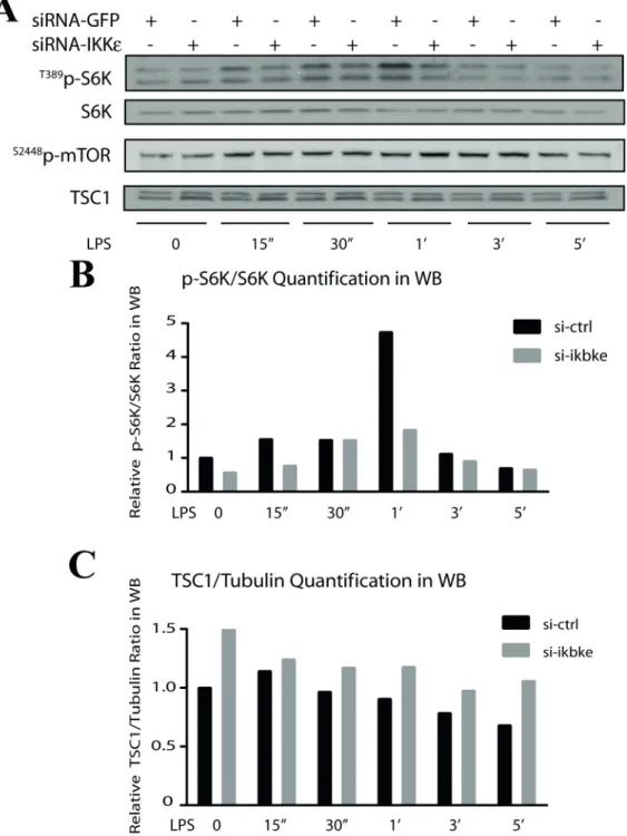
3.2. TSC1 is stabilized upon IKBKE loss in SW480 cells
To better understand the nature of regulation on S6K upon IKKε loss in SW480 cells, further analyses of upstream molecules controlling S6K were carried out.
For this purpose, IKBKE was silenced in SW480 cells but we only used LPS (10 ng/mL) stimulation (0–5 h) to activate mTOR and its downstream targets. As a result, we observed a strong reduction in S6K phosphoactivation (Figure 2A, lane 1, and Figure 2B) in the absence of IKKεâ while mTOR phosphorylation was not affected (Figure 2A, lane 3). Therefore, the potential IKKε regulation on S6K is not through mTORC1 but an alternative pathway. One such candidate could be mTOR inhibitor TSC1. To verify, we checked TSC1 protein levels upon IKKε silencing and we found that TSC1 was strongly stabilized in the absence of IKKε in SW480 cells (Figure 2A, lane 4, and Figure 2C). These observations collectively suggested the existence of a novel regulatory function for IKKε to control TSC1 or S6K activities.
As shown in previous studies, IKKβ (a related IκB kinase) controls TSC1/2 stability through direct phosphorylation (Lu et al., 2007). We therefore wanted to compare relative effects of IKBKE or IKBKB silencing on TSC1 stability. For this purpose, we transfected SW480 cells with siRNAs against IKBKE or IKBKB and GFP as the control, followed by LPS stimulations after 48 h. LPS (10 ng/mL) was used up to 1 h when peak activation of IKKε was observed. The depletion of IKKε or IKKα (Figure 3A, lanes 2 and 3) protein levels was confirmed by WB (Figure 3A; quantified in Figure 3B). Upon LPS stimulation in control samples, TSC1 levels were observed to be first increased upon 30 min of treatment but then reduced back to normal levels after 1 h (Figure 3A, lane 1, and its quantification in Figure 3B). In the absence of IKKβ or IKKε, however, TSC1 protein levels were observed
to be higher than in control samples at 30 min or 1 h of LPS treatment. In a sharp contrast to the TSC1 level of control samples, an increased TSC1 level was sustained even after 1 h of LPS treatment upon IKKε or IKKβ deficiency. Interestingly, TSC1 stabilization was even more pronounced in the absence of IKKε than that of IKKβ in SW480 cells (Figure 3A, lane 1, and its quantification in Figure 3B).
To understand if the regulation on TSC1 stability by IKKε is at a transcriptional or posttranslational level, we first checked TSC1 and TSC2 mRNA expression. From the analysis of the RNA-Seq data from our previous work (Göktuna et al., 2016), we found that the mRNA expressions of TSC1 or TSC2 (Figures 3C and 3D) were not different in intestinal epithelial samples of wt or
Ikbkeko mice in a Wnt-dependent tumor model (β-catc.a. –
or constrictively active â-catenin– is short for
Villin-Cre-ERT2/Ctnnb1+/lox(ex3) mice (Göktuna et al., 2014)). Then
we also checked the TSC1 mRNA levels via qRT-PCR in control versus IKBKE-silenced SW480 cells (Figure 3E). Concomitantly, we did not observe any change in TSC1 expression upon IKKε depletion. Therefore, we concluded that IKKε regulates TSC1 posttranslationally.
3.3. TSC1 directly interacts with IKBKE and is phosphorylated by this kinase
To elucidate the nature of interaction between TSC1 and IKKε, we ectopically expressed FLAG- or MYC-tagged proteins in HEK293 cells (Figure 4A) Expression levels of each protein were measured in WB by the use of anti-FLAG or anti-Myc antibodies (Figure 4A, lanes 1 and 2). Noticeably, ectopically expressed Myc-TSC1
Figure 1. S6K cannot be activated in the absence of IKKε. SW480 cells were first silenced with IKKε siRNA for 48 h and later treated with
either no serum, 10 ng/mL LPS, or 100 ng/mL TNFα for an additional 24 h for the activation of the mTOR/S6K pathway. Upon IKKε silencing, p70-S6K phosphorylations were seriously diminished in all stress conditions relative to the control. A) WB results for IKKε, TBK1, phospho-S6K, and S6K proteins by the use of specific antibodies. B) Quantification of phospho-S6K/S6K band intensities in WB (for p-S6K quantifications only heavy isoform has been used since total antibody only recognizes this isoform).
Figure 2. S6K inactivation and TSC1 stabilization correlate upon IKBKE silencing. SW480 cells were silenced with IKKε siRNA
(please refer to Göktuna et al., 2016 for depletion controls such as IKKε and α-tubulin western blots; data are obtained from a similar experimental setup) for 48 h and then they were treated with 10 ng/mL LPS for 0 to 5 h for the activation of mTOR/S6K and IKKε. According to WB results, only S6K (A) activation, not mTOR (B), was diminished in the absence of IKKε (as quantified by phospho-S6K/S6K densitometric band intensities) and S6K inactivation was inversely correlating with the stabilization of TSC1 (as quantified in (C)).
showed a clear upshift only when it was coexpressed with FLAG-IKKε (Figure 4A, lane 2). This upshift might be due to phosphorylation of TSC1 by IKKε. To
examine this hypothesis, we first tested the possibility of an interaction between these proteins by the use of coimmunoprecipitation (co-IP) assay after ectopically
expressing both proteins together in HEK293 cells (Figure 4B). As a result, we found that IKKε and TSC1 proteins physically interact when ectopically expressed (Figure 4B,
right and left panels).
Since no endogenous interaction between IKKε and TSC1 could be detected (results not shown), we thought
Figure 3. IKKε deficiency affects the posttranslational stabilization of TSC1. siRNAs targeting IKKε or IKKβ (siRNA-GFP used as
mock) were used for silencing respective genes in SW480 cells for 48 h, and then cells were treated with LPS (10 ng/mL) for 0 to 1 h for the activation of related signaling pathways (m: minutes). As a result of WB by the use of specific antibodies, TSC1 protein levels were observed to be stabilized especially upon IKKε silencing in SW480 cells (A) (as quantified by densitometric band intensities in ImageJ (B)). RNA-Seq data from our previous study (Göktuna et al., 2016) was analyzed for relative TSC1 (C) or TSC2 (D) mRNA expressions in wt (β-catc.a./Ikbke+/+) or Ikbkeko (β-catc.a./Ikbke-/-) intestinal epithelial tumor samples and we found no effect on their transcription. TSC1
mRNA expression was also checked by RT-PCR in IKBKE-silenced SW480 cells (by transfecting control vs. two different IKBKE siRNA constructs) (E) and still no differences in TSC1 mRNA expressions were noted as a result of IKKε deficiency.
that their interaction could be transient, as in the case of many other similar interactions involving a kinase and its substrate (Sommese and Sivaramakrishnan, 2016). Next, we tested if TSC1 is a direct substrate for IKKε by performing an in vitro kinase assay (Figure 4C). Briefly, we used a short fragment (amino acids 500–600, containing the putative S511 phosphorylation site) of recombinant human-GST-TSC1 as a substrate and ectopically expressed Myc-IKKεWT or Myc-IKKεKD (kinase dead, K38A) proteins
(harvested from HEK293 via IP) as kinase in the assay. As a result, only catalytically active IKKε (Myc-IKKεWT) could
phosphorylate the GST-TSC1500-600 substrate (Figure 4C,
lane 1), suggesting that TSC1 is a direct substrate for IKKε.
4. Discussion
In our previous studies aiming to understand IKKε involvement in colorectal tumorigenesis, we observed strong downregulation in phosphorylations of AKT and ERK1/2 kinases, whose activities were found to be essential for the survival of tumor cells upon genotoxic stress or oncogenic transformation (Göktuna et al., 2016). Indeed, we have confirmed a direct phosphoregulatory control on AKT by IKKε in CRC cell lines (data not shown). These observations were consistent with the previous findings
Figure 4. TSC1 interacts with IKKε and is phosphorylated by this kinase. A) FLAG-TSC1 and Myc-IKKε constructs were ectopically
expressed in HEK293 cells as indicated. TSC1 shows a phosphospecific shift (lane 2) upon coexpression with IKKε in HEK293 cells. B) FLAG-TSC1 and Myc-IKKε constructs were ectopically expressed in HEK293 cells as indicated. Then Myc-IKKε constructs were harvested from the cell lysates with anti-Myc antibody and coprecipitated proteins were analyzed by western blot. FLAG-TSC1 was observed to bind to IKKε in two separate trials (right and left panels). C) Myc-IKKεwt or Myc-IKKεKD proteins were ectopically expressed
in HEK293 cells and these kinases were harvested from cell lysates with immunoprecipitation by the use of anti-Myc antibody. Then GST-TSC1 (500-600) substrates were used for in vitro kinase assay. Only Myc-IKKεwt was observed to phosphorylate the TSC1 substrate
in inflammatory cells (Guo et al., 2011; Xie et al., 2011). We concluded that downregulation of AKT and ERK phosphoactivation may explain the reduced growth of mouse colorectal tumors through increased apoptosis in the absence of IKKε (Göktuna et al., 2016). However, our observations on IKKε involvement in CRC tumorigenesis were not limited to these survival kinases. IKKε loss has pleiotropic effects on colorectal cancer cells varying from increased apoptosis to loss of proinflammatory cytokine expression and reduced tumor growth in various tumor models (Göktuna et al., 2016). Therefore, in this study, we investigated if IKKε is also involved in other mechanisms related to oncogenic transformation by the use of IKKε loss-of-function models in cancer cell lines.
Results of the experiments showed a robust downregulation of downstream targets in the mTOR pathway upon IKKε loss. One plausible explanation could be that IKKε deficiency leads to downregulation of mTORC activity due to diminished AKT phosphoactivation (Xie et al., 2011; Huang and Fingar, 2014). Moreover, it is also possible that IKKε may directly phosphorylate the mTORC complex, similarly to the IKK-related kinase TBK1 upon growth factor or innate immunity agonist stimulation in immune cells (Bodur et al., 2018). However, direct interference by IKKε on mTORC or its upstream elements like AKT can be ruled out in this case, since mTOR
phosphorylation is not affected in SW480 cells. Moreover, the ability of mTOR to induce S6K activity does not solely depend on mTOR phosphorylation. The mTOR complex can also be regulated by inhibitory molecules like TSC1, AMPK, or GSK3α/β. As described earlier, TSC1/2 can also prevent mTORC-driven S6K activation by converting Rheb-GTP to its inactive form (Laplante and Sabatini, 2012). Numerous reports suggested that TSC1/2 can be directly phosphoregulated via different effectors such as AKT, ERK, RSK, and IKKα (Saxton and Sabatini, 2017), some of which can also regulate GSK3α/β activities. On the other hand, IKK-related kinase TBK1 was also shown to activate mTORC1 through phosphoregulating inhibitory AMPK in adipocytes (Zhao et al., 2018). Consequently, we hypothesized that IKKε may be directly or indirectly regulating TSC1/2 activity as suggested by stabilized TSC1 protein levels upon IKKε deficiency. The indirect regulation hypothesis was finally ruled out since we did not observe any difference in GSK3α/β phosphorylation upon IKKε deficiency (data not shown). Hence, we concentrated on IKKε-mediated regulations over S6K and its upstream inhibitor TSC1 in colorectal cancer cell lines. As a result, we identified a unique regulatory connection within mTOR/S6K signaling in which TSC1 stability can be controlled via its phosphorylation by IKKε. This new regulatory mechanism is most probably independent of
Figure 5. Proposed mechanism of TSC1 and IKKε interaction in tumor cells. Here we propose a new
regulatory connection for sustaining mTOR/S6K signaling via direct phosphoregulation of TSC1 by IKKε in transformed colorectal cancer cells (the new link is shown in bold).
References
AlQurashi N, Hashimi SM, Wei MQ (2013). Chemical inhibitors and microRNAs (miRNA) targeting the mammalian target of rapamycin (mTOR) pathway: potential for novel anticancer therapeutics. Int J Mol Sci 14: 3874-3900.
Barbie TU, Alexe G, Aref AR, Li S, Zhu Z, Zhang X, Imamura Y, Thai TC, Huang Y, Boyden M et al. (2014). Targeting an IKBKE cytokine network impairs triple-negative breast cancer growth. J Clin Invest 124: 5411-5423.
Bianchini A, Loiarro M, Bielli P, Busà R, Paronetto MP, Loreni F, Geremia R, Sette C (2008). Phosphorylation of eIF4E by MNKs supports protein synthesis, cell cycle progression and proliferation in prostate cancer cells. Carcinogenesis 29: 2279-2288.
Bodur C, Kazyken D, Huang K, Ekim Ustunel B, Siroky KA, Tooley AS, Gonzalez IR, Foley DH, Acosta-Jaquez HA, Barnes TM et al. (2018). The IKK-related kinase TBK1 activates mTORC1 directly in response to growth factors and innate immune agonists. EMBO J 37: 19-38.
Challa S, Guo JP, Ding X, Xu CX, Li Y, Kim D, Smith MA, Cress DW, Coppola D, Haura EB, et al. (2016). IKBKE is a substrate of EGFR and a therapeutic target in non-small cell lung cancer with activating mutations of EGFR. Cancer Res 76: 4418-4429. Chau TL, Gioia R, Gatot JS, Patrascu F, Carpentier I, Chapelle JP, O’Neill L, Beyaert R, Piette J, Chariot A (2008). Are the IKKs and IKK-related kinases TBK1 and IKK-epsilon similarly activated? Trends Biochem Sci 33: 171-180.
AKT or mTORC regulation by IKKε (Xie et al., 2011; Bodur et al., 2018) since mTOR or GSK3α/β phosphorylations were not affected. The proposed mechanism for this new regulatory circuit is summarized in Figure 5. We believe that this is the first report that demonstrates a direct interaction between IKKε and TSC1. The biological function of the observed results in tumor cell growth or survival are yet to be elucidated with more comprehensive studies.
Targeting mTORC and its downstream elements in cancer has long been thought to be a prominent strategy since mTORC acts as an environmental sensor of various stimuli (e.g., from cytokine, growth, or death receptors) to activate downstream molecules for oncogenic transformation (Zoncu et al., 2011). Currently there are numerous preclinical and clinical studies determining the therapeutic potential of usage of rapamycin and its analogs (rapalogs) for the inhibition of mTOR signaling or related pathways. However, the success and efficiency of such approaches are seriously limited due to the existence of negative feedback loops within mTOR signaling (Sun et al., 2015; Saxton and Laplante, 2017). For example, mTORC1 (directly) or S6K1 (indirectly) can mediate insulin receptor substrate-1 (IRS-1) phosphorylation and its subsequent degradation leads to desensitization of RTKs to growth factors as a feedback inhibition (cited in Laplante and Sabatini, 2012). Similarly, regulatory pathways can result in reduction of RTK expression like the one through platelet-derived growth factor receptors (PDGFRs), whose detailed mechanism is yet to be identified (Saxton and Sabatini, 2017). Therefore, we need a better understanding of mTOR signaling and its downstream regulatory circuits to develop superior therapeutic strategies in cancer therapy.
IKKε is known to have diverse substrates in many various signaling pathways, resulting in alternative or bypass mechanisms for continuation of transmitting the signals or providing cross-talk between others (Chau et
al., 2008; Shen and Hahn, 2011). IKKε-mediated control of mTORC1, AKT, or TSC1 suggests the existence of multiple simultaneous control steps within mTOR signaling. The results obtained in this report also illustrate tissue and cell specificity of certain regulatory circuits and help to widen our understanding of signaling networks, their cross-talk, and their regulation by effector molecules. Identification of signaling networks linked with IKK-related kinases may offer unique opportunities to find alternative therapeutic approaches for fighting against cancer. In a related study, it was shown that IKKε activity is essential for the survival of pancreatic cancer cells upon mTOR inhibition (Rajurkar et al., 2017). Additionally, another study pointed out that TBK1 may provide the activation of the AKT/ mTOR pathway in lung tumorigenesis depending on the context (Cooper et al., 2017). Therefore, the combination of mTOR and IKK-related kinase inhibition may prove a strong strategy to tackle cancers where cancer cell growth is primarily dependent on these pathways. These combinatorial approaches may circumvent the drawbacks associated with mTOR inhibition and can provide better therapeutic alternatives for the development of personalized cancer therapies.
Acknowledgments
I thank Dr Alain Chariot (GIGA and WELBIO, University of Liege, Belgium) for all the support he provided to make this study possible. I also thank Dr Mien-Chie Hung (MD Anderson Cancer Center, TX, USA) for providing TSC1 plasmid constructs, Dr Ivan Dikiç (Frankfurt Univ., Frankfurt, Germany) for providing pLK5-IKKε plasmid, and Dr Tieu Lan Chau for critical reading of the manuscript. This study was supported by F.N.R.S, FLF, and WELBIO funds in Belgium. The author is also supported by a TÜBİTAK ARDEB 116Z349 grant.
Cooper JM, Ou YH, McMillan EA, Vaden RM, Zaman A, Bodemann BO, Makkar G, Posner BA, White MA (2017). TBK1 provides context-selective support of the activated Akt/mTOR pathway in lung cancer. Cancer Res 77: 5077-5094.
Dowling RJ, Topisirovic I, Fonseca BD, Sonenberg N (2010). Dissecting the role of mTOR: lessons from mTOR inhibitors. Biochim Biophys Acta 1804: 433-439.
Feng Z, Zhang H, Levine AJ, Jin S (2005). The coordinate regulation of the p53 and mTOR pathways in cells. P Natl Acad Sci USA 102: 8204-8209.
Göktuna SI, Canli O, Bollrath J, Fingerle AA, Horst D, Diamanti MA, Pallangyo C, Bennecke M, Nebelsiek T, Mankan AK et al. (2014). IKKα promotes intestinal tumorigenesis by limiting recruitment of M1-like polarized myeloid cells. Cell Rep 7: 1914-1925.
Göktuna SI, Shostak K, Chau TL, Heukamp LC, Hennuy B, Duong HQ, Ladang A, Close P, Klevernic I, Olivier F et al. (2016). The prosurvival IKK-related kinase IKKε integrates LPS and IL17A signaling cascades to promote Wnt-dependent tumor development in the intestine. Cancer Res 76: 2587-2599. Guo JP, Coppola D, Cheng JQ (2011). IKBKE protein activates
Akt independent of phosphatidylinositol 3-kinase/PDK1/ mTORC2 and the Pleckstrin homology domain to sustain malignant transformation. J Biol Chem 286: 37389-37398. Hsieh AC, Costa M, Zollo O, Davis C, Feldman ME, Testa JR,
Meyuhas O, Shokat KM, Ruggero D (2010). Genetic dissection of the oncogenic mTOR pathway reveals druggable addiction to translational control via 4EBP-eIF4E. Cancer Cell 17: 249-261.
Hsu S, Kim M, Hernandez L, Grajales V, Noonan A, Anver M, Davidson B, Annunziata CM (2012). IKKε coordinates invasion and metastasis of ovarian cancer. Cancer Res 72: 5494-5504.
Huang J, Manning BD (2008). The TSC1–TSC2 complex: a molecular switchboard controlling cell growth. Biochem J 412: 179-190. Huang J, Manning BD (2009). A complex interplay between Akt,
TSC2, and the two mTOR complexes. Biochem Soc Trans 37: 217-222.
Huang K, Fingar DC (2014). Growing knowledge of the mTOR signaling network. Semin Cell Dev Biol 36: 79-90.
Inoki K, Corradetti MN, Guan KL (2005). Dysregulation of the TSC-mTOR pathway in human disease. Nat Genet 37: 19-24. Inoki K, Li Y, Xu T, Guan KL (2003a). Rheb GTPase is a direct target
of TSC2 GAP activity and regulates mTOR signaling. Genes Dev 17: 1829-1834.
Inoki K, Li Y, Zhu T, Wu J, Guan KL (2002). TSC2 is phosphorylated and inhibited by Akt and suppresses mTOR signalling. Nat Cell Biol 4: 648-657.
Inoki K, Ouyang H, Zhu T, Lindvall C, Wang Y, Zhang X, Yang Q, Bennett C, Harada Y, Stankunas K et al. (2006). TSC2 integrates Wnt and energy signals via a coordinated phosphorylation by AMPK and GSK3 to regulate cell growth. Cell 126: 955-968.
Inoki K, Zhu T, Guan KL (2003b). TSC2 mediates cellular energy response to control cell growth and survival. Cell 115: 577-590. Jastrzebski K, Hannan KM, Tchoubrieva EB, Hannan RD, Pearson
RB (2007). Coordinate regulation of ribosome biogenesis and function by the ribosomal protein S6 kinase, a key mediator of mTOR function. Growth Factors 25: 209-226.
Kim JY, Beg AA, Haura EB (2013). Non-canonical IKKs, IKKε and TBK1, as novel therapeutic targets in the treatment of non-small cell lung cancer. Expert Opin Ther Tar 17: 1109-1112. Laplante M, Sabatini DM (2012). mTOR signaling in growth control
and disease. Cell 149: 274-293.
Lee DF, Kuo HP, Chen CT, Hsu JM, Chou CK, Wei Y, Sun HL, Li LY, Ping B, Huang WC et al. (2007). IKKβ suppression of TSC1 links inflammation and tumor angiogenesis via the mTOR pathway. Cell 130: 440-455.
Lu J, Yang Y, Guo G, Liu Y, Zhang Z, Dong S, Nan Y, Zhao Z, Zhong Y, Huang Q (2017). IKBKE regulates cell proliferation and epithelial–mesenchymal transition of human malignant glioma via the Hippo pathway. Oncotarget 8: 49502-49514. Ma L, Chen Z, Erdjument-Bromage H, Tempst P, Pandolfi PP (2005).
Phosphorylation and functional inactivation of TSC2 by Erk implications for tuberous sclerosis and cancer pathogenesis. Cell 121: 179-193.
Manning BD, Tee AR, Logsdon MN, Blenis J, Cantley LC (2002). Identification of the tuberous sclerosis complex-2 tumor suppressor gene product tuberin as a target of the phosphoinositide 3-kinase/akt pathway. Mol Cell 10: 151-162. Niederberger E, Moser CV, Kynast KL, Geisslinger G (2013). The
non-canonical IκB kinases IKKε and TBK1 as potential targets for the development of novel therapeutic drugs. Curr Mol Med 13: 1089-1097.
Péant B, Gilbert S, Le Page C, Poisson A, L’Ecuyer E, Boudhraa Z, Bienz MN, Delvoye N, Saad F, Mes-Masson AM (2017). IκB-Kinase-epsilon (IKKε) over-expression promotes the growth of prostate cancer through the C/EBP-β dependent activation of IL-6 gene expression. Oncotarget 8: 14487-14501.
Potter CJ, Pedraza LG, Xu T (2002). Akt regulates growth by directly phosphorylating Tsc2. Nat Cell Biol 4: 658-665.
Rajurkar M, Dang K, Barrena MG, Liu X, Fernandez-Zapico ME, Lewis BC, Mao J (2017). IKBKE is required during KRAS-induced pancreatic tumorigenesis. Cancer Res 77: 320-329.
Roux PP, Ballif BA, Anjum R, Gygi SP, Blenis J (2004). Tumor-promoting phorbol esters and activated Ras inactivate the tuberous sclerosis tumor suppressor complex via p90 ribosomal S6 kinase. P Natl Acad Sci USA 101: 13489-13494.
Saitoh M, Pullen N, Brennan P, Cantrell D, Dennis PB, Thomas G (2002). Regulation of an activated S6 kinase 1 variant reveals a novel mammalian target of rapamycin phosphorylation site. J Biol Chem 277: 20104-20112.
Saxton RA, Sabatini DM (2017). mTOR signaling in growth, metabolism, and disease. Cell 168: 960-976.
She QB, Halilovic E, Ye Q, Zhen W, Shirasawa S, Sasazuki T, Solit DB, Rosen N (2010). 4E-BP1 is a key effector of the oncogenic activation of the AKT and ERK signaling pathways that integrates their function in tumors. Cancer Cell 18: 39-51. Shen RR, Hahn WC (2011). Emerging roles for the non-canonical
IKKs in cancer. Oncogene 30: 631-641.
Sommese RF, Sivaramakrishnan S (2016). Substrate affinity differentially influences protein kinase C regulation and inhibitor potency. J Biol Chem 291: 21963-21970.
Sun Z, Wang Z, Liu X, Wang D (2015). New development of inhibitors targeting the PI3K/AKT/mTOR pathway in personalized treatment of non-small-cell lung cancer. Colloq Inse 26: 1-14. Xie X, Zhang D, Zhao B, Lu MK, You M, Condorelli G, Wang CY,
Guan KL (2011). IκB kinase ε and TANK-binding kinase 1 activate AKT by direct phosphorylation. P Natl Acad Sci USA 108: 6474-6479.
Zhao P, Wong KI, Sun X, Reilly SM, Uhm M, Liao Z, Skorobogatko Y, Saltiel AR (2018). TBK1 at the crossroads of inflammation and energy homeostasis in adipose tissue. Cell 172: 731-743. Zoncu R, Sabatini DM, Efeyan A (2011). mTOR: From growth signal
integration to cancer, diabetes and ageing. Nat Revs Mol Cell Biol 12: 21-35.
Zubair H, Azim S, Srivastava SK, Ahmad A, Bhardwaj A, Khan MA, Patel GK, Arora S, Carter JE, Singh S et al. (2016). Glucose metabolism reprogrammed by overexpression of IKKε promotes pancreatic tumor growth. Cancer Res 76: 7254-7264.