OPTICAL GAIN AND LASING OF
COLLOIDAL SEMICONDUCTOR
QUANTUM WELLS INTIMATELY
INTEGRATED INTO OPTICAL CAVITIES
a thesis submitted to
the graduate school of engineering and science
of bilkent university
in partial fulfillment of the requirements for
the degree of
master of science
in
electrical and electronics engineering
By
Mustafa Sak
OPTICAL GAIN AND LASING OF COLLOIDAL SEMICONDUC-TOR QUANTUM WELLS INTIMATELY INTEGRATED INTO OPTICAL CAVITIES
By Mustafa Sak July 2019
We certify that we have read this thesis and that in our opinion it is fully adequate, in scope and in quality, as a thesis for the degree of Master of Science.
Hilmi Volkan Demir(Advisor)
Fatih ¨Omer ˙Ilday
Sergey Vasil’evich Gaponenko
Approved for the Graduate School of Engineering and Science:
Ezhan Kara¸san
Director of the Graduate School ii
iii
ABSTRACT
OPTICAL GAIN AND LASING OF COLLOIDAL
SEMICONDUCTOR QUANTUM WELLS INTIMATELY
INTEGRATED INTO OPTICAL CAVITIES
Mustafa Sak
M.S. in Electrical and Electronics Engineering Advisor: Hilmi Volkan Demir
July 2019
Colloidal semiconductor quantum wells, also known as nanoplatelets (NPLs), attract an increasingly greater deal of interest as a promising material platform for light-generating applications. The superior optical properties of NPLs including their ultra-large absorption cross-sections, purely homogeneous broadening, and suppressed Auger recombination make them highly attractive for solution-processable color convertors, LEDs and lasers. In this thesis, we studied the optical gain properties and performance levels of tailored heterostructures of such NPLs intimately integrated into various optical cavities. To do so, we systematically measured their amplified spontaneous emission under one- and two-photon absorption excitations. Also, with these hetero-NPLs as the gain media, we have proposed and demonstrated a new class of practical whispering gallery mode (WGM) NPL-fiber architecture with high stability and low lasing thresholds enabled by record low waveguide loss coefficients in its class. Moreover, we have developed a single-mode vertical-cavity surface-emitting laser (VCSEL) of these hetero-NPLs closely integrated into the wedge cavity of a pair of distributed Bragg reflectors, leading to a record low lasing threshold in its class. The findings obtained in these WGM laser and NPL-VCSEL structures indicate that these NPLs are excellent for high-performance colloidal lasing.
Keywords: Semiconductor nanocrystals, colloidal nanoplatelets, optical gain,
iv
ÖZET
OPTİK ÇINLAÇLAR İÇİNDE YAKİNEN TÜMLEŞTİRİLMİŞ
KOLOİDAL YARIİLETKEN KUANTUM KUYULARININ
OPTİK KAZANCI VE LAZER EYLEMİ
Mustafa Sak
Elektrik ve Elektronik Mühendisliği, Yüksek Lisans Tez Danışmanı: Hilmi Volkan Demir
Temmuz 2019
Nanolevhalar olarak da adlandırılan koloidal yarıiletken kuantum kuyuları ışık üreten uygulamalar için umut vadeden bir materyal platformu olarak artan miktarda yoğun ilgi görmektedir. Nanolevhaların çok yüksek optik soğurma katsayıları, tamamen homojen optik ışıma genişlemeleri ve bastırılmış Auger eksiton birleşimi gibi üstün optik özellikleri onları çözelti halinde işlenebilir renk dönüştürücüler, ışık yayan diyotlar ve lazerler için oldukça ilgi geçici yapmaktadır. Bu tez kapsamında, özel olarak tasarlanmış heteroyapıya sahip bu nanolevhaları farklı optik çınlaçlar içerisine yakinen tümleştirerek optik kazanç özellikleri ve performans seviyeleri üzerine çalıştık. Bu amaç doğrultusunda, nanolevhaların bir- ve iki-foton soğurma uyarımı altında yükseltilmiş kendiliğinden ışımalarını sistematik olarak ölçtük. Ayrıca, bu hetero-nanolevhaları optik kazanç ortamı olarak kullanarak, kendi sınıfında rekor düşüklükte dalga kılavuzu kayıp katsayısı sayesinde yüksek kararlılık ve düşük lazer eşiği değerine sahip pratik bir fısıldayan-galeri-kip (FGK) nanolevha-fiber mimarisi önerip ve gösterdik. Buna ek olarak, bu hetero-nanolevhaları bir çift dağıtılmış Bragg yansıtıcılar tabanlı kama çınlaç içinde tümleştirerek yapılan, sınıfında rekor düşüklükte lazer eşik değerine sahip tek modlu bir dikey çınlaçlı yüzey ışımalı lazer (DÇYIL) geliştirdik. Burda FGK lazer ve DÇYIL yapılarından elde edilen bulgular bu nanolevhaların yüksek performanslı koloidal lazer eylemi için mükemmel olduklarını göstermektedir.
Anahtar sözcükler: Yarıiletken nanokristaller, koloidal nanolevhalar, optik kazanç,
v
Acknowledgement
First, I would like to thank my advisor, Prof. Hilmi Volkan Demir, for being a great role model. With his supportive attitude and visionary scientific approach, he has taught me a lot.
I would like to thank the past and present members of Demir Research Group. I would also like to thank Dr. Savas Delikanli, Dr. Kivanc Gungor, Dr. Yemliha Altintas for helping and guiding me throughout my M.S. studies. Moreover, I would like to thank Nima Taghipour, Ibrahim Tanriover, Selim Bozdogan, Furkan Isik, Didem Dede, and Onur Erdem for their friendship and great times that we had within these two years.
I would like to thank and acknowledge TUBITAK-BIDEB for the financial support of my M.S. studies.
I would like to thank and express my profound gratitude to my family, my mother Gulter, my father Zekeriya, and my sister Emine, for their strong encouragement and invaluable support.
Finally, with all my heart, my very special thanks and deepest gratitude go to Mine Kerpicci. This would not be possible without her unfailing support and eternal love. There are not strong enough words to thank her for her presence, support and love.
vi
Contents
1 Introduction ... 1
1.1 Organization of the Thesis ... 4
2 Background ... 6
2.1 Colloidal Semiconductor Nanocrystals ... 6
2.1.1 Quantum Confinement ... 6
2.1.2 Synthesis of Colloidal Semiconductor Nanocrystals ... 9
2.1.3 Colloidal Semiconductor Quantum Wells ... 10
2.1.4 Optical Gain of Colloidal Semiconductor Nanocrystals ... 13
2.2 Optical Resonators ... 17
2.2.1 Fabry-Perot Resonator ... 17
2.2.2 Ring Resonator ... 21
3 Experimental Methods ... 23
3.1 Steady-State Absorption Spectroscopy ... 23
3.2 Steady-State Photoluminesce Spectroscopy ... 25
3.3 Electron Microscopies ... 25
3.4 Optical Gain Measurements ... 26
3.4.1 Optical Gain Threshold Measurement ... 26
vii
3.4.3 Waveguide Loss Coefficient Measurement ... 30
4 Whispering-gallery-mode Laser of Colloidal Nanoplatelets ... 31
4.1 Motivation ... 31
4.2 Experiments ... 33
4.2.1 Precursor Preparation ... 33
4.2.2 Colloidal Synthesis of 4 ML CdSe NPLs ... 33
4.2.3 Preparation of CdS Crown Growth Solution ... 33
4.2.4 CdSe/CdS Core/Grown NPL Synthesis ... 34
4.2.5 CdSe/CdS@Cd1-xZnxS Core/Crown/Shell NPL Synthesis ... 34
4.2.6 Sample Preparation ... 35
4.2.7 Simulation Methodology ... 35
4.3 Results and Discussion ... 35
4.4 Summary ... 48
5 Vertical-cavity Surface-emitting Laser of Colloidal Nanoplatelets ... 49
5.1 Motivation ... 49
5.2 Experiments ... 50
5.2.1 Colloidal Synthesis ... 50
5.2.2 Sample Preparation ... 50
5.3 Results and Discussion ... 51
viii
6 Conclusion ... 56
6.1 Concluding Remarks ... 56
6.2 Outlook ... 57
ix
List of Figures
Figure 2.1: Schematic illustration showing the continuous bands of bulk semiconductors
along with the effect of size among nanocrystals on energy band gap. Decreasing NC size
effectively increases the energy band gap. ... 8
Figure 2.2: The nucleation and growth model of nanocrystals. Reprinted with permission
from [41]. ... 9
Figure 2.3: (a) Illustration of 4 ML CdSe NPLs, (b) HAADF-TEM image showing 4 ML
CdSe NPLs, (c) HAADF-TEM image showing 5 ML CdSe NPLs [44]. ... 11
Figure 2.4: PL and absorption spectra of CdSe NPLs with 3, 4 and 5 ML thicknesses [44].
... 12
Figure 2.5: Schematic illustration of core/crown, core/shell core/crown@shell
heterostructures of NPLs. ... 13
Figure 2.6: Schematics of the basic light-matter interactions for a two-level atomic system.
... 13
Figure 2.7: Photon flux in a medium along +z axis. Change in the flux in a volume with
an infinitesmall length is defined as 𝑑Φ. ... 15 Figure 2.8: Raditive recombination and nonradiative Auger recombination of biexcitons.
In the latter case, the exciton energy is transferred to a third carrier, and then is lost through
lattice vibrations. ... 17
Figure 2.9: Schematic illustration of a Fabry-Perot resonator with two reflecting flat
x
Figure 2.10: Structure of a 3-bilayer distributed bragg reflector (DBR) fabricated over a
glass-substrate. ... 20
Figure 2.11: Reflectivity of 11-bilayer SiO2-Si3N4 distributed bragg reflecter (DBR)
fabricated over a quartz substrate. ... 21
Figure 2.12: Schematics of a) an optical waveguide resonator, b) a three-mirror ring
resonator, c) a whispering-gallery-mode (WGM) resonator. ... 21
Figure 3.1: a) Schematic of the experimental setup behind steady-state absorption
spectroscopy. b) An exemplary absorbance spectrum of 4 ML thick CdSe core NPL
dissolved in hexane. ... 24
Figure 3.2: a) Schematic of the experimental setup behind steady-state photoluminescence
spectroscopy. b) An exemplary photoluminescence spectrum of 4 ML thick CdSe core
NPL dissolved in hexane. ... 25
Figure 3.3: a) SEM image of a SiO2 fiber with 250 µm diameter. b) HAADF-STEM image
of CdSe/CdS@Cd1-xZnxS core/crown shell NPLs. ... 26
Figure 3.4: Schematic of experimental setup to measure ASE spectrum of NCs. ... 27
Figure 3.5: Experimental optical setup for ASE measurement under one-photon
absorption. ... 28
Figure 3.6: a) Exemplary emission spectra of CdSe/Cd0.25Zn0.75S core/shell NPLs under
one-photon absorption. b) Exemplary integrated intensity of CdSe/Cd0.25Zn0.75S core/shell
NPLs showing the optical gain threshold... 28
Figure 3.7: a) Schematic of the experimental setup of variable strip length measurement.
xi
Figure 3.8: Schematic of the experimental setup to measure waveguide loss coefficient.
... 30
Figure 4.1: HAADF-STEM images of CCGAS NPLs with 3 monolayers of gradient alloy
shell showing a) their lateral sizes, b) their vertical thicknesses... 36
Figure 4.2: a) Absorption and PL spectra of our CCGAS NPLs. b) SEM image of the fiber
showing the coating of NPLs with the film thickness. ... 36
Figure 4.3: For NPL-coated fiber with 125 µm diameter, a) emission spectra at various
pump fluences, b) integrated intensity as a function of the pump fluence under one-photon
absorption excitation. ... 37
Figure 4.4: For NPL-coated fiber with 250 µm diameter, a) emission spectra at various
pump fluences, b) integrated intensity as a function of the pump fluence under one-photon
absorption excitation. ... 38
Figure 4.5: For NPL-coated fiber with 125 µm diameter, a) emission spectra at various
pump fluences, b) integrated intensity as a function of the pump fluence under two-photon
absorption excitation. ... 38
Figure 4.6: For NPL-coated fiber with 250 µm diameter, a) emission spectra at various
pump fluences, b) integrated intensity as a function of the pump fluence under two-photon
absorption excitation. ... 39
Figure 4.7: Variable stripe length (VSL) measurement of our NPL-coated fiber with 250
µm core diameter at various excitation fluences. ... 40 Figure 4.8: Net modal gain coefficient as a function of the pump fluence along with the
numerical fitting. ... 41
Figure 4.9: Waveguide loss coefficient measurement along with exponential fits for a) 125
xii
Figure 4.10: a) Illustration of the cross-section of NPL-WGM resonator. b) Image of the
NPL-WGM under optical excitation with the resulting laser emission. c) Illustration of
experimental setup . ... 42
Figure 4.11: PL spectra of 250-µm fiber acts as an NPL-WGM resonator under 1PA
excitation. ... 43
Figure 4.12: PL intensity of NPL-WGM resonator at various pump fluences under 1PA
pumping. ... 43
Figure 4.13: PL spectra of 250-µm NPL-coated fiber, which acts as an NPL-WGM
resonator under 2PA excitation. ... 44
Figure 4.14: PL intensity of NPL-WGM resonator at various pump fluences under 2PA
pumping. ... 44
Figure 4.15: E-field distribution of NPL-WGM resonator with 125 µm diameter fiber..
... 45
Figure 4.16: Lasing peaks of the NPL-WGM resonator with 125 µm dimeter, where
experimental and numerical peak locations spectrally overlap... 46
Figure 4.17: Normalized PL intensity of our laser as a function of the polarizer angle and
fitted quadratic cosine function. ... 47
Figure 4.18: Normalized laser intensity our NPL-WGM resonator under a fixed pumping
for stability measurement. ... 48
Figure 5.1: a) Absorption and PL spectra of our CCGAS NPLs. b)HAADF-STEM images
of CCGAS NPLs with 4 monolayers of gradient alloyed shell. ... 51
Figure 5.2: Reflectivity of the fabricated DBR, where near perfect reflection was achieved
xiii
Figure 5.3: a) Schematic illustration of our wedge VCSEL cavity. b) Image of our
NPL-VCSEL cavity pumped with a pump fluence above lasing threshold with the resulting
lasing spot on the paper. ... 52
Figure 5.4: PL spectra of NPL-VCSEL under 1PA excitation, demonstrating a
single-mode lasing. ... 53
Figure 5.5: Emission intensity of NPL-VCSEL under 1PA excitation as a function of the
pump fluences, where the record low lasing threshold and saturation at higher pump can
be observed... 54
Figure 5.6: Laser output as a function of the analyzer angle and its fit to a quadratic cosine
function. ... 54
xiv
List of Tables
Table 2.1 Exciton Bohr radii of II-VI semiconductors that are frequently used in this
1
Chapter 1
Introduction
Semiconductors have dramatically revolutionized daily lives since the invention of the first semiconductor transistor, which was realized on a germanium crystal by Walter Brattain, John Bardeen and William Shockley at Bell Labs in 1947 [1]. In the last half century, semiconductor technologies have altered a wide range of fields including computing, military electronics, communication and healthcare by offering a robust and power efficient systems. Besides, although Einstein theoretically explained the mechanisms behind the light-matter interactions such as optical absorption, spontaneous emission and stimulated emission phenomena in 1917 [2], the first successful demonstration of stimulated emission was achieved in 1955 [3] and the first practical laser (light amplification by stimulated emission of radiation) was built by Theodore H. Maiman in 1960, of which gain medium is made out of ruby [4]. However, one needs to wait until 1962 to see the first presentation of semiconductor laser made of gallium arsenide [5], which has an emission at 850 nm corresponding to the near-infrared region of the electromagnetic spectrum. From this date forward, semiconductors have taken an unsubstituted part in the field of photonics.
Energy band gap of a semiconductor material is defined as the energy difference between the top of fully-occupied valence band and the bottom of unoccupied conduction band.
2
Thanks to their relatively small energy band gaps, semiconductors are irreplaceable candidates to be used in optoelectronics. The strong and controllable interaction between semiconductors and light (electromagnetic wave) enables the utilization of light-harvesting and light-emitting devices for a broad range of applications including lighting, displays and information technologies. This crucial wide-scale role of semiconductors makes the cost of their growth method more important and impactful commercially. However, traditional semiconductor growth techniques like czochralski method and epitaxial growth require ultra-high vacuum and high temperature conditions, resulting in high-cost fabrication and difficulty in large-scale integration. Hence, the development of low-cost and large-area methods to grow semiconductors presents strategic importance. To this end, colloidal semiconductor nanocrystals have emerged as a promising material platform owing to their solution processable nature and their wet chemistry synthesis, which makes them a cost-effective alternative to conventional thin-film semiconductors.
Besides the cost-related issues, colloidal semiconductor nanocrystals (NCs) have attracted great attention owing to their favorable characteristics. Thanks to the controllable quantum confinement realized in these nanometer-sized semiconductor nanocrystals, their optical and electronic features can be conveniently tuned simply by altering their shape, size and compositions [6], [7]. To date, various shapes of colloidal semiconductor NCs have been synthesized via wet chemistry including zero-dimensional quantum dots [8], [9], one-dimensional nanorods[10], [11] and recently, two-one-dimensional quantum wells [12], [13]. Also, different heterostructures have been engineered to specifically alter the excitonic features of NCs to obtain desired characteristics [6], [14], [15]. So far, owing to their remarkable photophysical properties, NCs have been used in different light-emitting and light-harvesting applications including photovoltaic cells [16], photodetectors [17], luminescent solar concentrators [18], light-emitting diodes (LEDs) [19] and lasers [20].
Among various classes of nanocrystals, colloidal semiconductor quantum wells (CQWs), the so-called colloidal nanoplatelets (NPLs), stand out as an auspicious member of nanocrystal family owing to their superior electronic and optical properties. Their characteristics such as giant oscillator strength [21], extraordinarily large linear and nonlinear absorption cross-sections [22], ultralarge modal optical gain performances [23]
3
and purely homogeneous broadening [24] make them great candidates to achieve low-threshold optical gain and high-performance colloidal lasers. In fact, with their color tunability, any laser that operates in visible to infrared regions can be built using them [25].
Today, various shapes of colloidal nanocrystals are utilized as gain media for lasing and optical gain application including quantum dots [20], nanorods [26], quantum wells [27], [28] and perovskite nanocrystals [29], [30]. Nonetheless, the prevalence of nanocrystals lasers is restrained by a dominant process, the nonradiative Auger recombination, which makes them impractical for real-life applications. Auger is a rapid recombination process annihilating the active carriers, limiting their performance of colloidal lasers. For this reason, different heterostructures (core/crown [31], [32] and core/shell [28], [33]) of NPLs have been introduced to tune their excitonic features to overcome this issue, which thus enables highly efficient optical gain performance [34]. Hence, by virtue of heterostructures along with aforementioned characteristics, NPLs offer a high-performance media for optical gain and lasing applications.
The realization of a practical NC laser requires the use of the optical cavities (resonators) to confine the emission of optical gain media and provide optical feedback with the help of amplifying in the gain media for lasing action. Previously, different types of NCs have been utilized in colloidal laser applications in close-packed films or in solution phase integrated to various optical cavities. These include vertical cavity surface-emitting lasers [35], which have been implemented by using specialized Fabry-Perot resonators, whispering gallery mode lasers [36], which utilized by ring resonators. Depending on the specific applications, resonator types can be meticulously designed, and the phase (in solution or solid film) of the NC gain media can be adjusted. Furthermore, the excitation regime of the NC laser (single photon or multi photon) can be chosen to address a unique application such as bioimaging under-skin treatment. Hence, it is crucially essential to systematically investigate the optical gain and lasing performances of various heterostructures of colloidal NPLs coupled to different optical cavities.
To investigate these issues, in this thesis, we have designed, developed and analyzed novel nanocrystal-based lasers integrated to different optical resonators. For this purpose, we
4
have proposed and synthesized engineered-heterostructures of solution-processed colloidal NPLs including core/crown@gradually-alloyed-shell architecture. Then, we have examined their electronic and optical features to obtain desired characteristics for our optoelectronic devices. Here, we have systematically investigated single- and multi-photon pumped optical gain properties of CdSe/CdS@Cd1-xZnxS
core/crown@gradually-alloyed-shell (C/C@GAS) NPLs. Furthermore, we have employed this advanced heterostructure to obtain a novel fiber-based whispering gallery mode (WGM) laser and vertical-cavity surface-emitting laser (VCSEL). Our record low waveguide loss coefficients and record low lasing thresholds reveal the true potential of the advanced heterostructures of these NPLs for optical gain and lasing studies. These findings indicate that this new generation of nanocrystals may play a vital role to overcome long-standing challenges of traditional colloidal nanocrystal lasers and maybe even surpass the performances of their conventional epitaxially-grown counterparts.
1.1 Organization of the Thesis
In Chapter 2, we present the scientific background about the colloidal semiconductor nanocrystals including their quantum behavior, synthesis, excitonic and optical properties. We also explained the characteristics and different types of the optical resonators in this chapter.
In Chapter 3, we describe experimental methods that are frequently used throughout the thesis such as steady-state absorption and PL spectroscopy. In addition, optical gain characterization methods covering gain threshold, variable stripe length and waveguide loss coefficient measurements are introduced.
In Chapter 4, we propose, simulate and demonstrate multi-mode lasing behavior of colloidal nanoplatelets in solid film phase enabled by fiber-based whispering-gallery-mode optical cavity. We present record low waveguide loss coefficients. Furthermore, by this study, we unleash the potential of close-pack NPL films to be used for multi-mode lasers.
5
In Chapter 5, we propose and show vertical-cavity surface-emitting laser based on advanced heterostructures of NPLs. Here we report record low lasing threshold among all colloidal nanocrystals.
In Chapter 6, we conclude our thesis with concluding remarks and future outlook along with a summary of the contributions of this thesis.
6
Chapter 2
Background
2.1 Colloidal Semiconductor Nanocrystals
In this part, we present the scientific background on the colloidal semiconductor nanocrystals. Before explaining the synthesis methodology of NCs, the quantum confinement effect and the influence of heterostructures on this effect are discussed. Then, we explain size- and composition-dependent excitonic and optical characteristics of NCs. Finally, we conclude this part by reviewing the mechanism behind the optical gain phenomenon among colloidal NCs.
2.1.1 Quantum Confinement
Colloidal semiconductor nanocrystals are nanometer-sized crystalline semiconductors that contain a few hundreds to thousands of atoms. As a result of their nanometer-sized dimensions, generated electrons and holes in NCs can easily feel the boundaries of this nano-structure as potential barriers. Before going into details about quantum confinement phenomenon, it is essential to explain the Bohr radius and the formation of the exciton within nanocrystals.
7
The exciton Bohr radius of a material can be written as follows: [37]
𝑎𝐵 = ε𝑎0𝑚𝑒 𝑚∗
where ε is the dielectric constant of the medium, 𝑎0 is the Bohr radius of hydrogen atom, 𝑚𝑒 is the rest mass of the electron and 𝑚∗is the electron-hole reduced mass. As a result of
this material-dependent behavior, exciton Bohr radius depends on the material choice. Exciton Bohr radii of frequently used semiconductors in this thesis are provided in Table 2.1 [38].
Table 2.1 Exciton Bohr radii of II-VI semiconductors that are frequently used in this thesis.
Semiconductor Type Exciton Bohr Radius
CdTe 7.3 nm
CdSe 5.6 nm
CdS 2.9 nm
ZnS 2.5 nm
If the size of nanocrystal is comparable to or smaller than its exciton Bohr radius, quantum confinement effect will be observed. Due to this strong confinement in a truly small volume, Coulombic attraction between electron and hole bounds them to form an exciton. Moreover, quantum confinement regime of NCs defines their photophysical behavior, resulting in atomic-like discrete energy levels. In the literature, 3D-confined nanocrystals are called as quantum dots, where 2D- and 1D-confined ones are named as nanorods and nanoplatelets, respectively. For instance, in an 1D-confined nanoplatelet, only the thickness of this NC is smaller than the exciton Bohr radius and excitons can freely move in the lateral directions. In addition, owing to discrete energy levels of these quantum-confined NCs, their band gaps can be decreased by increasing their size, resulting a red
8
shift in their PL emission spectrum. Hence, the band gap of NCs can be simply tuned by changing their size along the way of quantum confinement by using the same material composition (Figure 2.1). The band gap of a nanocrystal is expressed by:
𝐸𝑔(𝑁𝐶) = 𝐸𝑔(𝑏𝑢𝑙𝑘) + 𝐸𝑐𝑜𝑛𝑓+ 𝐸𝑒𝑥𝑐
where the confinement energy and the exciton energy are given as:
𝐸𝑐𝑜𝑛𝑓= ћ𝜋 2𝑟2( 1 𝑚ℎ+ 1 𝑚𝑒) 𝐸𝑒𝑥𝑐 =−1.8𝑒2 4𝜋𝜀𝑟
Here ћ is the reduced Plank constant, r is the radius of nanocrystal, 𝑚ℎ is the effective mass of hole, 𝑚𝑒 is the effective mass of electron, and 𝜀 is the dielectric constant. As given
above, the band gap of NCs is strongly size-dependent. The exciton energy is inversely proportional to radius r while the confinement energy is inversely proportional to 𝑟2. Thus,
the confinement energy plays a more critical role to determine the band gap of NCs if the particle is strongly confined.
(2.2) (2.2) (2.3) (2.3) (2.4) (2.4) Figure 2.1: Schematic illustration showing the continuous bands of bulk semiconductors along with the effect of size among nanocrystals on energy band gap. Decreasing NC size effectively increases the energy band gap.
9
2.1.2 Synthesis of Colloidal Semiconductor Nanocrystals
The successful demonstration of semiconductor nanocrystals was reported in early 1980s in a dielectric matrix where size-dependent absorption features have been observed [39]. Within a half-decade, electrical and optical properties of various nanometer-sized semiconductors were explored [40]. However, the first uniform and nearly monodisperse nanocrystals of semiconductors including CdS, CdTe, and CdSe were reported by Bawendi Group in early 1990s [8]. From then on, great efforts have been made to synthesize different colloidal NCs. The process of colloidal synthesis requires three main factors: ligands, solvents and precursors. Solvent creates the suitable environment for colloidal synthesis. In high temperature environment, precursors are dissolved into monomers and form the NCs by nucleation. Ligands such as oleic acid and oleylamine are used to control the final size as well as to obtain stable, monodisperse and uniform NCs [41], [42].
Figure 2.2: The nucleation and growth model of nanocrystals. Reprinted with permission from [41].
10
According to the nucleation and growth model of nanocrystals, which are introduced by La Mer and Dinegar [43], nucleation and growth steps should be separated to synthesize highly monodisperse NCs. A rapid injection of precursors into the hot solution that generates supersaturated solution initiates the nucleation of precursor monomers, resulting in nanometer-sized crystals. This fast nucleation depletes the concentration of the precursors and pulls it down to a level lower than the nucleation threshold, restraining the formation of new nuclei as given in Figure 2.2. After that, previously formed NCs start to grow with small variations in their size. The growth time is a deterministic factor to control the size of NCs. As a result of the-so-called Ostwald ripening, NCs with bigger sizes start to grow and smaller ones dissolve as the growth time increases. Hence, with the control over Ostwald ripening and the growth time, it is possible to determine the size of NCs effectively. Nowadays, considering this relatively basic approach, various shapes of NCs can be synthesized. Since all NCs used throughout this thesis are the atomically flat ones, commonly known as colloidal quantum wells, we will mostly focus on them.
2.1.3 Colloidal Semiconductor Quantum Wells
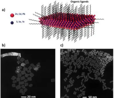
The first synthesis of colloidal semiconductor quantum wells was reported by Ithurria and Dubertret in 2008 [12]. Since the shape of these atomically-thin NCs seems like platelets, they are alternatively coined as colloidal nanoplatelets (NPLs). One of the characteristic features of NPLs is their vertical thickness, which can be precisely controlled with slight changes in their synthesis procedure. Illustration of a 4 monolayer (ML) thick CdSe NPL is given in Figure 2.3a, where 4 layers of Se atoms are sandwiched within 5 consecutive layers of Cd atoms. High-angle annular dark field transmission electron microscopy (HAADF-TEM) images of 4 ML and 5 ML CdSe NPLs are shown in Figures 2.3b and 2.3c, respectively [44]. Since the quantum confinement effect occurs in the vertical direction, controlling the number of monolayers significantly affects the absorption and photoluminescence (PL) spectra of the resulting NPLs. By increasing the number of MLs, quantum confinement of the carriers becomes more relaxed, causing a red shift in the emission and absorption spectra, which can be also interpreted from Equation 2.3 (see
11
Figure 2.4). Moreover, the emerging step-like absorption behavior of NPLs indicates their quantum-well-like density of states.
For the 4 ML case, the first excitonic peak appears at 510 nm, which corresponds to the upward transition from heavy-hole of the valence band to the conduction band, and the second peak at 484 nm corresponds to the one that belongs to the light-hole. NPLs exhibit strong quantum confinement in only one dimension since their magic-sized thicknesses are significantly smaller than their exciton Bohr radius, while their lateral thicknesses are much larger [21]. Different than other classes of NCs, high monodispersity of NPLs allows to suppress the inhomogeneous broadening, and they thus exhibit purely homogenous broadening [24]. This results in a very sharp PL line with a full-width-at-half-maximum (FWHM) typically narrower than 10 nm.
Even though monodispersity and high control on thickness is achieved, only CdSe NPLs in a simple core architecture yield photoluminescence quantum efficiency (PL-QY) around 25% due to the quenching of photogenerated carriers via nonradiative decay processes or surface traps. To improve and tune their optical and electronic properties, Figure 2.3: (a) Illustration of 4 ML CdSe NPLs, (b) HAADF-TEM image showing 4 ML CdSe NPLs, (c) HAADF-TEM image showing 5 ML CdSe NPLs [44].
12
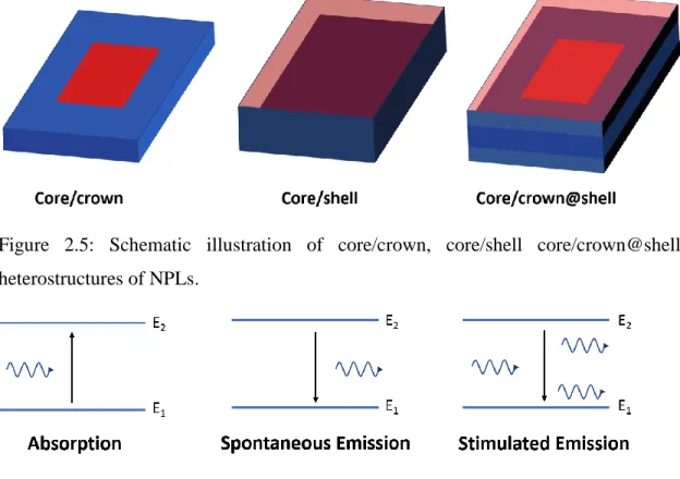
various heterostructures of NCs have been developed. Growing a crown around the core of an NPL only in the lateral direction without changing its vertical thickness was previously used to improve their stability and PL-QY [32]. Moreover, coating a shell around the core was used to control the quantum-confinement and the localization of the hole and electron in the NC [45]. Finally, advanced heterostructures like core/crown@shell was used to further engineer the core NPLs [46]. The schematic illustrations of core/crown, core/shell and core/crown@shell are depicted in Figure 2.5.
With proper choices of semiconductor materials in the heterostructures, Type-I, Type-II or quasi Type-II band alignment can be achieved in NPLs. For Type-I band alignment, both electron and hole wavefunctions are confined in the core region of the NPL. Since the band gap of ZnS is wider than CdSe, CdSe/ZnS core/shell heterostructure of NPL, Type-I can be achieved with them [47]. Besides, in Type-II band alignment, electron and hole confined in different regions of the NC. This delocalization of electron and hole reduces the overlap of their wavefunctions, resulting in stronger suppression of nonradiative Auger recombination. CdSe/CdTe core/crown NPLs reported by Dubertret Figure 2.4: PL and absorption spectra of CdSe NPLs with 3, 4 and 5 ML thicknesses [44].
13
group is a good example of this band alignment type [48]. Finally, in quasi type-II alignment, one of the carriers is relaxed through the whole NC while the other one is confined in a region, typically the core, as in the case of CdSe/CdS core/crown NPLs.
2.1.4 Optical Gain of Colloidal Semiconductor Nanocrystals
In this part, we will discuss the light-matter interactions and fundamentals of optical gain in atomic systems to explain the lasing behavior among semiconductor nanocrystals.
Three basic light-matter interactions might occur in an atomic system. Let’s consider a two-level atomic system. If the system is excited by a photon with an energy larger than the energy difference between two states (correspondingly, the bandgap in semiconductors), atom will be excited to the upper state. This process is called upward transitions, leading to absorption (see Figure 2.6). Then, the excited atom in the upper state relaxes and turns back to the lower state. This relaxation (or recombination in semiconductors) can be radiative or nonradiative. In the radiative case, a photon will be emitted, and this process is known as spontaneous emission. However, this type of Figure 2.5: Schematic illustration of core/crown, core/shell core/crown@shell heterostructures of NPLs.
14
emission is not coherent, which means that each of the emitted photons has a different direction and a different phase even though it has the same energy. On the contrary, in the stimulated emission case, if the emission is triggered with a photon having ℎ𝜈0 energy,
more than one photon with the same phase, direction and energy are emitted. This phenomenon is essential for achieving lasing action.
Now, let’s elaborate these processes using the rate equations. The rate of change in the population of excited atoms in a two-level system is:
𝑁2′ = −𝑁2
𝜏21
where 𝑁2 is the number of excited atoms in the second level, 𝑁2′ is its time derivative and
𝜏21 is the lifetime of this transition. In fact, this decay might be radiative or nonradiative. Thus, 𝜏21 has both radiative and nonradiative components, and can be expressed as:
1 𝜏21= 1 𝑡𝑠𝑝+ 1 𝜏𝑛𝑟
Here, 𝑡𝑠𝑝 is the spontaneous emission lifetime and 𝜏𝑛𝑟 is the nonradiative emission lifetime. If we only consider the radiative part of the relaxation, we can rewrite Equation 2.6 for spontaneous emission case as:
(𝑁2′)
𝑠𝑝 = −
𝑁2 𝑡𝑠𝑝
and the solution of this differential equation can be expressed as:
𝑁2(𝑡) = 𝑁2(0)𝑒−
𝑡 𝑡𝑠𝑝
Moreover, the spontaneous emission rate (𝑃𝑠𝑝) can be written as:
𝑃𝑠𝑝 =
1 𝑡𝑠𝑝 =
𝑐
𝑉𝜎21(𝜈)
where V is the volume of the optical resonator, c is the speed of light and 𝜎21(𝑣) is the transition cross-section. On the other hand, stimulated emission (absorption rate) is given as: (2.5) (2.5) (2.5) (2.5) (2.6) (2.6) (2.6) (2.6) (2.7) (2.7) (2.7) (2.7) (2.8) (2.8) (2.8) (2.8) (2.9) (2.9) (2.9) (2.9)
15 𝑃𝑠𝑡( 𝑜𝑟 𝑃𝑎𝑏𝑠) = 𝑛𝑐
𝑉𝜎21(𝜈)(𝑜𝑟 𝜎12(𝜈)) = 𝜙𝜎21(𝜈)(𝑜𝑟𝜎12(𝜈))
Here n is the number of photons, c is the speed of light, 𝜎21(𝜈)(𝜎12(𝜈)) is the stimulated emission (absorption) cross-section, and 𝜙 is the photon flux density. This expression is called as the rate of induced transition (𝑊𝑖). In addition, these cross-sections can be related as follows:
𝜎12(21)(𝜈) = 𝑆12(21)𝑔12(21)(𝜈)
Here 𝑆12(21) is the transition strength and 𝑔12(21)(𝑣) is frequency-dependent Lorentzian
lineshape function.
Now let’s consider a medium with a of flux input of Φ as depicted in Figure 2.7. Depending on the states of atoms in the medium, the flux changes. In the case of optical absorption, flux will decrease along the way of propagation through the medium and 𝑑𝜙 will be negative. If the absorption and stimulated emission rate are equal, no change happens in the flux, which is called as the transparency condition. Finally, the flux may be amplified through the propagation meaning the population of the excited atoms is larger than the population of ground state atoms. To quantify this net gain behavior, we can express the photon flux change along the z-axis as:
𝑑Φ 𝑑𝑧 = (𝑁2− 𝑁1)Φ𝜎(𝜈) = 𝑁Φ𝜎(𝜈) (2.10) (2.10) (2.10) (2.10) (2.11) (2.11) (2.11) (2.11) (2.12) (2.12) (2.12) Figure 2.7: Photon flux in a medium along +z axis. Change in the flux in a volume with an infinitesmall length is defined as 𝑑Φ.
16
Here N is defined as the population difference between level-2 atoms (𝑁2) and level-1 atoms (𝑁1). Positive N (𝑁2− 𝑁1) means that the population inversion condition is satisfied, and the medium induces net optical gain. Solution of Equation 2.12 gives space depending behavior of the photon flux as:
Φ(𝑧) = Φ(0)𝑒𝑁𝜎(𝜈)𝑧 = Φ(0)𝑒𝑔𝑚𝑎𝑡(𝜈)𝑧
where 𝑁𝜎(𝜈) expression is defined as the gain coefficient of material (𝑔𝑚𝑎𝑡(𝜈)). However, for most of the cases, spatial distribution of the flux does not entirely fall inside the gain medium within the cavity. To take into account incomplete overlap, the material gain coefficient requires a correction factor for the fractional confinement leakage as:
g𝑚𝑜𝑑𝑎𝑙(𝜈) = 𝛤𝑔𝑚𝑎𝑡(𝜈)
Here g𝑚𝑜𝑑𝑎𝑙 is the measurable modal gain coefficient of medium and 𝛤 is the confinement
factor of the optical mode inside the cavity, which is defined as:
𝛤 = ∫ |𝐸̅| 2𝑑𝑣 +𝑉 −𝑉 ∫ |𝐸̅|∞ 2𝑑𝑣 −∞
where V is the volume of the cavity. It is worth noting that 𝛤 can only take values between 0 and 1.
Upon optical excitation, semiconductors generate electron and hole pairs instead of excited atoms. On the other hand, for quantum confined semiconductor NCs, electron and hole form bounded pairs known as excitons. In fact, NCs are highly promising for optical gain studies due to their spectral tunability ranging from ultraviolet to infrared spectrum [7]. However, their optical gain performance is critically limited by the dominant nonradiative Auger recombination [49]. This very rapid process (typically with a lifetime of ~100 ps) annihilates the biexcitons (see Figure 2.8). Instead of emitting a photon, the energy of recombination is transferred to a third (hole or electron) carrier to generate a hot carrier. Then, this hot carrier relaxes via carrier-phonon interactions. Thus, it is
(2.13) (2.13) (2.13) (2.13) (2.14) (2.14) (2.14) (2.14) (2.15) (2.15) (2.15) (2.15)
17
significantly important to suppress this Auger recombination to achieve high-performance optical gain behavior among NCs.
2.2 Optical Resonators
In this section, we will briefly discuss the basics of the optical resonators including Fabry-Perot and ring resonators. In addition, we will explain the physics behind some specialized resonators, which were used throughout in this thesis study. Optical resonators are the optical analogues of electronic resonating circuit. They store and confine the electromagnetic radiation, by reflecting it back and forth at specific frequencies. Owing to the configurability of their resonance frequencies, they might be used as optical filters as well. Optical resonators are essential components of the lasers since they provide optical feedback for stimulated emission.
2.2.1 Fabry-Perot Resonator
An ideal Fabry-Perot resonator consists of two reflecting parallel mirrors, which are placed as depicted in Figure 2.9. Electromagnetic wave (light) bouncing back and forth from these partial mirrors determines the resonant frequencies of the standing wave along with its spatial distribution within the cavity.
Figure 2.8: Raditive recombination and nonradiative Auger recombination of biexcitons. In the latter case, the exciton energy is transferred to a third carrier, and then is lost through lattice vibrations.
18
In fact, the reflecting surfaces do not have to be planar. They can be spherical (concave or convex) to support specific modes (e.g., Gaussian beams). To comprehend the nature of a Fabry-Perot resonator, let’s analyze the behavior of the circulating light within it. Since light is an electromagnetic wave, its nature is governed by Maxwell equations. Here let us consider the following wave equation as a simplified version of Maxwell’s equation:
𝛻2𝑢 − 1
𝑐2
𝜕2𝑢
𝜕𝑡2 = 0
Here u is a scalar wavefunction, c is the speed of light and 𝛻2 is the Laplacian operator.
A possible solution of Equation 2.16 is a sinusoidal wave given as follows:
𝑢(𝑧, 𝑡) = 𝐴(𝑧)cos (2𝜋𝜈𝑡 + 𝜙)
Since there is no nonlinear component within the system, we can express u in its phasor domain as:
𝑢(𝑧, 𝑡) = 𝑅𝑒{𝑈(𝑧)𝑒𝑗2𝑛𝜈𝑡}
Substituting Equation 2.18 into 2.17 yields the time independent Helmholtz equation as follows: (𝛻2 + (2𝜋𝜈 𝑐 ) 2 ) 𝑈(𝑧) = 0 (2.16) (2.16) (2.16) (2.16) (2.17) (2.17) (2.17) (2.17) (2.18) (2.18) (2.18) (2.18) (2.19) (2.19) (2.19) Figure 2.9: Schematic illustration of a Fabry-Perot resonator with two reflecting flat mirrors.
19
Solution of this equation can take the following form:
𝑈(𝑧) = 𝐴1sin(𝛼𝑧) + 𝐴2cos (𝛼𝑧)
Replacing this expression to Helmholtz equation yields 𝛼 =2𝜋𝜈𝑐 . Moreover, the solution should satisfy the boundary conditions of 𝑈(𝑧 = 0) = 0 and 𝑈(𝑧 = 𝑑) = 0. Hence, these boundary conditions yield 𝐴2 = 0 and, U must satisfy:
𝑈(𝑧 = 𝑑) = 𝐴1sin(𝛼𝑑) = 0
To satisfy both Equation 2.21 and Helmholtz equation, 𝛼 should be: 𝛼 =𝜋𝑞
𝑑 = 2𝑛𝜈
𝑐
where q is an integer number. Equation 2.22 gives us the allowed modes of a Fabry-Perot cavity as:
𝜈𝑞 =
𝑐 2𝑑𝑞
Hence, a Fabry-Perot resonator can only support the discrete frequencies that satisfy Equation 2.23 by forming standing wave patterns within the cavity mirrors. It is worth noting that it is required to overlap the spectral band of the gain medium with the supported modes of the Fabry-Perot cavity to possibly construct a laser from it.
The reflectivity of the mirrors is significantly important for the quality a Fabry-Perot cavity. Most of the daily mirrors use aluminum or silver coating to create the reflection. However, those metal layers are highly lossy in the visible regime, decreasing the quality factor (Q-factor) of the resulting cavity. Thus, it is required to construct a low-loss mirrors to use in Fabry-Perot cavities. Distributed Bragg reflectors (DBRs) are excellent candidates for this purpose. Alternating layers of dielectrics are coated to fabricate DBRs. The illustration of a 3-bilayer DBR is depicted in Figure 2.10. For convenience, quarter-wave dielectrics are usually chosen to fabricate DBRs.
(2.20) (2.20) (2.20) (2.20) (2.21) (2.21) (2.21) (2.21) (2.22) (2.22) (2.22) (2.22) (2.23) (2.23) (2.23) (2.23)
20
The thickness of high (𝑛ℎ) and low (𝑛𝑙) refractive index medium may be chosen to be
quarter-wavelength within that medium. Quantitatively, their thicknesses are then set to 𝑡ℎ =4𝑛𝜆0
ℎ and 𝑡𝑙 =
𝜆0
4𝑛ℎ. Here 𝑛ℎ is the high dielectric medium, 𝑛𝑙 is the low-index dielectric
medium and 𝜆0 is the desired operating wavelength. Through the back and forth propagation in 𝜆/4 thicknesses, wave accumulates 𝜋 phase. In addition to that, each reflection from low to high index adds 𝜋 phase too. Hence, the reflections from each interface constructively interfere and collectively a high reflection from the overall structure can be achieved. The reflection of an m-bilayer DBR can be calculated as: [50]
𝑅 = [𝑛𝑎𝑖𝑟(𝑛2)
2𝑁− 𝑛
𝑔𝑙𝑎𝑠𝑠(𝑛1)2𝑁
𝑛𝑎𝑖𝑟(𝑛2)2𝑁+ 𝑛𝑔𝑙𝑎𝑠𝑠(𝑛1)2𝑁] 2
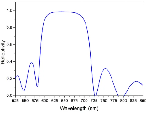
The experimental reflection result of a fabricated DBR, at 645 nm, on a quartz substrate is shown in Figure 2.11, where 11-bilayer of SiO2 (n=1.46) and Si3N4 (n=2.04) are
deposited.
(2.24)
(2.24)
(2.24)
(2.24) Figure 2.10: Structure of a 3-bilayer distributed bragg reflector (DBR) fabricated over a glass-substrate.
21
2.2.2 Ring Resonator
Ring resonators confine the light by waveguiding or reflecting it through their structures. Apart from Fabry-Perot cavities, optical isolators can be placed within them to inhibit one component of the standing waves. This prevents the formation of standing waves, and they can be designed to support only traveling waves. For that reason, they are also called as travelling-wave resonators.
Figure 2.11: Reflectivity of 11-bilayer SiO2-Si3N4 distributed bragg reflecter (DBR)
fabricated over a quartz substrate.
Figure 2.12: Schematics of a) an optical waveguide resonator, b) a three-mirror ring resonator, c) a whispering-gallery-mode (WGM) resonator.
22
Various types of ring resonators can be constructed. Three common examples, which are in the forms of an optical waveguide ring resonator, a three-mirror ring resonator and a whispering-gallery-mode (WGM) resonator, are provided in Figure 2.12. Since they support only their resonant frequencies, they may be used as optical filters (e.g., by coupling them to nearby optical fibers).
The allowed wavelengths of a ring resonator are directly related to the optical path that light takes in one round around the cavity. For example, for an optical waveguide ring resonator, the resonant wavelengths can be calculated as:
𝑚𝜆𝑚 = 2𝜋𝑛𝑅
Here m is the mode number, 𝜆𝑚 is the wavelength of the mth mode, n is the refractive
index, and R is the radius of the cavity.
WGM resonators make a special type of ring resonators that can support whispering gallery modes. The first discussions on WGMs date back to early 1900s, where the propagation of sound waves was examined on a curved gallery of a cathedral [51]. In the last decades, WGM-based resonators have attracted great attention in photonics for their low loss and high-Q natures. In order to support WGMs, the electromagnetic wave should be able to perform total internal reflection at the outer interface of the cavity. Thus, the refractive index of the cavity medium should be higher than the outer media. Since they follow the physics of ring resonators, their resonance frequencies can be calculated via Equation 2.25. Schematic illustration of a typical WGM resonator is given in Figure 2.12c, where the refractive index of cavity (n1) is higher than outer medium (n2). Owing to their
superior optical properties, WGM resonators have been used in various applications including biosensors [52], optofluidic lasers [53] and opto-electronic oscillators [54].
(2.25)
(2.25)
(2.25)
23
Chapter 3
Experimental Methods
In this chapter, we will explain the characterization techniques that are used to study optical and structural properties of colloidal NPLs.
3.1 Steady-State Absorption Spectroscopy
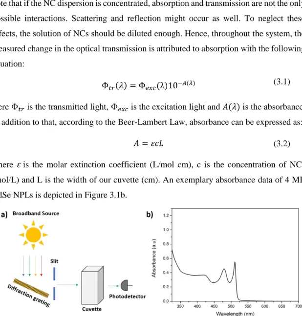
Steady-state absorption spectroscopy is a powerful technique to characterize the thickness and structure of NPLs. Figure 3.1a illustrates the working principle behind the absorption spectroscopy. A broadband source (white light) is used along with a rotating diffraction grating to excite samples at the desired wavelength. Moreover, a small slit is placed to select only a specific wavelength. The whole system acts as a monochromator that sweeps the excitation wavelength from UV to visible region of electromagnetic spectrum by rotating the diffraction grating. Then, the light is transmitted through our sample inside the quartz cuvette, which is dissolved in a suitable solvent (e.g., hexane, toluene, water). Some portion of the light is absorbed, and transmitted light is collected via a photodetector.
To analyze the data, the collected transmitted light is compared with a reference, which is obtained by a cuvette filled by solvent without any nanocrystals. An important point to
24
note that if the NC dispersion is concentrated, absorption and transmission are not the only possible interactions. Scattering and reflection might occur as well. To neglect these effects, the solution of NCs should be diluted enough. Hence, throughout the system, the measured change in the optical transmission is attributed to absorption with the following equation:
Φ𝑡𝑟(𝜆) = Φ𝑒𝑥𝑐(λ)10−𝐴(𝜆)
Here Φ𝑡𝑟 is the transmitted light, Φ𝑒𝑥𝑐 is the excitation light and 𝐴(𝜆) is the absorbance. In addition to that, according to the Beer-Lambert Law, absorbance can be expressed as:
𝐴 = 𝜀𝑐𝐿
where 𝜀 is the molar extinction coefficient (L/mol cm), c is the concentration of NCs (mol/L) and L is the width of our cuvette (cm). An exemplary absorbance data of 4 ML CdSe NPLs is depicted in Figure 3.1b.
Since optical properties of NPLs are strongly size-dependent, absorption spectroscopy is used to determine the thickness of NPLs after their synthesis. Moreover, formation of heterostructures can be also verified by this method. For instance, it is expected to observe a red-shift in the excitonic features of the absorption spectrum if CdS shell is coated around a CdSe core. Hence, the absorption spectrum helps us to understand and estimate the formation and thickness of the heterostructures.
(3.1) (3.1) (3.1) (3.1) (3.2) (3.2) (3.2) (3.2)
Figure 3.1: a) Schematic of the experimental setup behind steady-state absorption spectroscopy. b) An exemplary absorbance spectrum of 4 ML thick CdSe core NPL dissolved in hexane.
25
3.2 Steady-State Photoluminesce Spectroscopy
Complementing absorption spectroscopy, stead-state photoluminesce (PL) spectroscopy is also an important characterization technique for colloidal NCs. Its setup typically consists of two monochromators. Similar to absorption spectroscopy, the first one is used for excitation purpose. Following the optical excitation, spontaneous emission of the NCs is collected via the second monochromator. The schematic showing the experimental setup of photoluminesce spectroscopy is given in Figure 3.2a. Spontaneous emission of the NPLs exhibits thickness dependent characteristics. 5 ML thick CdSe core has a photoluminesce emission peak centered at ~553 nm, while 4 ML and 3 ML thick CdSe cores have photoluminescence emission at ~513 and ~465 nm, respectively. Thus, by examining the photoluminesce spectrum of an NPL solution, the thickness of the NCs can be determined. An example case showing the photoluminesce spectrum of 4 ML thick CdSe core NPLs is shown in Figure 3.2b.
3.3 Electron Microscopies
Although optical microscopy is a robust and reliable imaging technique, its resolution is limited by the optical diffraction. Considering a visible light at 400 nm, the resolution (R) of an optical microscope can be at most 200 nm by using the rough relation of R=λ/2. Hence, to resolve features smaller than this limit, electron beams are utilized. Electrons Figure 3.2: a) Schematic of the experimental setup behind steady-state photoluminescence spectroscopy. b) An exemplary photoluminescence spectrum of 4 ML thick CdSe core NPL dissolved in hexane.
26
accelerated under high voltage can have De-Broglie wavelength around a few nanometers and thus can be used to image nanocrystals.
Scanning electron microscopy (SEM) is a strong method that is used to image in µ-meter range. A highly focused electron beam is used to scan the surface. Backscattered and secondary electrons are collected to obtain the material information and to construct the image. An example SEM image of a bare fiber with 250 µ𝑚 diameter is given in Figure 3.3a. Transmission electron microscopy (TEM) is also a powerful tool to characterize shape, structure and size of nanocrystals. Moreover, TEM can be used to determine the crystalline structure of NCs. In this thesis, high-angle annular dark field scanning transmission electron microscopy (HAADF-STEM) was commonly used to image our 10-40 nm NPLs. An exemplary HAADF-STEM image of core/crown@shell NPLs is shown in Figure3.3b.
3.4 Optical Gain Measurements
3.4.1 Optical Gain Threshold Measurement
Amplified spontaneous emission (ASE) can be observed in colloidal NCs when they are excited with a pulsed laser with high enough pump fluences. ASE occurs when the population inversion condition in the semiconductor material is satisfied, meaning that optical gain threshold is exceeded. Hence, the presence of ASE is a must to achieve lasing Figure 3.3: a) SEM image of a SiO2 fiber with 250 µm diameter. b) HAADF-STEM image
27
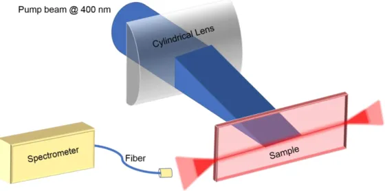
in nanocrystals, and the ASE performance is directly related to the lasing performance of a gain medium. Schematic illustration of the experimental setup for ASE measurement is shown in Figure 3.4
To characterize the ASE performance, NPLs are usually drop-casted or spin-coated on a quartz substrate. Then, the samples are excited with a stripe-shaped pulsed laser beam. As a result of the internal reflection between the NPL film and upper surface of the quartz substrate, waveguiding effect occurs, which guides the emission to the edge of the sample. This emission is then collected with a fiber connected to a spectrometer.
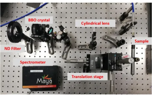
As previously mentioned, nonradiative Auger recombination is the rapid process that annihilates biexcitons. Hence, it is required to generate biexcitons before Auger recombination takes place. To ensure this, we use Spectra Physics (Spitfire Pro XP) laser with 120 fs pulse width and 1 kHZ repetition rate at 800 nm as an optical pumping source. For one-photon absorption (1PA) regime, frequency is doubled via a BBO (beta barium borate) crystal and the remaining of 800-nm beam is filtered out by employing a bandpass filter. To adjust the amplitude of pump fluences, a rotatable ND (neutral density) filter is placed right before the BBO crystal. Finally, the stripe shape is achieved by using a cylindrical lens. Emission of the sample is collected by using Maya2000 Pro spectrometer. Photograph of the experimental setup used for our ASE measurements is given in Figure 3.5.
28
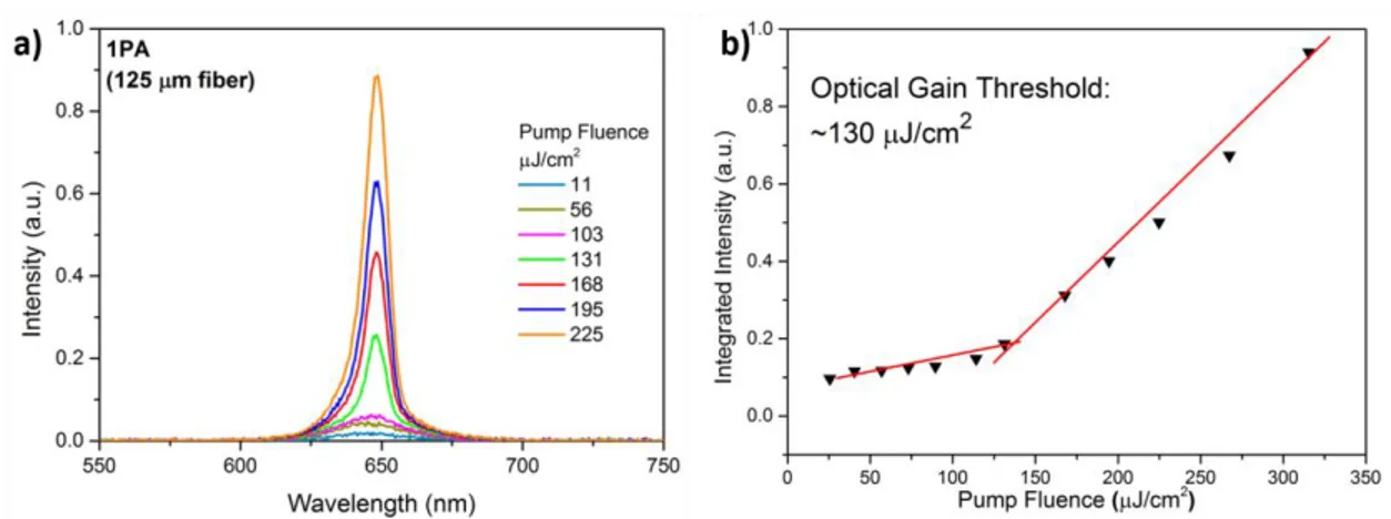
Figure 3.6a depicts exemplary emission spectra of CdSe/Cd0.25Zn0.75S core/shell NPLs under one-photon absorption. As pump intensity is increased, a transition from spontaneous emission to ASE occurs with a narrow red-shifted emission after the optical gain threshold. To quantify the exact threshold, integrated intensity of the emission is plotted as a function of the pump intensity as given in Figure 3.6b. After the optical gain threshold (2.45 µJ/cm2 in this case), ASE starts to dominate overall emission with a super
linear increase in the integrated intensity.
Figure 3.6: a) Exemplary emission spectra of CdSe/Cd0.25Zn0.75S core/shell NPLs under one-photon absorption. b) Exemplary integrated intensity of CdSe/Cd0.25Zn0.75S core/shell NPLs showing the optical gain threshold.
Figure 3.5: Experimental optical setup for ASE measurement under one-photon absorption.
29
3.4.2 Variable Stripe Length Measurement
We employed variable stripe length (VSL) measurement to characterize the net peak modal gain coefficients of colloidal NPLs. Similar to the optical gain threshold characterization technique, we excite our samples with a stipe-shaped femtosecond laser beam with a pump intensity larger than ASE threshold. However, in this case, an adjustable slit is placed right before our samples as can be seen in Figure 3.7a. By increasing the length of the excitation stripe, emission spectra of an NPL-coated quartz sample are collected. After a certain stripe length, ASE peak starts to appear, resulting in an exponential increase in the integrated intensity data. An exemplary VSL measurement data is plotted in Figure 3.7b.
Then, to quantify net modal gain coefficients, empirical data is fitted to the following equation:
𝐼(𝑙) =𝐼𝑠
𝐺(𝑒𝐺𝑙− 1)
where 𝐼(𝑙) is the integrated intensity, G is the net modal gain coefficient, l is the stripe length and 𝐼𝑠 is a constant. By this fit, one G value for a specific pump intensity can be obtained. Later, this measurement is repeated for various pump intensities and multiple G values are obtained. Finally, G is plotted as a function of the pump intensity. As the pump intensity is increased, G increases as well. However, a saturation of the net modal gain of the NPLs is observed as the pump intensity is further increased. Such gain saturation is a typical characteristic seen in all gain materials.
(3.3)
(3.3)
(3.3)
(3.3) Figure 3.7: a) Schematic of the experimental setup of variable strip length measurement. b) Exemplary VSL data after measurement showing the exponential fit.
30
3.4.3 Waveguide Loss Coefficient Measurement
Measurement of waveguide loss coefficients is frequently used for optical gain characterization. Since amplified spontaneous emission is waveguided through the edge of the sample, a waveguiding loss (α) attenuates this emission during the propagation. To measure this coefficient, the sample along with fiber is connected to a high precision translation stage. Then, the sample and the fiber are moved together on an axis perpendicular to the optical excitation axis. Hence, the distance between the edge of the sample and the stripe is changed. After a certain distance, an unexcited part on the sample is revealed, where ASE intensity is attenuated via waveguide loss. This intensity is collected from the edge and plotted as a function of the distance from the edge of the sample. The schematic showing this measurement is given in Figure 3.8.
Then, the obtained data is fitted to the following expression to quantify α:
𝐼(𝑙) = 𝐼𝑝𝑒−𝛼𝑑
where 𝐼𝑝 is the peak ASE intensity, α is the waveguide loss coefficient, and d is the
distance between the edge of the sample and the end of excitation beam.
(3.4)
(3.4)
(3.4)
(3.4) Figure 3.8: Schematic of the experimental setup to measure waveguide loss coefficient.
31
Chapter 4
Whispering-gallery-mode Laser
Colloidal Nanoplatelets
This chapter is based on our manuscript M. Sak, N. Taghipour, S. Delikanli, S. Shendre, I. Tanriover, Y. Gao, J. Yu, Z. Yanyan, S. Yoo, C. Dang, H. V. Demir, in submission (2019).
4.1 Motivation
Colloidal semiconductor nanocrystals recognized as promising candidates for optical gain studies [26], [55], [56] owing to their cost-effective synthesis and solution processable nature. Moreover, they can be spectrally tuned by modifying their size, shape and composition [57]. Up to now, different classes of NCs including colloidal quantum dots [55], colloidal quantum wells [35] and nanorods [58] have been studied for optical gain and lasing applications. Colloidal quantum wells, or semiconductor nanoplatelets (NPLs), earn an incomparable spot among these NCs thanks to their large absorption cross-sections [22], fully homogeneous broadening [24], huge oscillator strength [21], and large optical gain coefficients [23]. Moreover, by utilizing heterostructures including core/shell [28], [33] and core/crown [31], [32], the excitonic properties of NPLs can be adjusted to achieve
32
high performance in optical gain. To date, different heterostructures of NPLs used as gain medium coupled to various optical resonators including photonic-crystal nanobeam cavities [59], vertical-cavity surface-emitting lasers [35], and multiphoton-pump Fabry-Pérot cavities [60].
Lately, whispering-gallery-mode resonators have emerged as a promising optical resonator thanks to their very low optical losses [36]. As a result of cutting-edge nanofabrication technologies, various forms of WGM cavities were made including micro rings [61], micro disks [62] and micro spheres [63]. As a practical WGM resonator, fiber-based structures have been utilized for lasing in the solution phase of various materials such as nanorods [36], colloidal quantum dots [53] and dyes [64]. Nonetheless, the performances of these structures are mostly limited as a result of the low packing-density of the gain media. This causes poor lasing stability, high optical gain and lasing thresholds. To address these issues, close-packed films of nanocrystals can be used in fiber-based WGM resonators as a gain medium.
Here, we propose and demonstrate a fiber-based WGM laser made of close-packed NPLs films which addresses the issues of instability and large lasing thresholds by the first WGM-NPL lasing structure. We show ASE and lasing from close-packed films of core/crown/gradually-alloyed-shell (CCGAS) NPLs coated around fibers under one-photon- (1PA) and two-one-photon-absorption (2PA) regimes. The laser cavity is made of a unique fiber-based WGM resonator. To do so, we integrated bare glass fibers with close-packed CCGAS NPLs films by utilizing adhesion. Then we systematically investigated their 1PA and 2PA ASE performances using two different fiber diameters (125 µm and 250 µm). A record low waveguide loss coefficient (~7.1 cm-1) and high net modal gain
coefficient (~485 cm-1) were achieved by this novel structure, resulting in low ASE thresholds of ~90 µJ/cm2 and ~3.0 mJ/cm2 for 1PA and 2PA regimes, respectively. Moreover, we demonstrated an NPL-WGM laser with ~190 µJ/cm2 lasing threshold under 1PA pumping with a linearly polarized emission that has a stable emission under 150 minutes of optical excitation.
33
4.2 Experiments
4.2.1 Precursor Preparation
Cadmium precursor (cadmium myristate) was synthesized according to a previous article [32]. 3.13 g of Na myristate and 1.23 g of Cd(NO3)2·4H2O were dissolved in 250 and 40
mL methanol, respectively. After that, the solution was stirred for three hours. Then, the white powder was collected and washed by methanol to wipe out the remaining unreacted precursors. After three consecutive washing process, the powder was left in vacuum for twelve hours to dry.
4.2.2 Colloidal Synthesis of 4 ML CdSe NPLs
To synthesize 4 ML thick CdSe core NPLs, a recipe from a previous publication was followed with some slight changes [32]. Inside a three-neck glass flask, 28 mL of octadecene, 26 mg of Se and 340 mg of cadmium myristate was placed. Then, this mixture was kept under vacuum for 30 minutes at room temperature. After that, the solution was heated up to 240 °C within inert gas environment. At 200 °C, 140 mg of Cd(CH3COO)2·2H2O was injected to this solution. After reaching the temperature of 240
°C, the solution was kept at this temperature for 180 seconds. Then, 1 mL of oleic acid was injected to the solution and the temperature is decreased to room temperature. Finally, ethanol and hexane were added to precipitate CdSe NPLs, which were then dissolved and kept within the hexane.
4.2.3 Preparation of CdS Crown Growth Solution
To prepare the growth solution, we followed a slightly changed recipe from a previous publication [32]. 8 mL of octadecene, 680 µL of oleic acid and 960 mg of Cd(CH3COO)2·2H2O were put into a glass beaker. Then, this solution was sonicated for
60 minutes. After that, the temperature was raised to 180 °C and frequently sonicated while stirring to obtain a whitish gel. Finally, 6 mL of 0.1 M S-octadecene was added to cadmium precursor for the crown growth.
34
4.2.4 CdSe/CdS Core/Crown NPL Synthesis
To synthesize CdSe/CdS core/crown NPLs, a recipe from previously published article was followed after some slight changes [32]. 15 mL of octadecene and 4 ML thick CdSe NPLs dissolved in hexane were placed into a glass flask. Then, this solution was left under vacuum at a temperature of 80 °C to evaporate hexane. Under inert atmosphere, the solution was heated up to 240 °C and anisotropic growth mixture was injected with a rate of 10 mL/h. After that, we waited for NPLs to reach the desired crown size and we decreased the temperature to room temperature. As-synthesized core/crown NPLs were centrifuged for precipitation after adding ethanol. Finally, by using methanol and ethanol, NPLs were washed three times to eliminate excess precursors and obtained NPLs were dissolved in hexane.
4.2.5 CdSe/CdS@Cd
1-xZn
xS Core/Crown/Shell NPL Synthesis
For this, a published recipe was followed with some slight modifications [33]. 0.4 M of Cd(NO3)2·4H2O and 0.4 M of Zn(NO3)2·6H2O solutions were prepared in N-methyl
formamide. For S precursor, 40-48 wt% water-based solution of (NH4)2S was used. 40 µL
of (NH4)2S was injected to 4 mL of N-methyl formamide along with 1 mL of CdSe/CdS
core/crown NPLs under stirring. After 120 seconds, acetonitrile and toluene were added and NPLs were centrifuged. After that, NPLs were dissolved in N-methyl formamide (NMF) and this cleaning process was performed two more times to eliminate excess S precursors. Then, NPLs was dispersed in 4 mL of NMF. 1 mL solution of X% Cd(NO3)2
and (100-X)% of Zn(NO3)2 within NMF was added to these NPLs for cation deposition.
Under ambient conditions, the solution was stirred for 45 minutes. Then, by adding acetonitrile and toluene, NPLs were precipitated. This cleaning process was undertaken two more times to separate unreacted cation precursors. This growth steps were then repeated to obtain desired number of shells. For the first Cd1-xZnxS shell layer X value
was selected as 50, while for the second and the third ones they were selected as 10 and 5, respectively. Finally, CCGAS NPLs were dissolved and kept in hexane.
![Figure 2.2: The nucleation and growth model of nanocrystals. Reprinted with permission from [41]](https://thumb-eu.123doks.com/thumbv2/9libnet/5927659.123196/23.918.238.753.560.924/figure-nucleation-growth-model-nanocrystals-reprinted-permission.webp)
