A NOVEL ROLE FOR 5-hmC IN THE REGULATION OF
CANCER TESTIS GENE EXPRESSION IN CANCER AND
MESENCHYMAL TO EPITHELIAL TRANSITION
A THESIS SUBMITTED TO
THE DEPARTMENT OF MOLECULAR BIOLOGY AND GENETICS AND THE GRADUATE SCHOOL OF ENGINEERING AND SCIENCE
OF BILKENT UNIVERSITY
IN PARTIAL FULFILLMENT OF THE REQUIREMENTS FOR THE DEGREE OF
DOCTOR OF PHILOSOPHY
OVER PAGE
0.1COVER PAGE
BY
SİNEM YILMAZ ÖZCAN
DECEMBER, 2014
ii I dedicated my thesis to my mum and dad for the endless love they gave, my husband and son for being the meaning of my life.
iii I certify that I have read this thesis and that in my opinion it is fully adequate, in scope and in quality, as a thesis for the degree of Doctor of Philosophy.
____________________ Assis. Prof. Dr. Ali O. Güre (Advisor) I certify that I have read this thesis and that in my opinion it is fully adequate, in scope, and in quality, as a thesis for the degree of Doctor of Philosophy.
____________________ Assoc. Prof. Dr. Işık Yuluğ
I certify that I have read this thesis and that in my opinion it is fully adequate, in scope, and in quality, as a thesis for the degree of Doctor of Philosophy.
__________________ Assis. Prof. Dr. Stefan Fuss
I certify that I have read this thesis and that in my opinion it is fully adequate, in scope, and in quality, as a thesis for the degree of Doctor of Philosophy.
____________________ Assoc. Prof. Dr. Sreeparna Banerjee I certify that I have read this thesis and that in my opinion it is fully adequate, in scope, and in quality, as a thesis for the degree of Doctor of Philosophy.
____________________ Prof. Dr.Can Akçalı
Approved for the Graduate School of Engineering and Science __________________
Prof. Dr. Levent Onural
iv
ABSTRACT
A NOVEL ROLE FOR 5-hmC IN THE REGULATION OF CANCER TESTIS GENE EXPRESSION IN CANCER AND MESENCHYMAL TO EPITHELIAL
TRANSITION BS TRACT
Sinem YILMAZ ÖZCAN
Ph.D. in Molecular Biology and Genetics Supervisor: Assist. Prof. Dr. Ali O. Güre
December 2014
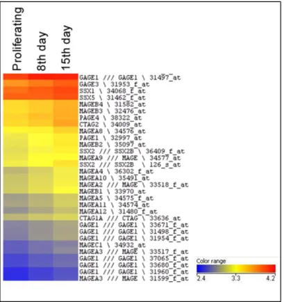
Cancer/testis (CT) genes show highly restricted expression among normal tissues, limited to germ cells in the testis and ovary, and to trophoblast cells, , but are frequently expressed in various cancers. Other than a clear association with promoter-specific demethylation and histone deacetylation, the specific mechanisms by which these genes are expressed are currently unknown. In this study, we tested various mechanisms including promoter- and region-specific epigenetic mechanisms to gain a better understanding of CT gene expression.
To better study the epigenetic mechanisms regulating CT gene expression, we searched for a model that dynamically expresses CT genes. As a result of preliminary bioinformatic efforts and literature search, we chose to study CT gene expression in Caco-2 spontaneous differentiation model. We showed that PAGE-2,-2B and SPANX-B genes were up-regulated significantly as Caco-2 cells differentiated. Differentiation was also characterized as a mesenchymal to epithelial transition as evidenced by the decrease in mesenchymal markers (Fibronectin1, Vimentin and Transgelin) and the concomitant increase in epithelial markers (E-cadherin, Claudin 4 and Cdx2). CT protein (SPANX-B and PAGE-2,-2B) positive cells were positive for epithelial protein (Cdx2), and negative for mesenchymal proteins (Fibronectin1, Vimentin). Although we could not find a significant difference in promoter proximal DNA demethylation of CT genes, we identified that promoter proximal DNA was hydroxymethylated with a gradual increase in hydroxymethylation as cells differentiated. The change in hydroxymethylation level was concordant with an increase in TET enzyme levels and co-localization of TET2 protein with CT proteins in the corresponding cells. Besides, we found that promoters of CT genes lost EZH2, H3K27me3 and HP1 marks as CT genes were up-regulated. Reversal of differentiation resulted in loss of CT and TET gene expression and EMT induction. Thus, for the first time, we describe dynamic expression of CT genes in association with DNA hydroxymethylation in mesenchymal to epithelial transition.
In addition to promoter-proximal alterations, we thought that epigenetic alterations leading to CT gene expression in cancer could occur within larger regions containing CT
v genes, but with clear boundaries. As genes that do not show an expression pattern
similar to CT genes can be located within their proximity, we hypothesized that there could be clear boundaries between neighbouring regions containing CT genes and those with non-CT type expression patterns. We, therefore, identified 2 genes; ALAS2 and
CDR1, in close proximity to two different CT genes (PAGE-2,-2B and SPANX-B), which
were downregulated in cancer, and thus showed an expression pattern opposite to that of these two CT genes. ALAS2 and CDR1 were downregulated in lung and colon cancer cell lines compared to healthy counterparts. We found that the downregulation of ALAS2 and CDR1 in cancer cell lines, in contrast to CT genes, was independendent of DNA hypomethylation. We also found that ALAS2 and CDR1 downregulation in cancer was possibly related to decreased levels of hydroxymethylation in promoter proximal regions. As the upregulation of PAGE-2,-2B and SPANX-B genes was associated with increased hydroxymethylation at promoter-proximal regions, these two groups of genes, despite their close proximity were found to be controlled inversely albeit possibly by the same mechanisms. We tested if ectopic upregulation of ALAS2 and CDR1 in cancer cell lines would result in a tumor-suppressive effect, but were unable to find any. As both genes are located about 200 and 50 kbs from SPANX-B and PAGE-2, we propose that the there might be a boundary within these regions that could possibly have an insulator-like function to help distinguish the two very different epigenetic events occuring in tumorigenesis.
As almost all CT genes map within highly homologous inverted repeats it is possible that 3 dimensional chromosomal structures formed around these repeats underlie the common epigenetic mechanism responsible for coordinate CT gene expression. To test for this hypothesis, we analyzed expression of various transcripts identified within and outside the NY-ESO-1 repeat region. However, we could not find a correlation between the presence of such transcripts and CT gene expression patterns.
Keywords: Cancer testis genes, PAGE-2,-2B, SPANX-B, DNA hydroxymethylation,
vi
ÖZET
KANSERDE VE MEZENKİMALDEN EPİTELE GECİŞ SÜRECİNDE 5-hmC’NİN KANSER TESTİS GEN İFADESİNDEKİ ÖZGÜN ROLÜ
0.1ABSTRACT Sinem YILMAZ ÖZCAN
Moleküler Biyoloji ve Genetik Doktora Tezi Tez Yöneticisi: Yard. Doç. Dr. Ali O. Güre
Aralık 2014
Kanser testis (KT) genleri normal dokular içinde sınırlı şekilde sadece yumurtalık ve testislerdeki eşey üreme hücreleri ve trofoblast hücrelerinde ifade
edilirken, pek çok kanserde sıklıkla ifade edildiği gözlenmiştir. Promotor bölgesine özgü DNA demetilasyonu ve histon deasetilasyonuyla ilgili açık bir ilişki haricinde bu
genlerin ifade edilmesindeki spesifik mekanizmalar bilinmemektedir. Bu calışmada KT gen ifadesini daha iyi anlayabilmek icin KT genlerini bulunduran promoter bölgelerine özgü ve alana sınırlı epigenetik mekanizmaları test ettik.
KT gen ifadesi varliginda ve yokluğundaki epigenetik mekanizmalari daha iyi calışabilmek için, KT genlerini dinamik olarak ifade eden bir model aradık. Öncü biyoinformatik analizler ve literatür araştırması sonucunda, KT genlerini Caco-2 farklılaşmasında incelemeyi seçtik. PAGE-2,-2B ve SPANX-B gen ifadelerinin
farklılaşan Caco-2 hücrelerinde anlamlı şekilde arttığını gösterdik. Mezenkimal belirteç gen ifadeleri (Fibronectin1, Vimentin ve Transgelin) azaldığı ve eş zamanlı olarak epitel belirteç gen ifadeleri (E-cadherin, Claudin 4 ve Cdx2) arttığı için farklılaşma
mezenkimalden epitele geçiş süreci olarak tanımlanmıştır. Bunun yanısıra, KT proteinlerini (SPANX-B ve PAGE-2,-2B) bulunduran hücrelerin epitel belirteç
proteinini (CDX2) de bulundurduğu, mezenkimal belirteç proteinlerini (Fibronectin1 ve Vimentin) de bulundurmadığını gösterdik. Farklılaşmış Caco-2 hücrelerinde artan KT genlerinin promotor yakınındaki DNA bölgelerinde anlamlı demetillasyon
gözlemlenmemekle birlikte bu bölgelerde hidroksimetilasyon seviyelerinde aşamalı bir artış tespit edilmiştir. Promotor yakını bölgelerdeki hidroksimetilasyon seviyesindeki artış aynı zamanda TET enzim seviyesindeki artış ile ve de TET2 proteini ve KT proteinlerinin aynı hücrelerdeki konumlanması ile uyumlu bulunmuştur. Bunların yanısıra, KT gen ifadesi ile birlikte KT genlerinin promotor bölgelerindeki EZH2 ve HP1 proteinlerinin işgali ve H3K27me3 işareti azalmıştır. Caco-2 farklılaşması tersine döndürüldüğünde KT ve TET gen ifadelerinin azalması ve epitelden mezenkimale geçiş ile sonuçlanmıştır. Böylece bu calışma ile ilk defa KT genlerinin dinamik ifadesi
gösterilmiş, bu süreç mezenkimalden epitele geçiş ve DNA hidroksimetilasyonu ile açıklanmıştır.
vii Promotor yakını bölgelerdeki değişimlerin yanısıra, kanserde KT gen ifadesine sebep olan epigenetik değişikliklerle alternatif epigenetik değişikliklerin aynı anda sınırları belli farklı bölgelerde meydana geldiğini düşündük. KT genleri ve KT genlerine komşuluk eden ama KT gen ifade paterninden farklı gen ifadesine sahip olan genler arasında sınırlar olabileceğini hipotezledik. Boylece, KT genlerine (PAGE-2,-2B ve
SPANX-B) komşuluk eden ve kanserde gen ifadesi azalan 2 geni, ALAS2 ve CDR1’i,
bulduk. ALAS2 ve CDR1 gen ifadelerinin sağlıklı dokulara kıyasla kolon ve akciğer kanseri hücre hatlarında azaldığını gösterdik. ALAS2 ve CDR1 gen ifadelerinin kanser hücre hatlarındaki azalışlarının KT gen ifadesinden farklı olarak DNA metilasyonundan bağımsız oldugunu bulduk. Kanserde ALAS2 ve CDR1 gen ifadesi azalışlarının promotor yakını bölgelerdeki artan hidroksimetilasyon seviyesi ile kuvvetli ihtimal ilişkili
olabileceğini gözlemledik. PAGE-2,-2B ve SPANX-B gen ifadelerindeki artış da DNA hidroksimetillasyonu ile ilişkili bulunmustur, bu iki grup genlerinin farklı ifade
paternlerine rağmen. Kanser hücre hatlarındaki ALAS2 ve CDR1 gen ifadelerinin çarpıcı azalışına rağmen, bu genlerin kanser hücrelerindeki ektopik ifadeleri sonucu hücre canlılığı ölçümlerinde anlamlı bir değişiklik bulunmamıştır. ALAS2 ve CDR1, sırasıyla
PAGE-2,-2B ve SPANX-B genlerine 200 ve 50 kbs uzaklıkta konumlandığından, bu
bölgeler arasında farklı epigenetik olayların meydana gelmesinde etkili yalıtkan bir sınır bulunduğunu önermekteyiz.
KT genlerinin neredeyse tamamı yüksek homolojik benzerliği bulunan tekrar bölgelerinde konumlandığından, bu tekrar bölgelerinin katlanarak 3 boyutlu yapılar oluşturması ve bu yapıların eş güdümlü KT gen ifadesinden sorumlu mekanizma olması muhtemeldir. Bu hipotezi test etmek icin, NY-ESO-1 genini içeren tekrar bölgesinin içindeki ve dışındaki genlerin ifadesini inceledik. Ancak anlamlı bir ilişki tespit edemedik.
Anahtar sözcükler: Kanser testis genleri, PAGE-2,-2B, SPANX-B, DNA hidroksimetilasyonu, mezenkimalden epitele geçiş
viii
ACKNOWLEDGEMENT
I would like to owe my sincere gratitude to my advisor Assist. Prof. Dr. Ali O. Güre for not only guiding me in this work but also improving my thinking in this journey and helping me to find my way in the scientific area. He helped with his wide knowledge and experience but also let me to express myself, try and sometimes even fail in this period.
I would like to express my very great appreciation to Assoc. Prof. Dr. Sreeparna Banerjee and Dr. Aslı Sade Memişoğlu for their mental and physical support in this nice collaboration. It was such an important chance for me to work with them since it taught me a lot about being in a productive collaboration.
I would like to extend my appreciation to my thesis follow up committee members Assoc. Prof. Dr. Işık Yuluğ and Assis. Prof. Dr. Stefan Fuss. They always participated with their important critics and motivated me to do further during my PhD study.
I am grateful to Kerem Mert Şenses, Barış Küçükkaraduman, Dilan Çelebi and Yasemin Kaygusuz for their joyful and indispensable contributions to this study. This work could not be completed without their help.
I also would like to thank all past and present members of Gure lab; Kerem Mert Şenses, Şükrü Atakan, Barış Küçükkaraduman, Alper Poyraz, Seçil Demirkol , Waqas Akbar, Mehdi Ghassemi, Murat İşbilen, Duygu Akbaş, Derya Dönertaş, Aydan Bulut Karslıoğlu since we altogether generated a perfect and amusing working environment.
I am especially grateful to my friends in Bilkent, Kerem Mert Şenses, Şükrü Atakan, Emre Yurdusev, Ceyhan Ceran, Gizem Ölmezer, İrem Gürbüz, Eylül
Harputlugil, Merve Aydın, Ece Akhan Güzelcan since they were always with me and this journey was meaningful with them. Gurbet Karahan, Nilüfer Sayar and Dilan Çelebi were more than a friend and became a part of my family during these six years, though I am thankful for their companionship.
ix
I would like to express my very great appreciation to all Bilkent MBG family.
Bilge Kılıç, Füsun Elvan, Sevim Baran, Yıldız Karabacak, Yavuz Ceylan and Abdullah Ünnü made this department as a home for all of us.
Undoubtedly, I am grateful to my mother Nuray Yılmaz and father Duran Yılmaz for being beside me and believing me in all stages of my educational life. I do not know how to thank my husband Tahsin Özcan since he always supported me with patience and standed by me during though times. Finally, I would like to thank my baby boy Ege to being my little sunshine at the end of this long journey.
I was supported by TÜBİTAK BİDEB 2211 scholarship during my PhD study; thereby I would like to thank TÜBİTAK to giving me this opportunity.
x
Table of Contents
ABSTRACT ... iv
ÖZET ... vi
ACKNOWLEDGEMENT ... viii
List of Figures: ... xiv
List of Tables: ... xvii
Abbreviations: ... xviii
1 INTRODUCTION ...1
1.1 CANCER TESTIS GENES ...1
1.1.1 Expression Patterns of Cancer Testis Genes ...1
1.1.2 Genomic Structure of Cancer Testis Genes ...3
1.1.3 Epigenetics of Cancer Testis Genes ...3
1.1.4 Cancer Testis Gene Expression in Stem Cells ...5
1.1.5 Cancer Testis Gene Expression during Epithelial to Mesenchymal Transition ...5
1.2 CACO-2 SPONTANEOUS DIFFERENTIATION MODEL ...6
1.3 DNA HYDROXYMETHYLATION AS EPIGENETIC CONTROL MECHANISMS OF GENE EXPRESSION...8
1.4 AIM AND HYPOTHESIS ...12
2 MATERIALS AND METHODS ...13
2.1 MATERIALS...13
2.1.1 General Chemicals ...13
2.1.2 Instruments ...24
2.1.3 Cell Lines and Tissue Culture Reagents ...25
2.2 SOLUTIONS AND MEDIA ...27
2.2.1 General Solutions ...27
2.2.2 Cell Culture Solutions ...29
2.3 METHODS ...30
2.3.1 Cancer cell culture techniques ...30
2.3.2 RNA isolation with TRIzol ...30
xi
2.3.4 Identification of CT-proximal down-regulated genes in cancer compared to healthy
counterparts by Cancer Genome Anatomy Project ...31
2.3.5 Q RT-PCR Experiments with Taqman Probe Chemistry ...32
2.3.6 Sequencing of sodium bisulphite treated tumor cell lines’ and normal tissues’ DNAs ...32
2.3.7 Plasmid constructions ...33
2.3.8 Generation of stable cell lines ...34
2.3.9 β-Galactosidase Staining Assay ...35
2.3.10 MTT Cell Viability Assay ...35
2.3.11 Spontaneous differentiation and dedifferentiation of Caco-2 cell line ...36
2.3.12 In silico analysis of CT gene expression during Caco-2 spontaneous differentiation ...36
2.3.13 Total RNA isolation and DNaseI treatment ...36
2.3.14 cDNA synthesis ...37
2.3.15 Q RT-PCR of CT genes ...37
2.3.16 In silico identification of differentially expressed mesenchymal and epithelial genes during Caco-2 spontaneous differentiation ...37
2.3.17 Q RT-PCR of mesenchymal and epithelial marker genes ...38
2.3.18 Promoter methylation analysis ...38
2.3.19 Hydroxymethylated DNA Immunoprecipitation ...39
2.3.20 Immunofluorescence microscopy ...39
2.3.21 Protein Isolation ...40
2.3.22 Western Blotting ...40
2.3.23 Chromatin Immunoprecipitation (ChIP) ...40
2.3.24 In vitro treatment of tumor cell lines with 5-aza-2-deoxycytidine ...41
2.3.23 Q RT-PCR Experiments with SYBR Green chemistry for noncoding RNA expression in NY-ESO-1 repeat region ...42
xii
3.1 EPIGENETIC MECHANISMS LEADING CANCER TESTIS GENE EXPRESSION IN
CACO-2 SPONTANEOUS DIFFERENTIATION MODEL ...43
3.1.1 Cancer Testis gene expression in Caco-2 spontaneous differentiation model: ...43
3.1.2 Characterization of Caco-2 spontaneous differentiation in association with MET and CT gene expression: ...45
3.1.3 Epigenetic mechanisms underlying CT gene expression in Caco-2 spontaneous differentiation: ...57
3.2 REGION SPECIFIC EPIGENETIC MECHANISMS OF CANCER TESTIS (CT) AND CT PROXIMAL GENE EXPRESSION ...66
3.2.1 Analysis of CT proximal genes down-regulated in cancer by CGAP: ...66
3.2.2 The down-regulation in mRNA expression of CT proximal genes; ALAS2 and CDR1 and CT gene expression in cancer: ...67
3.2.3 The promoter proximal DNA methylation of ALAS2 and CDR1 genes in normal tissues and colon and lung cancer cell lines:...71
3.2.4 The response of CT proximal genes; ALAS2, CDR1 and CT genes; PAGE-2,-2B, SPANX-B to 5-aza-2’-deoxycytidine treatment in cancer cell lines: ...76
3.2.5 The promoter proximal DNA hydroxymethylation of ALAS2 and CDR1 genes in normal tissues, colon and lung cancer cell lines: ...77
3.2.6 The gene expression of TET enzymes in normal tissues and colon and lung cancer cell lines: ...78
3.2.7 The result of ectopic expressions of ALAS2 and CDR1 genes in cancer cell lines in cell viability manner: ...79
3.2.8 ALAS2 and CDR1 expression in Caco-2 spontaneous differentiation model ...82
3.3 GENE EXPRESSION INSIDE AND OUTSIDE OF A CANCER TESTIS GENE-CONTAINING REPEAT REGION ...84
3.3.1 Uncoordinated expression of 1, IκBG and the noncoding RNAs in NY-ESO-1 containing repeat region:...84
4 DISCUSSION AND CONCLUSION ...86
5 FUTURE PERSPECTIVES ...92
6 REFERENCES ...93
APPENDIX A ...101
xiii
APPENDIX C ...109 7 PUBLICATION ...120
xiv
List of Figures:
FIGURE 1.1.1 1: THE EXPRESSION PATTERN OF CT-X GENES IN NORMAL TISSUES AND CANCER ... 2 FIGURE 1.2 1: DIFFERENTIALLY EXPRESSED GENES IN CACO-2 SPONTANEOUS
DIFFERENTIATION. ... 8 FIGURE 1.3 1: ACTIVE DNA DEMETHYLATION ... 10 FIGURE 1.3 2: PASSIVE DNA DEMETHYLATION ... 10 FIGURE 3.1.1 1: CT GENE EXPRESSION INCREASES DURING CACO-2 SPONTANEOUS
DIFFERENTIATION. ... 43 FIGURE 3.1.1 2: CACO-2 CELLS DIFFERENTIATE DURING 30 DAYS POST CONFLUENCE
CULTURING. ... 44 FIGURE 3.1.1 3: AMONG 6 CT GENE FAMILIES; THE GENE EXPRESSIONS OF PAGE-2,-2B AND
SPANX-B INCREASE IN CACO-2 SPONTANEOUS DIFFERENTIATION. ... 45
FIGURE 3.1.2 2: CT GENE MRNA LEVELS INCREASE CONCOMITANT WITH MESENCHYMAL TO EPITHELIAL TRANSITION. ... 47 FIGURE 3.1.2 3: INCREASE IN CDX-2 (34 KDA, EPITHELIAL PROTEIN) AND SPANX-B (12 KDA,
CT PROTEIN), THE DECREASE IN TRANSGELIN (23 KDA), FIBRONECTIN (263 KDA) AND VIMENTIN (54 KDA) (MESENCHYMAL PROTEINS) AT THE PROTEIN LEVEL BY
WESTERN BLOT EXPERIMENT IN 3 DIFFERENT DIFFERENTIATION SETS. ... 48 FIGURE 3.1.2 4: DECREASE IN MRNA LEVELS OF MESENCHYMAL MARKER GENES (VIM,
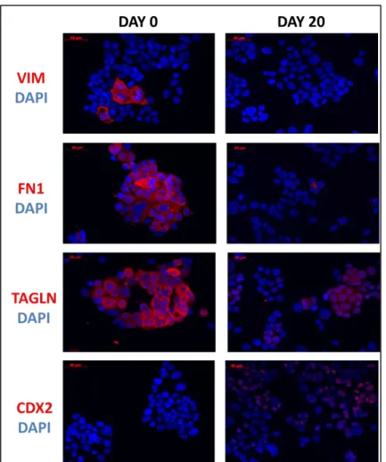
FN1 AND TAGLN) AND INCREASE IN MRNA LEVEL OF EPITHELIAL MARKER GENE (CDX2) EXIST IN PROTEIN LEVEL AS WELL. ... 49 FIGURE 3.1.2 5: INCREASE IN MRNA LEVELS OF CT GENES (SPANX-B AND PAGE-2,-2B)
EXISTS IN PROTEIN LEVEL AS WELL. ... 50 FIGURE 3.1.2 6: DOUBLE IMMUNOFLUORESCENCE STAINING AT DAY 0 SHOWS THAT
VIMENTIN POSITIVE CELLS ARE NEGATIVE FOR SPANX-B PROTEIN AND SPANX-B POSITIVE CELLS ARE NEGATIVE FOR VIMENTIN PROTEIN. ... 51 FIGURE 3.1.2 7: DOUBLE IMMUNOFLUORESCENCE STAINING AT DAY 0 SHOWS THAT
VIMENTIN POSITIVE CELLS ARE NEGATIVE FOR PAGE-2,-2B PROTEIN AND PAGE-2,-2B POSITIVE CELLS ARE NEGATIVE FOR VIMENTIN PROTEIN. ... 52 FIGURE 3.1.2 8: DOUBLE IMMUNOFLUORESCENCE STAINING SHOWS THAT CDX2 PROTEIN
CO-LOCALIZES WITH SPANX-B PROTEIN IN THE CORRESPONDING CELLS DURING DIFFERENTIATION. ... 53 FIGURE 3.1.2 9: DOUBLE IMMUNOFLUORESCENCE STAINING SHOWS THAT CDX2 PROTEIN CO-LOCALIZES WITH PAGE-2,-2B PROTEIN IN THE CORRESPONDING CELLS DURING DIFFERENTIATION. ... 54 FIGURE 3.1.2 10: DOUBLE IMMUNOFLUORESCENCE STAINING AT DAY 0 SHOWS THAT
FIBRONECTIN POSITIVE CELLS ARE NEGATIVE FOR SPANX-B PROTEIN AND SPANX-B POSITIVE CELLS ARE NEGATIVE FOR FIBRONECTIN PROTEIN. ... 55 FIGURE 3.1.2 11: DOUBLE IMMUNOFLUORESCENCE STAINING AT DAY 0 SHOWS THAT
FIBRONECTIN POSITIVE CELLS ARE NEGATIVE FOR 2,-2B PROTEIN AND PAGE-2,-2B POSITIVE CELLS ARE NEGATIVE FOR FIBRONECTIN PROTEIN. ... 56
xv
FIGURE 3.1.3 1: THE PROMOTER PROXIMAL DNA REGIONS OF 3 CT GENES ARE HEAVILY HYPERMETHYLATED. ... 58 FIGURE 3.1.3 2: THE AMOUNT HYDROXYMETHYLATED DNA INCREASES IN THE
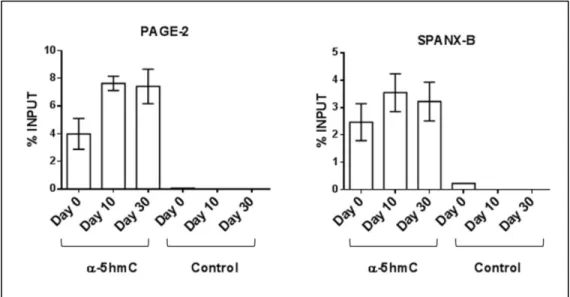
PROMOTER PROXIMAL REGIONS OF PAGE-2 AND SPANX-B GENES DURING CACO-2 SPONTANEOUS DIFFERENTIATION. ... 59 FIGURE 3.1.3 3: MRNA LEVELS OF TET GENES (TET1, TET2, TET3), RESPONSIBLE FOR 5HMC
RESIDUE GENERATION, INCREASE DURING CACO-2 SPONTANEOUS
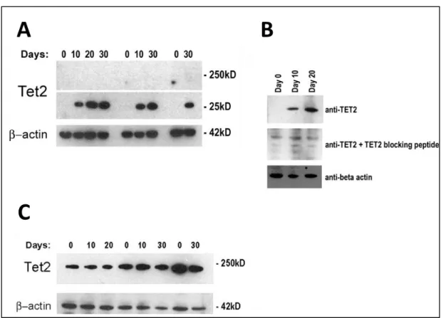
DIFFERENTIATION. ... 60 FIGURE 3.1.3 4: A POSSIBLE SMALL VARIANT OF TET2 PROTEIN IS FOUND TO BE
INCREASED IN CACO-2 DIFFERENTIATION CONSISTENTLY IN 3 DIFFERENT
DIFFERENTIATION SETS. ... 61 FIGURE 3.1.3 5: A POSSIBLE TRUNCATED FORM OF TET2 IS GENERATED WITH CA+2
DEPENDENT CALPAINS. ... 62 FIGURE 3.1.3 6: DOUBLE IMMUNOFLUORESCENCE STAINING SHOWS THAT TET2 PROTEIN
CO-LOCALIZES WITH SPANX-B AND PAGE-2,-2B PROTEINS IN CACO-2 CELLS DURING DIFFERENTIATION. ... 63 FIGURE 3.1.3 7: BINDING OF (A) EZH2, H3K27ME3 AND (B) HP1 TO THE PROMOTERS OF
PAGE-2B, PAGE-2 AND SPANX-B DECREASES DURING DIFFERENTIATION SHOWN BY
CHIP WITH THE INDICATED ANTIBODIES OR CONTROL IGG. ... 64 FIGURE 3.1.3 8: AS THE DEDIFFERENTIATION OCCURS, CELLS GAIN MESENCHYMAL
CHARACTER, DOWN-REGULATE CTS AND TET ENZYMES AT THE SAME TIME. ... 65
FIGURE 3.2.1 1: THE GENOMIC LOCATION OF ALAS2 AND CDR1 GENES WITH RESPECT TO PROXIMAL CANCER-TESTIS ANTIGENS; PAGE-2,-2B AND SPANX-B RESPECTIVELY. ... 67
FIGURE 3.2.2 1: THE MRNA EXPRESSION OF ALAS2 GENE IS SHOWN IN NORMAL TISSUES AND A PANEL OF COLON AND LUNG CANCER CELL LINES BY TAQMAN PROBE BASED Q RT-PCR. GAPDH GENE WAS USED AS ENDOGENOUS CONTROL. ALTHOUGH
ALAS2 EXPRESSION WAS DETECTED IN VARIABLE AMOUNTS IN ALL HEALTHY
TISSUES, IN NONE OF THE TESTED LUNG AND COLON CANCER CELL LINES ALAS2 MRNA EXPRESSION EXISTED. ... 68 FIGURE 3.2.2 2: THE MRNA EXPRESSIONS OF PAGE-2,-2B GENES PROXIMAL TO ALAS2 ARE
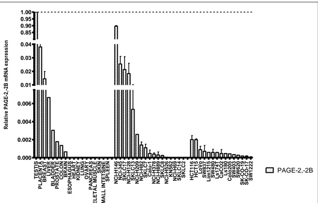
SHOWN IN NORMAL TISSUES AND A PANEL OF COLON AND LUNG CANCER CELL LINES BY TAQMAN PROBE Q RT-PCR. ... 69 FIGURE 3.2.2 3: THE MRNA EXPRESSION OF CDR1 GENE IS SHOWN IN NORMAL TISSUES
AND A PANEL OF COLON AND LUNG CANCER CELL LINES BY TAQMAN PROBE Q RT-PCR. ... 70 FIGURE 3.2.2 4: THE MRNA EXPRESSION OF SPANX-B GENE PROXIMAL TO CDR1 IS SHOWN
IN NORMAL TISSUES AND A PANEL OF COLON AND LUNG CANCER CELL LINES BY TAQMAN PROBE Q RT-PCR. ... 71
FIGURE 3.2.3 1: THE ORGANIZATION OF ALAS2 GENE; PROMOTER, EXON-INTRON STRUCTURE AND BISULPHITE SEQUENCING RESULT OF THE ANALYZED CPG
xvi
FIGURE 3.2.3 2: THE ORGANIZATION OF CDR1 GENE; PROMOTER, EXON-INTRON STRUCTURE AND BISULPHITE SEQUENCING RESULT OF THE ANALYZED CPG
RESIDUES ARE SHOWN. ... 75
FIGURE 3.2.4 1: THE RESPONSE OF CT GENES AND CT PROXIMAL GENES TO 5-AZA-2’-DEOXYCYTIDINE TREATMENT IS SHOWED BY Q RT-PCR. ... 77
FIGURE 3.2.5 1: THE AMOUNT HYDROXYMETHYLATED DNA IN THE PROMOTER
PROXIMAL REGIONS OF CT GENES AND CT-PROXIMAL GENES ARE SHOWN. ... 78
FIGURE 3.2.6 1: THE MRNA LEVELS OF TET GENES (TET1, TET2, TET3) IN COLON CANCER AND LUNG CANCER CELL LINES WITH RESPECT TO NORMAL COUNTERPARTS. ... 79
FIGURE 3.2.7 1: 3 CANDIDATE CLONES ARE IDENTIFIED AS A RESULT OF
Β-GALACTOSIDASE STAINING EXPERIMENT PERFORMED AFTER PCDNA4/TO/LACZ TRANSFECTION TO STABLE CLONES EXPRESSING TET REPRESSOR. ... 80 FIGURE 3.2.7 2: ALAS2 GENE EXPRESSION IN TETRACYCLINE TREATED CLONES
COMPARED TO UNTREATED AND UNTRANSFECTED CLONES. ... 81 FIGURE 3.2.7 3: THE CELL VIABILITY RESULTS ARE MEASURED BY MTT ASSAY AFTER
ECTOPIC EXPRESSION OF ALAS2 AND CDR1 GENE. ... 82
FIGURE 3.3.1 1: THE LOCALIZATION OF PRIMERS HITTING IΚBG, NY-ESO-1 AND 3
NONCODING RNA GENES IN NY-ESO-1 REPEAT REGION. ... 85 FIGURE 3.3.1 2: THE EXPRESSIONS OF NONCODING RNAS IN REPEAT REGION, IΚBG AND
NY-ESO-1 IN NY-ESO-1 POSITIVE (MAHLAVU, SK-LC-17 AND MDA-MB157) AND
NEGATIVE CELL LINES (HCT116, SW20 AND MCF-7) ARE ANALYZED BY Q RT-PCR. ... 85
FIGURE 4 1: THE PROPOSED WINDOW OF EMT AND THE SUGGESTED EXPRESSION
xvii
List of Tables:
TABLE 2.1.1 1: LIST OF CHEMICALS ENZYMES KITS ... 13
TABLE 2.1.1 2: LIST OF CONSTRUCTS AND VECTORS ... 17
TABLE 2.1.1 3: LIST OF POSITIVE AND NEGATIVE CONTROLS ... 18
TABLE 2.1.1 4: PRIMERS USED ... 19
TABLE 2.1.1 5: LIST OF ANTIBODIES USED IN IF STAINING AND WESTERN BLOTTING ... 22
TABLE 2.1.1 6: LIST OF ANTIBODIES USED IN CHIP ... 23
TABLE 2.1.2 1: LIST OF INSTRUMENT ... 24
TABLE 2.1.3 1: LIST OF CELL CULTURE REAGENTS ... 25
TABLE 3.2.3 1: THE METHYLATION STATUS OF ALAS2 PROMOTER IN ANALYZED NORMAL TISSUES AND CANCER CELL LINES SHOWN AS PERCENT METHYLATION ... 74
TABLE 3.2.3 2: THE METHYLATION STATUS OF CDR1 PROMOTER IN ANALYZED NORMAL TISSUES AND CANCER CELL LINES SHOWN AS PERCENT METHYLATION ... 76
xviii
Abbreviations:
CT Cancer testis
5-AZA 5-aza-2-deoxycytidine
LINE1 Long interspersed elements
EZH2 Enhancer of zeste homolog 2
DNMT DNA methyltransferase
LSD1 Lysine-specific demethylase 1
BORIS Brother of the Regulator of Imprinted Sites
LAGE L antigen family member 1
NY-ESO-1 New York esophageal squamous cell
carcinoma 1
MAGE-A Melanoma-associated antigens
SSX Synovial sarcoma, X breakpoint 2
SPAN-X Sperm protein associated with the nucleus
PAGE P antigen family
MSC Mesenchymal stem cell
hESC Human embryonic stem cell
IL13RA Interleukin 13 receptor, alpha 1
SOX2 Sex-determining region Y (SRY)-Box2
CSC Cancer stem cell
EMT Epithelial to mesenchymal transition
HMLE Human mammary epithelial cell
TGF-β Transforming growth factor beta
ECM Extracellular matrix
DKO Double knockout
GAPDH Glyceraldehyde 3-phosphate
dehydrogenase
TAGLN Transgelin
FN1 Fibronectin 1
VIM Vimentin
CDX2 Caudal type homeobox 2
CLDN4 Claudin 4
CDH1 E-Cadherin
TET Ten-eleven translocation methylcytosine
dioxygenase IκBG
ALAS2 Aminolevulinate, delta-, synthase 2
CDR1 Cerebellar degeneration-related protein 1
HP-1 Heterochromatin protein 1
PBS Phosphate buffered saline
SAGE Serial analysis of gene expression
EST Expressed sequence taq
SDS Sodium dodecyl sulphate
PVDF Polyvinylidene fluoride
xix
ChIP Chromatin immunoprecipitation
hMEDIP Hydroxymethylated DNA
immunoprecipitation
CGAP Cancer genome anatomy project
DNMTi DNA methyltransferase inhibitor
IF Immunofluorescence
H3K27me3 Histone 3 Lysine 27 trimethylation
5Mc 5-methylcytosine
1 INTRODUCTION
1.1 CANCER TESTIS GENES
1.1.1 Expression Patterns of Cancer Testis Genes
Cancer testis (CT) antigens are tumor-associated antigens that are activated in various human tumors from different origins. Cancer testis antigen gene expression is restricted to germ cells in testis and ovary and in trophoblast cells among healthy tissues [1]. Due to the aberrant gene expression in cancer and the restricted expression pattern in healthy tissues, CT genes are considered as a model to study epigenetic mechanisms behind gene expression and complex gene regulation processes.
Up to now, more than 140 CT genes belonging to at least 70 gene families have been identified by different methodologies such as T-cell epitope cloning, serological analysis of recombinant cDNA expression libraries and representational difference analysis [2,3].
CT genes have variable expression in different types of cancer. Melanoma, ovarian, bladder and non-small cell lung cancers have high CT gene expression, whereas breast and prostate cancers have moderate CT gene expression. Hematological
malignancies such as lymphomas and leukemia, renal, colon and pancreatic cancers were identified as low CT expressing cancers[4] [5]. By using genome wide survey expression for 153 CT genes in normal and cancer expression libraries, Hofmann et al. classified CT genes in 3 groups; testis-restricted, testis/brain restricted and testis selective [6].
2
Figure 1.1.1 1: The expression pattern of CT-X genes in normal tissues and cancer
CT genes on chromosome X are mainly expressed in testis and placenta among normal tissues. CT-X gene expression is detected in brain and in some of the normal tissues at very low levels albeit. Various CT genes are induced and detected in cancer tissues. Adopted from [6]. (Hofmann et al. PNAS, 105, 51, 2008 Genome-wide analysis of cancer/testis gene expression. Copyright (2008) National Academy of Sciences, U.S.A.)
Gure et al. found that the analyzed nine CT genes (LAGE1, NY-ESO-1,
MAGE-A1, MAGE-A3, MAGE-A4, MAGE-A10, CT7, SSX2 and SSX4) were coordinately
expressed in non-small cell lung cancer. According to the study, the frequency of expression of a second CT antigen by a tumor already expressing a CT antigen was higher than the expression frequency if these two events were independent. The study also showed significant correlation between CT expression and larger tumours’ and later stages of disease [7]. The associations between CT expression and different variables such as metastatic disease, poor survival and advanced tumour type have been
established in many studies [5].
It has been previously shown that CT genes except SPAN-X were expressed in earlier stages of spermatogenesis such as spermatogonia and primary spermatocytes. CT gene expression seen at oogonia was similarly in the earlier stages of oogenesis. In the later stages of both spermatogenesis and oogenesis, CT gene expression was diminished [8].
3
1.1.2 Genomic Structure of Cancer Testis Genes
Cancer testis genes can be classified as the ones encoded from Chromosome X, CT genes and the ones encoded from chromosomes other than Chromosome X, non-CT genes. Most of the CT genes are members of multigene families and each family is composed of proximally located and highly homologous genes that vary from 3
(NY-ESO) to more than 12 (MAGE) genes. The multigene families exist on well-defined
clusters that either form a direct or inverted repeat on Chromosome X [4,5]. In 2004, Warburton and his colleagues published the first genome wide inverted repeat structure of human genome identified by a software package named Inverted Repeat Finder program. The most dramatic result they obtained was the abundance of large and highly homologous inverted repeat regions containing CT genes on chromosome X. 10 of 20 inverted repeats that they identified on X-chromosome contained a gene expressed in testis tissue [9]. Since inverted repeats have the capacity to form self organizing loops and is a common property of majority of CT genes, the possible roles of repeats and loop formation on gene expression is an important question.The effect of the repeat structure on gene expression was proposed by Bredenbeck et al. They showed that gene
expression inside the repeat region was coordinated compared to the gene expression outside the repeat region coding MAGEA and CSAG genes [10]. A direct physical link between gene expression and repeat structure, however, is still to be identified.
1.1.3 Epigenetics of Cancer Testis Genes
CT gene expression regulation is primarily epigenetic in nature. As well as
promoter DNA hypermethylation in normal somatic tissues lacking CT gene expression, promoter DNA hypomethylation of CT genes in various CT expressing tumors have been shown in various studies. Induction of CT genes with the DNA hypomethylating agent, 5-aza-2`-deoxycytidine (5-AZA), in cancer cell lines is an important evidence of DNA methylation as a leading epigenetic mechanism controlling CT gene expression [1,11-15]. In addition to promoter and proximal promoter DNA hypomethylation, CT gene expression is associated with global DNA hypomethylation when MAGEA11 expression and LINE1 methylation are studied [16]. The DNA methylation status of CT
4 genes shows intra- and inter-tumour heterogeneity [14,15]. Thus the clinical usages of DNA methylation inhibitors have importance in CT based immunotherapy studies. Role of histone acetylation in CT gene expression was revealed with synergistic effects of histone deacetylase inhibitors with DNA hypomethylating agent [11,17]. Recently role EZH2 and histone methylation on the expressions of GAGE and MAGE genes was presented in breast cancer cell lines. S-adenosylhomocysteine hydrolase inhibitor named 3-deazaneplanocin was previously shown to disrupt EZH2 complex. The combination of DNA hypomethylating agents with 3-deazaneplanocin resulted with enhancing
expressions of GAGE and MAGE type CT genes [18]. In another study the inhibition of histone methyltransferase (KMT6) and histone demethylases (KDM1 and KDM5B) improved the effect of DNA hypomethylating agent deoxyazacytidine on expressions of
NY-ESO-1, MAGE-A1 and MAGE-A3 genes in lung cancer cell lines. Thereby,
incorporation of histone methylation in epigenetic regulation of CT genes was verified [19]. The synergistic effect of histone methylation and DNA demethylation was shown recently with the combined treatment of DNMT inhibitor and LSD1 inhibitor. LSD1 inhibitor inhibited demethylation of H3K4me2 and H3K4me1 and could synergistically activate CT gene expression when used with DNMT inhibitor[20]. The dominant function of DNA methylation in CT gene expression mechanisms compared to histone marks was suggested in works of De Smet et al. They generated a methylated
MAGEA1/hph construct that was resistant to hygromycin upon stable re-activation. By either treating the generated clone with histone acetyltransferase inhibitor or by
depleting DNA methyltransferase-1, they showed hygromycin resistant cells developed DNA hypomethylation and active histone marks[21]
The transcription factor BORIS was established as a candidate for the regulation of CT genes. The occupancy of BORIS in NY-ESO-1 promoter was associated with gene expression [22]. The induction of BORIS resulted in the induction of MAGE-A3 and
MAGE-A1 genes [23,24]. However in another study, BORIS overexpression with an
adenoviral system did not induce CT genes, neither were promoter and global DNA demethylation levels altered [25]. Thus the role of BORIS in CT gene expression is controversial.
5
1.1.4 Cancer Testis Gene Expression in Stem Cells
CT gene expression in stem cells and whether CT genes have a role in stem cell differentiation pathways are interesting questions. Expression of various CT genes (NRAGE, NY-ESO-1, MAGE-1 and SSX) has been detected in undifferentiated
mesenchymal stem cells [26,27]. In addition to the CT gene expression in mesenchymal stem cells, melanoma and glioma cancer stem cells also express many CT genes
(MAGE, GAGE, NY-ESO-1 and SSX families) according to different studies[28-30]. CT gene expression (SSX, NY-ESO-1 and N-BAGE) was observed in
undifferentiated mesenchymal stem cells (MSC). CT gene expression was attenuated as MSCs differentiate to adipocytes and osteocytes [31]. In addition to MSCs, expressions of MAGE-D1,-D2 genes in human embryonic stem cells (hESC) and expressions of
GAGEs, MAGE-A3,-A6,-A4,-A8 genes in human embryoid body cells were described by
Lifantseva et al.[32]. In contrast, Loriot et al. showed that various CT genes either had very low or no expression in human embryonic cell lines compared to melanoma cell lines and testis by Q RT-PCR[33]. Whether CT gene expression being present in hESCs is controversial, the expression of CT genes in cancer stem cells (CSC) was
demonstrated by showing the expression of MAGED3, -D1, IL13RA, SPANXA and
SPANXC in CD133 and SOX2 positive CSCs derived from glioma cells lines and tissues
[30].
1.1.5 Cancer Testis Gene Expression during Epithelial to Mesenchymal Transition
The relation between CSCs and epithelial to mesenchymal transition (EMT) process was characterized by Mani et al. EMT was induced in non-transformed immortalized human mammary epithelial cells (HMLEs) either by ectopically expressing Snail or Twist or TGFβ1 exposure. These cells started to resemble mesenchymal cells by expressing mesenchymal markers (N-cadherin, Vimentin,
Fibronectin) and down-regulating epithelial markers (E-cadherin) and gained the ability
to form mammospheres which was highly observed in human breast CSCs. EMT
induced HMLEs was enriched in CD44high/CD24low population showing a strong
6 study was further improved by establishing the high expression levels of mesenchymal markers and the low expression levels of epithelial markers in CD44high/CD24low cells isolated from patient tissue samples. [34]
According to these two observations; CT gene expression in glioma CSCs and the association of EMT with CSCs, there might be a connection between CT gene expression and EMT phenotype.
In literature, there were two papers confirming the relation of CT gene
expression with EMT process. Contrary to the expectation, both of them claimed that CT gene expression was associated with the epithelial phenotype but not the mesenchymal phenotype in EMT. In the study conducted by Gupta et al., the gene expression in transformed HMLE in response of either salinomycin (shown to be effective on mesenchymal type of cells in tumor) or paclitaxel (shown to be effective on epithelial type of cells in tumor) was analyzed. MAGE-A1 gene was up-regulated in salinomycin treatment which eliminated the mesenchymal cells and down-regulated in paclitaxel treatment which eliminated the epithelial cells [35]. Similarly, the result of another study done by Thomson et al. claimed that CT (SPANXA1,-A2, SPANX-B1,-B2, SPANXC,
MAGEA8, SOX2) gene expression was diminished in two EMT models generated by
stable transfection of Snail gene and TGF-β exposure[36].
The up-regulation of MAGE-A1 gene in epithelial enriched population and the down-regulation of various CT genes in two important EMT models proved that CT gene expression might be related with epithelial phenotype in EMT.
1.2 CACO-2 SPONTANEOUS DIFFERENTIATION MODEL
To associate CT gene expression with epithelial phenotype of a cell, we used Caco-2 spontaneous differentiation model. Caco-2 and HT29 cell lines were derived from colorectal tumors and well known with their capabilities to differentiate into mature intestinal cells, such as enterocyte, mucus and M cells. Because of their differentiation potential, they became important tools for in vitro structural and
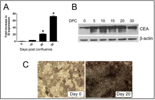
7 functional studies of the intestine cells [37]. At low seeding density, Caco-2 cells exhibit proper cell division and generate normal unpolarized and undifferentiated cells [38]. Under standard culture conditions and upon cell to cell contact formation, in 20 to 30 days differentiation process starts. Caco-2 cells stop dividing and become a
monolayer of polarized epithelial cells. In addition to tight junctions, apical and basal-lateral membranes appear and cells resemble to polarized enterocytes both in structural and functional manner. Even though Caco-2 cells are derived from colon, they express hydrolyase enzymes and transport ions and water similar to enterocytes do [39]. It is important to note that although Caco-2 cells are valuable model for mimicking the differentiation taking place from crypts to villus, these cells are malignant and belonging to colon tissue but not small intestine [37].
In order to identify this model in depth and identify the transcriptional regulation during the enterocytic differentiation, two important studies have been done by Halbleib and Sääf et al. Caco-2 differentiation was generated by growing the cells on permeable filter supports for 26 days in this study. As well as the microscopic investigation, microarray experiments performed with RNAs isolated at different time points during differentiation were used for clarification of the differentiation process. Halbleib et al. showed that as well as apical brush border assembly (such as myosin 1A), many other proteins required for epithelial junctional complexes (Occludin, Claudin 1,
protocadherins and desmosomal cadherins,) were regulated in transcriptional levels. The transcriptional regulation in some of extracellular matrix proteins (1, Laminin-5), and intermediate filament proteins (Keratin-20, Keratin-18) were also established. This study claimed that the transcriptional regulation had an important role on Caco-2 cells being differentiate into a fully functional enterocyte in vitro without the effect of stromal cells and signals normally present in vivo[40]. In the other study carried by Sääf et al., according to microarray data there were two distinct clusters named
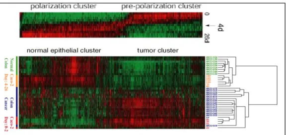
prepolarization (samples from 0 to 4 days) and polarization cluster (samples from 4 to 26 days) which were formed by the difference in gene regulation at day 4. When the gene expression pattern during Caco-2 differentiation was compared with normal colon and colorectal cancer samples, there were two clusters; tumor and normal epithelial cluster. While prepolarized cells remained in tumor clusters, polarized cells remained
8 normal epithelial cluster. Several genes belonging to cell cycle checkpoints, ECM
components, Wnt pathway were shown to be differentially regulated between tumor and normal epithelial cluster. As a result of this study, it has been established that
differentiated Caco-2 cells resemble more to a normal epithelial cell than a tumor cell [41].
Figure 1.2 1: Differentially expressed genes in Caco-2 spontaneous differentiation.
When the gene expression analysis is performed in Caco-2 spontaneous differentiation, there is a dramatic switch in gene expression pattern at day 4 time point. The
undifferentiated Caco-2 cells (Day0-2) cluster with colon cancer and the differentiated Caco-2 cells (Day4-26) cluster with normal colon tissue. Adopted from [41].
1.3 DNA HYDROXYMETHYLATION AS EPIGENETIC CONTROL MECHANISMS OF GENE EXPRESSION
With the identification of 5-hydroxymethylcytosine (5-hmC) residue in Purkinje neurons and embryonic stem cells, 5-methylcytosine (5-mC) has become no more the only epigenetic modification of DNA [42,43]. This was groundbreaking information in epigenetics. 5-hyroxymethylcytosine was established as an epigenetic mark on DNA due to its binding partners and unique distribution patterns affecting gene expression with the following studies [43-45] . 5-hmC was shown to inhibit binding of methyl CpG binding protein 2 (MeCP2) to DNA thereby acting oppositely to 5-mC [46]. In addition
9 to inhibiting MeCP2 binding, 5-hmC has its own binding partners. 5-hmC was found to interact with Mbd3, a chromatin regulator, and this interaction was necessary for Tet1 binding to chromatin and interacting with Mbd3. It was also shown that Mbd3
preferably binds to hmC compared to mC [44]. When the genomic distribution of 5-hmC was investigated in ESCs, it was identified that 5-5-hmC mainly localized at gene rich, low to moderate CpG containing regions specifically transcription start sites, promoters and gene bodies. The bivalent domains containing both permissive
H3K4me3 and repressive H3K27me3 marks were also enriched for 5hmC residues. The role of 5hmC on gene expression was described as activating mainly [43,45,47].
Although recent study claimed that 5hmC could also act as inhibiting transcription in the case of being on distal regulatory sites such as enhancers [48].
Ten eleven translocation family proteins (TET1,-2 and -3) produce
5-hydroxymethylcytosine from 5-methylcytosine. [42,49]. TET enzymes further oxidize
and generate 5-formylcytosine and 5-carboxycytosine and 2-oxoglutarate and Fe+2 are
necessary cofactors for the oxidation reaction [50].
Though 5-hmC is a newly identified residue, its level in human tissues is more than the expected levels. Brain, kidney, liver and colorectal tissues have higher 5-hmC levels and the abundance of 5-hmC declines in the cancerous state [51,52]. Proper functioning of TET enzymes and the level of 5hmC on DNA are very crucial since various mutations of TET2 were reported in hematological malignancies [53].
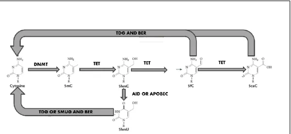
In addition to its epigenetic functions, 5hmC also act as an intermediate player in DNA demethylation. It exerts its function via two different paths, passive and active DNA demethylation. In passive mechanism, the generated 5hmC from 5mC cannot be recognized by DNMT1, thereby after each cell division cycle DNA become more demethylated [54]. Though in some circumstances cells need to demethylate DNA immediately such as after fertilization or in primordial germ cells [55]. In this case 5-hydroxymethylated cytosine residues are either further oxidized to 5-carboxylcytosine by TET enzymes or deaminated to 5-hyroxymethyluracil by AID/APOBEC enzymes. Then both 5-carboxylcytosine and 5-hydroxymethyluracil are removed from DNA by base excision repair mechanism (BER) [55,56].
10
Figure 1.3 1: Active DNA demethylation
TET enzymes oxidize 5-methylcytosine to 5-hydroxymethylcytosine, 5-formylcytosine and 5-carboxycytosine sequentially. 5-formylcytosine and 5-carboxylcytosine is excised by thymine DNA glycosylase (TDG) and cytosine is added with base excision repair (BER) mechanism. In addition, 5-hydroxyuracil can be generated from 5-hydroxymethyl with AID and APOBEC mediated deamination. 5-hydroxyuracil is further be excised with TDG or SMUG and cytosine is added via BER. Adopted from [45].
Figure 1.3 2: Passive DNA demethylation
5-hydroxymethylated cytosine inhibits binding of UHRF1 or DNMT1 or both UHRF1 and DNMT1, thereby by each cell division methylated cytosines are lost. Adopted from [45].
All TET enzymes have the ability to hyroxymethylate DNA; however their differential roles were established. In a study done in embryonic germ cell lines (EGCs),
11 TET1 was essential for the proper methylation of imprinted control regions whereas TET2 was indispensable for the precise reprogramming when ECGs were fused with B cells [57]. The activities of TET1 on transcription start sites and TET2 on gene bodies were established with the gene depletion study performed in mouse embryonic stem cells [58]. TET1 and TET2 deficient double knockout mouse model established TET3 have redundant functions since double knockout animals were viable though having decreased 5hmC levels and impaired imprinting [59].
New functions and novel interactions about the transcriptional role of 5hmC are emerging. Recently, enrichment of 5hmC on the gene bodies of neuronal genes in accompany with loss of H3K27me3 was shown to be essential for neurogenesis [60]. TET2 and TET3 were shown to directly interact with O-GlcNAc transferase (OGT). TET2-OGT and TET2/TET3-OGT interactions resulted with O-GlcNAcylation of H2B Ser112 and HCF1 which is a component of H3K4 methyltransferase SET1/COMPASS complex respectively [61,62]. OGT was shown to interact with TET3 and
O-GlcNAcylated TET3. The modified TET3 exported to nucleus and its catalytic activity was inhibited [63].
12 1.4 AIM AND HYPOTHESIS
Because of their unique cancer-specific expression pattern, studying the
regulation of CT gene expression can help reveal the deregulated epigenetic mechanisms during carcinogenesis. Additionally, CT genes can be used as perfect biomarkers for DNA hypomethylation known to occur in cancer. They might be useful in the detection and prognosis of cancer as well.
In this study, we aimed to study both region specific as well as promoter-proximal epigenetic alterations in CT gene expression in cancer. To be able to study the epigenetic basis of CT gene expression, we had three different approaches. In the first approach, we used a model dynamically expressing CT genes; thereby we studied and verified the responsible epigenetic mechanisms relevant to the transition from CT negative to positive gene expression. In the second approach, we studied CT and CT proximal regions with opposite expression patterns in healthy and cancer conditions. Finally we tried to find out an explanation to coordinate CT gene expression in cancer by analyzing CT encoding repeat regions on DNA since repeat demethylation was a known epigenetic event during carcinogenesis.
13
2 MATERIALS AND METHODS
2.1 MATERIALS
2.1.1 General Chemicals
General laboratory chemicals such as; Methanol, Ethanol, Isopropanol,
Chloroform, Formaldehyde, NaCl, tris-base, glycine are analytical grade and purchased from either Sigma-Aldrich (St. Louis, USA) or Calbiochem Merck Millipore
(Darmstadt, Germany). The detailed list of the chemicals and kits were shown below.
Table 2.1.1 1: List of chemicals enzymes kits
Name Catalog
number Company
TRIzol reagent 15596018 Ambion by Life Sciences
(CA, USA)
Nuclease free water AM9930 Ambion by Life Sciences (CA, USA)
DNA-free™ Kit
DNase Treatment and Removal Reagents
AM1906 Ambion by Life Sciences
(CA, USA)
RevertAid First Strand cDNA
Synthesis Kit # K1622
Thermo Scientific Inc. (IL, USA)
DyNAzyme II DNA Polymerase # F-501S Thermo Scientific Inc. (IL, USA)
OneTaq Hot Start DNA Polymerase M0481S New England BioLabs Inc.
14 Sciences (CA, USA)
TaqMan® Universal PCR Master
Mix 4364338
Applied Biosystems by Life Sciences (CA, USA)
Proteinase K P2308 Sigma Aldrich (St. Louis,
USA)
Phenol:Chloroform:IAA, 25:24:1, pH
6.6 AM9730
Ambion by Life Sciences (CA, USA)
EZ DNA Methylation-Gold™ Kit
(Bisulphite Conversion kit) D5006 Zymo Research (CA, USA) TA Cloning® Kit, with pCR™2.1
Vector, without competent cells K2020-40 Invitrogen by Life Sciences
(CA, USA)
Phusion® High-Fidelity DNA
Polymerase M0530S New England BioLabs Inc.
BamHI R0136S New England BioLabs Inc.
NotI R0189S New England BioLabs Inc.
XbaI R0145S New England BioLabs Inc.
HindIII R0104S New England BioLabs Inc.
EcoRI R0101S New England BioLabs Inc.
T-REx™ System K1020-01 Invitrogen by Life Sciences
Lipofectamine® 2000 Transfection
Reagent 11668-027
Invitrogen by Life Sciences (CA, USA)
15
TransIT-LT1 Transfection Reagent MIR 2304 Mirus Bio (Madison, USA)
β-Gal Staining Kit
K1465-01 Invitrogen by Life
Sciences(CA, USA)
Cell Proliferation Kit I(MTT) 11 465 007 001 Roche Applied Science (Basel, Switzerland)
FspI R0135S New England BioLabs Inc.
EpiSeeker hydroxymethylated DNA
Immunoprecipitation (hMeDIP) Kit ab117134 Abcam (UK)
UltraCruz™ Mounting Medium
sc-24941 Santa Cruz Biotechnology
(Texas, USA)
NuPAGE® Novex® 4-12% Bis-Tris
Protein Gels, 1.5 mm, 15 well NP0336BOX NuPAGE® Novex® by
Life Sciences (CA, USA)
Immobilon-P Membrane, PVDF,
0.45 µm, 26.5 cm x 3.75 m roll IPVH00010 Merck Millipore (MA,
USA)
Clarity™ Western ECL Substrate 170-5060 Bio Rad (CA, USA)
5-aza-2-deoxycytidine A3656 Sigma Aldrich (St. Louis, USA)
Protease inhibitor cocktail P8340 Sigma Aldrich (St. Louis, USA)
16
QIAquick Gel Extraction Kit 28706 QIAgen (CA, USA)
QIAGEN Plasmid Mini Kit 12125 QIAgen (CA, USA)
QIAGEN Plasmid Midi Kit 12145 QIAgen (CA, USA)
Precision Plus Protein™ Dual Color
Standards #161-0374 Bio Rad (CA, USA)
Gene Ruler 1 kb DNA Ladder #SM0311 Thermo Scientific Inc. (IL, USA)
Gene Ruler 100 bp DNA Ladder # SM0241 Thermo Scientific Inc. (IL, USA)
Kanamycin 60615 Sigma Aldrich (St. Louis,
USA)
Carbenicillin C1613 Sigma Aldrich (St. Louis,
USA)
β-Galactosidase G5635 Sigma Aldrich (St. Louis,
17
Table 2.1.1 2: List of constructs and vectors
Name
pcDNA2.1
3.9 kbp vector which bisulphite pcr
products were cloned in it with TA cloning procedure.
pcDNA.6TR 6662 bp vector coding tet repressor gene
pcDNA 4/TO/ lacZ
8224 bp control vector containing the gene for β-galactosidase under the control of tet repressor
pcDNA3.1/His/lacZ
Control vector constitutively expressing the gene for β-galactosidase to calculate transfection efficiency
pcDNA 4/TO/ ALAS2 ALAS2 expressing expression vector
18
Table 2.1.1 3: List of positive and negative controls
Name Catalog number Company
Human non-methylated DNA HCT116 DKO cells [DNMT1 (-/-) / DNMT3b (-/-)
D5014-1 Zymo Research (CA, USA)
Human methylated DNA SssI methylated
HCT116 DKO cells [DNMT1 (-/-) / DNMT3b (-/-)
D5014-2 Zymo Research (CA, USA)
Human Normal Adult Colon Male DNA
D1234090 (Lot
no: A805046) BioChain (CA, USA)
Human Normal Adult Colon Female
19
Table 2.1.1 4: Primers used
Primer Sequence Product
Length Tm RT-PCR & Q-RT-PCR GAPDH F 5’-TTCTTTTGCGTCGCCAGCCG -3’ 78 61.4 GAPDH R 5’-CGACCAAATCCGTTGACTCCGACC -3’ 66.1 TAGLN F 5’-ACGGCGGCAGCCCTTTAAACC -3’ 122 60.24 TAGLN R 5’-GGCCATGTCTGGGGAAAGAAGGC -3’ 59.74 FN1 F 5’-TGTGATCCCGTCGACCAATGCC -3’ 131 59.23 FN1 R 5’-TGCCACTCCCCAATGCCACG -3’ 59.62 VIM F 5’-CCAAGACACTATTGGCCGCCTGC -3’ 167 60.36 VIM R 5’-GCAGAGAAATCCTGCTCTCCTCGC -3’ 59.42 CDX2 F 5’-CGCTTCTGGGCTGCTGCAAACG -3’ 262 61.65 CDX2 R 5’-TAGCTCGGCTTTCCTCCGGATGG -3’ 60.11 CLDN4 F 5’- ACCTGTCCCCGAGAGAGAGTGC- 3' 157 59.4 CLDN4 R 5’ -GATTCCAAGCGCTGGGGACGG - 3' 60.11 CDH1 F 5’ - TGGGCCAGGAAATCACATCCTACA - 3' 91 57.57 CDH1 R 5’- TTGGCAGTGTCTCTCCAAATCCGA - 3' 57.8 TET1 F 5’- ACCTGCAGCTGTCTTGATCG- 3’ 186 60.39 TET1 R 5’- ACACCCATGAGAGCTTTTCCC- 3’ 60.27 TET2 F 5’- CGCTGAGTGATGAGAACAGACG- 3’ 187 61.29 TET2 R 5’- GCTGAATGTTTGCCAGCCTCG- 3’ 62.72 TET3 F 5’- GCATGTACTTCAACGGCTGC- 3’ 187 60.18 TET3 R 5’- ATTTCCTCGTTGGTCACCTGG- 3’ 60.27 IκBG F 5’- AGCACAGCGTGCAGGTGGAC- 3’ 209 66.55 IκBG R 5’- GAGATCTTCCAGCTGCATTCC- 3’ 62.57 NY-ESO-1 F 5’- CAGGGCTGAATGGATGCTGCAGA- 3’ 365 66.33 NY-ESO-1 R 5’- GCGCCTCTGCCCTGAGGGAGG- 3’ 72.33
Noncoding RNA1 F 5’- CACTGGCCCCAATTAGGAAGAAC- 3’ 275 64.55
Noncoding RNA1 R 5’- GAAGGCCTCATATCCCAATTCTAGC- 3’ 64.58
Noncoding RNA2 F 5’- TGCATACCCTTCCAGCTGTAGG- 3’ 387 64.54
Noncoding RNA2 R 5’- GGAGAAACCTTGGACAATACCCG- 3’ 64.55
Noncoding RNA3 F 5’- GTTAAATTAGAGCGCATTCATATTGCG- 3’ 176 61.57
Noncoding RNA3 R 5’- CTCACCCACTGCAAACATTCAATG- 3’ 62.86
BISULPHITE SEQUENCING PAGE-2 1A 5’- TGGTGGTTTATTTTATAGAGGTAGG -3’ 342 50.1 PAGE-2 1B 5’- ACCCTTTTCCCTCAAAAACCA -3’ 51.87 PAGE-2 2A 5’- TGTTGGTGTTTATGTTTGTTGTTAT -3’ 216 57.58 PAGE-2 2B 5’- ACCAACTAACTCCTCCACACATT -3’ 58.96 PAGE-2B 1A 5’- TGGAAGTGAAAGAAAGGGTGGG - 3’ 398 54.44 PAGE-2B 1B 5’- CAAAACCTATCCAAAACCAACTAACTC -3’ 53.2 PAGE-2B 2A 5’- TTGTTGTTGTATTTGTTTGTTGTTA -3’ 238 56.55 PAGE-2B 2B 5’- CTATCCAAAACCAACTAACTCCTC -3’ 57.33 SPANX-B 1A 5’- TGGGTTGAAATTTGTTTGGTAGTAGTT -3’ 523 53.81
20
SPANX-B 1B 5’- ACCCTCCCTATACATACCCTCC -3’ 53.50
SPANX-B 2A 5’- ATTGTAGGAGGGAAATG-3’ 432 52.54
SPANX-B 2B 5’- AAAACAAAACCACACCCT -3’ 57.39
ALAS2 1st region 1A 5’- AGATTATATTGTTTTATAAAAAGGTGAG-3’ 395 51.5
ALAS2 1st region 1B 5’- CAACTTACTAACAAAAATCTAAAACC-3’ 51.8
ALAS2 1st region 2A 5’- TTTTTAAAGGAGAGGAGATATTAGG-3’ 273 55.4
ALAS2 1st region 2B 5’- CTATTACATTCAAATACATTTCC-3’ 54.5
ALAS2 2nd region 1A 5’- GGGTTTTATTTTTAGTAAGGAAGG-3’ 225 54.6
ALAS2 2nd region 1B 5’- CCTAAAAAACCAACTAACAAACC-3’ 56.1
ALAS2 2nd region 2A 5’- GATATTTTTGGGGTTAATGTAGG-3’ 152 56
ALAS2 2nd region 2B 5’- AAAACAACTCTTACCTATTACCC-3’ 55.5
ALAS2 3rd region 1A 5’- ATGTATTAGTTTTTTGATTTAGATAGG-3’ 244 51.1
ALAS2 3rd region 1B 5’- AATTCTTATCCCAATCCTATTAC-3’ 52.7
ALAS2 3rd region 2A 5’-TTTTATTATTATAGGGTTGATATGAG-3’ 156 51.1
ALAS2 3rd region 2B 5’- TAAACTTAAACTCTATAATTCCC-3’ 52.7
CDR1 1st region 1A 5’- TGGTTTTTTAGATTAGTATGTTGG-3’ 341 52.5 CDR1 1st region 1B 5’- AAATAAATACAAACACTTTCTAATACC-3’ 50 CDR1 1st region 2A 5’- ATTTAAGGAGTTGTAGTTATTATTAG-3’ 234 51 CDR1 1st region 2B 5’- CTTCAAAATCATATTCATAACTCC-3’ 50.7 CDR1 2nd region 1A 5’- TTTAAGGGAATGGTAGTAGTTGG-3’ 343 52 CDR1 2nd region 1B 5’- CCATTAAAACTAAATACCATCATTATCC-3’ 51 CDR1 2nd region 2A 5’- AAATAGATTTTGGTAGTGATAGG-3’ 203 52.2 CDR1 2nd region 2B 5’- CTAAATAATAAAACCAAATTTAAACCC-3’ 50.4 CDR1 3rd region 1A 5’- GGATTATAGAATATGTTAGAATATTTGG-3’ 245 50.5 CDR1 3rd region 1B 5’- ATCTTCCTATATCTCCAAATCTTCC-3’ 51.9 CDR1 3rd region 2A 5’- GAATGTTAGAAGATTAGTATATTGGAG-3’ 166 50 CDR1 3rd region 2B 5’- ATCTCCAAAACTTCCAACATCTAC-3’ 52.6 hMEDIP-Q PCR
PAGE-2 Primer #1 F 5’- GACTCAGCCGGTAGGTCTGC-3’
152 62.3
PAGE-2 Primer #1 R 5’- CTGGGAGGAGCTGGATGACG-3’ 62.0
PAGE-2 Primer #2 F 5’- GAGCGCTGGTGGTTTACTCC-3’
173 61.0
PAGE-2 Primer #2 R 5’- TCCTTGCAGACCTCTGTGCG-3’ 62.4
PAGE-2B Primer #1 F 5’- AGTCACGAGGCGAATGTCCC-3’
214 62.2
PAGE-2B Primer #1 R 5’- GACCTACCGGCTGAGTCTCG-3’ 61.7
PAGE-2B Primer #2 F 5’- AGGTTCTCCACAGACGCAGG-3’
166 61.8
PAGE-2B Primer #2 R 5’- TGTGTGTGGACAGAAGGCGG-3’ 62.6
SPANX-B Primer#1 F 5’- AACCTACTGTAGACATCGAAGAACC-3’
125 60.1
SPANX-B Primer#1 R 5’- CGTCTTGTTGGCCTCATTGGC-3’ 62.4
ALAS2 Primer#1 F 5’- GAACACGGCCTGGCACA-3’
229 60.26
ALAS2 Primer#1 R 5’- ATGAACGTACAGCCAAGGG-3’ 57.45
CDR1 Primer#1 F 5’- TGCTGGAAGACCTGGAGATA-3’
330 57.45
CDR1 Primer#1 R 5’- CCCTCAAATCCATAGCTTCCG-3’ 58.5
21 ALAS2 Primer #1 F (((BAmHI 5'-ATTATTGGATCCACTTTAGGTTCAAGATGGTGACTGC- 3' 1650 62. 5
ALAS2 Primer #1 R 5'-ATTATTGCGGCCGCTGGCTTCTCAGGCATAGGTGG- 3' 65.
5 CDR1 Primer #2 F 5'-ATTATTGGATCCTGGAAGACATGGCTTGGTTGG- 3' 830 62. 4 CDR1 Primer #2 R 5'-ATTATTGCGGCCGCTGGCTTCTCAGGCATAGGTGG- 3' 65. 5 SEQUENCING OF pcDNA2.1
M13 reverse primer 5'- CAGGAAACAGCTATGAC -3' 51.
22
Table 2.1.1 5: List of antibodies used in IF staining and western blotting
Name of the antibody Supplier Catalog number
PRIMARY ANTIBODY
Anti-fibronectin antibody Abcam (UK) ab23750
Anti-vimentin antibody (EPR3776) Abcam (UK) ab92547
Anti-transgelin (SM22 alpha) antibody Abcam (UK) ab14106
Anti-CDX2 antibody (AMT28) Abcam (UK) ab15258
Anti-PAGE-2,-2B antibody (C-13) Santa Cruz Biotechnology (Texas, USA) sc-168892 Anti-SPANX-B antibody (N-13) Santa Cruz Biotechnology (Texas, USA) sc-162267
Anti-TET2 antibody Abcam (UK) ab-94580
Anti-TET2 antibody Active motif 61389
SECONDARY ANTIBODY
Alexa Fluor 488 donkey anti-goat IgG (H+L)
Invitrogen by Life
Sciences(CA, USA) A11055
Alexa Fluor 568 donkey anti-rabbit IgG (H+L)
Invitrogen by Life
Sciences(CA, USA) A10042
Alexa Fluor 568 donkey anti-mouse IgG (H+L)
Invitrogen by Life
23
Table 2.1.1 6: List of antibodies used in ChIP
Name of the antibody Supplier Catalog number
PRIMARY ANTIBODY
Anti-EZH2 Abcam (UK) ab3748
Anti-HP1 Abcam (UK) ab77256
24
2.1.2 Instruments
Table 2.1.2 1: List of instrument
Name Company
Applied Biosystem 7500 Q RT PCR Machine
Applied Biosystems by Life Sciences (CA, USA)
Applied Biosystem PCR Machine Applied Biosystems by Life Sciences (CA,
USA)
AutoFlow NU-8500 Water Jacket CO2
Incubator NuAire (MN, USA)
The STANDARD CO2 incubator Binder (Tuttlingen, GERMANY)
Centrifuges 5810 and 5810 R Eppendorf (Hamburg, GERMAY)
Electrophoresis Equipment XCell SureLock™ Mini-Cell
Electrophoresis System Life Sciences (CA, USA)
25
2.1.3 Cell Lines and Tissue Culture Reagents
The Caco-2 cell line was obtained from the SAP Enstitusu (Ankara, Turkey). HCT116, SW620, LoVo (colorectal cancer), MDAMB-157, MCF-7 (breast cancer) and Mahlavu (hepatocellular cancer) cancer cell lines were obtained from LGC Standards (Middlesex, UK). A lung cancer cell line, SK-LC-17, was from the Memorial Sloan Kettering Cancer Center (NY, USA).
Plastic cell culture materials such as; petri dishes, T-75 and T-25 cm flasks, multi-well plates, cryotubes were purchased from Greiner Bio-One (Austria) and serological pipettes were purchased from Costar Corporation (Cambridge, England). Other materials were listed below.
Table 2.1.3 1: List of cell culture reagents
Name Catalog number Company
RPMI 1640 medium F 1215 Biochrom AG )Berlin,
Germany)
DMEM medium FG 0415 Biochrom AG (Berlin,
Germany)
EMEM medium BE12-125F Lonza (USA)
Trypsin-EDTA SV3003101 HyClone (IL, USA)
L-Glutamine SH3003401 HyClone (IL, USA)
Penicillin/Streptomycin SV30010 HyClone (IL, USA)
Non-essential amino acids SH3023801 HyClone (IL, USA)
Fetal Bovine Serum S1620 Biowest (Nuaille,
FRANCE)
Blasticidin S HCl A11139-02 Invitrogen by Life Sciences(CA, USA)
26
Zeocin Selection reagent R25001 Invitrogen by Life Sciences(CA, USA)
Opti-MEM® I Reduced
Serum Medium 31985-062
Invitrogen by Life Sciences(CA, USA)
Tetracycline 87128 Sigma Aldrich (St. Louis,
USA)
Tetracycline reduced Fetal
27 2.2 SOLUTIONS AND MEDIA
2.2.1 General Solutions 10X PBS 25.6 g Na2HPO4·7H2O 80 g NaCl 2 g KCl 2 g KH2PO4
Bring to 1 liter with H2O.
Lysis Solution for DNA isolation (300 µl):
150 µl TE
Add 150 µl Proteinase K (from 200 µM stock)
15 µl SDS (from %10 SDS stock).
LB (500 ml):
5 g Tryptone
5 g NaCl
2.5 g Yeast Extract in 500 ml ddH2O
LB Agar + Carbenicillin (or Amphicillin)+ IPTG+ X-Gal (500 ml):
5 g Tryptone
5 g NaCl
2.5 g Yeast Extract
12.5 g Bacto Agar in 500 ml ddH2O
After autoclaving and cooling down the solution
28 o Add 250 µl IPTG from 1M stock (Final conc= 0.5 mM)
o Add 1000 µl X-Gal from 40 mg/ml stock (Final conc= 80 µg/ml) o Pour the agar in plates
SOC medium (100 ml):
2 g Tryptone
0.5 g Yeast extract
1000 µl 1M NaCl solution
250 µl 1M KCl solution
Add 97 ml ddH2O then autoclave
After autoclaving
o Add 1000 µl 2M Mg+2 stock solution (1M MgCl2.6H2O and 1M MgSO4.7H2O) (previously filter sterilized)
o Add 1000 µl 2M Glucose (previously filter sterilized)
RIPA Buffer (5 ml):
750 µl NaCl from 1M stock (final concentration 150 mM)
50 µl Triton-X (final concentration 1%)
50 µl from 10% Sodium DOC (final concentration 0.5%)
25 µl from 20 %SDS (final concentration 0.1%)
250 µl from 1M Tris-HCl at pH:8.0 (final concentration 50mM)
Protease cocktail from 100X to 1X
29
2.2.2 Cell Culture Solutions Complete DMEM and RPMI
10 % FBS 1 % L-Glutamine 1 % Penicillin/Streptomycin 500ml Medium Complete EMEM 20 % FBS 1 % L-Glutamine 1 % Penicillin/Streptomycin
1 % Non-essential amino acid solution
1 % Sodiumbicarbonate
1 % Sodiumpyruvate
500ml Medium
Blasticidin
10mg/ml of stock Blasticidin solution was prepared in sterile water, aliquoted then stored at -200C.
Tetracycline
5 mg/ml stock solution of tetracycline was prepared in 70% ethanol
Freezing mix
90% FBS
30 2.3 METHODS
2.3.1 Cancer cell culture techniques
Human colorectal cancer cell lines; HCT116, SW620, LoVo and human small cell lung cancer cell line; SK-LC-17 were grown in RPMI medium supplemented with 10% (v/v) heat-inactivated FBS, 1% L-glutamine and 1%penicillin/streptomycin. MCF-7 cells were grown in high glucose DMEM medium supplemented with 10% (v/v) heat-inactivated FBS, 1% L-glutamine , 1%penicillin/streptomycin, 1% insulin, 1% sodium pyruvate and MDA-MB-157 cells were cultured in DMEM medium supplemented with 10% (v/v) heat-inactivated FBS, 1% L-glutamine and 1%penicillin/streptomycin. HCT116.6TR and SK-LC-17.6TR clones were cultivated in complete RPMI medium containing 2 μg/ml and 1.2 μg/ml Blasticidin respectively. All cell lines were maintained in a 5 % CO2 atmosphere at 370C.
All cells were cultured by renewing the medium for every 2-3 days. When cells reached confluency, they were washed 1X PBS then harvested with trypsin-EDTA incubation and reseeded with complete medium. Stocks were prepared with 90%
DMSO and 10% FBS containing freezing mix by freezing at -20 and -80 0C
respectively. Then stocks were maintained at liquid nitrogen.
2.3.2 RNA isolation with TRIzol
One confluent T-75 flask of cells was used for RNA isolation. Cells were washed with 1X PBS, and then scraped with 1X PBS. After centrifugation, supernatant was removed and 1ml TRIzol was added. Cells were pipetted and homogenized in TRIzol by 5 minutes incubation at room temperature. 200 µl of chloroform was added. The mixture was mixed vigorously, incubated at 10 minutes at room temperature then centrifuged at
13000 rpm 15 minutes at 4 0C. Upper phase was removed, 500 µl isopropanol was
added. The mixture was inverted gently and incubated at 10 minutes at room
temperature then centrifuged at 13000 rpm 15 minutes 4 0C. The precipitate was washed
