NITROOLEFIN FUNCTIONALIZED BODIPY DYES
FOR PROTEIN LABELING
A THESIS
SUBMITTED TO THE MATERIALS SCIENCE AND
NANOTECHNOLOGY PROGRAM OF THE GRADUATE SCHOOL OF ENGINEERING AND SCIENCE OF BILKENT UNIVERSITY IN PARTIAL FULFILLMENT OF THE REQUIREMENTS
FOR THE DEGREE OF MASTER OF SCIENCE
By
HATİCE TURGUT January, 2013
i
I certify that I have read this thesis and that in my opinion it is fully adequate, in scope and in quality, as a thesis of the degree of Master of Science.
………. Prof. Dr. Engin U. Akkaya (Principal Advisor)
I certify that I have read this thesis and that in my opinion it is fully adequate, in scope and in quality, as a thesis of the degree of Master of Science.
………. Asst. Prof. Dr. Ayşe Begüm Tekinay
I certify that I have read this thesis and that in my opinion it is fully adequate, in scope and in quality, as a thesis of the degree of Master of Science.
………. Asst. Prof. Dr. Serdar Atılgan
Approved for the Institute of Engineering and Science:
………. Prof. Dr. Levent Onural
ii
ABSTRACT
NITROOLEFIN FUNCTIONALIZED BODIPY DYES FOR PROTEIN LABELING
Hatice Turgut
M.S. in Material Science and Nanotechnology Supervisor: Prof. Dr. Engin U. Akkaya
January, 2013
Protein labeling has significant importance in terms of visualizing dynamics of proteins, cell-cell interactions, mechanisms of life cycles of proteins, etc. Proteins are labeled by either synthetic or natural molecules with purposes such as analysis of 3D structures, determination of turnover number, covalent modifications and tracking protein-protein interactions. In addition to this, sensing and signalling thiol groups have gained popularity recently. Nitroolefin groups on dyes are good Micheal acceptors which undergo fast and selective reaction with thiol moieties. With this knowledge, in this study, we aimed to obtain derivatives of BODIPY dyes having nitroolefin substituents on its different positions. Nitroolefin functionalization of BODIPY dyes was targeted to result in conjugation of nitroolefins with thiol groups such as those belonging to cysteine residues on proteins. Three different nitroolefin functionalized BODIPY dyes have been designed, synthesized and characterized successfully. Incorporating triethylene glycol (TEG) units onto BODIPYs increased water-solubility of the molecules. To prove bioconjugation of the dyes with proteins, absorbance and emission changes were recorded after reaction with both L-cysteine and Bovine Serum Albumin (BSA) and large spectral changes were obtained. The result suggests that nitroolefin functionalization of BODIPY dyes is a promising way to sense biological thiols and hence labeling proteins having thiol groups.
Keywords: BODIPY, protein labeling, nitroolefin, thiol, cysteine, Bovine Serum
iii
ÖZET
PROTEİN ETİKETLEME AMAÇLI
NITROOLEFIN FONKSIYONLANDIRILMIŞ BODIPY BOYALARI
Hatice Turgut
Yükek Lisans, Malzeme Bilimi ve Nanoteknoloji Programı Tez Yöntecisi: Prof.Dr. Engin U. Akkaya
Ocak, 2013
Protein etiketleme, protein dinamiklerini gözlemleme, hücrelerin birbirleriyle etkileşimleri, proteinlerin yaşam döngülerinin mekanizmaları ve benzeri konular açısından oldukça önem taşımaktadır. Proteinler, yapay veya doğal moleküllerle, 3 boyutlu yapı analizleri, kovalent modifikasyonlar, protein-protein etkileşimlerini belirleme, devir sayısı tayini gibi amaçlarla etiketlenirler. Buna ek olarak, tiyol gruplarının algılanması ve sinyalizasyonu da son zamanlarda popülerlik kazanmıştır. Boyaların üzerindeki nitroolefin grupları, tiyol gruplarıyla hızlı ve seçici reaksiyon veren iyi Micheal akseptörleridir. Bu bilgiye dayanarak, bu çalışmada, değişik pozisyonlarında nitroolefin bulunan BODIPY türevleri elde etmeyi hedefledik. BODIPY boyalarının nitroolefin fonksiyonlandırılmasında, nitroolefin gruplarının proteinlerin üzerindeki sistein gibi tiyol içeren gruplarla konjugasyon yapmasını amaçladık. Üç farklı nitroolefin fonksiyonlandırılmış BODIPY boyaları başarıyla dizayn edildi, sentezlendi ve karakterize edildi. BODIPY üzerine trietilen glikol ünitelerinin eklenmesi de bu moleküllerin suda çözünülürlüğünü artırdı. Boyaların proteinle biyokonjugasyon yaptığını kanıtlamak için, hem L-sistein hem de Bovin Serum Albumin (BSA) ile boyaların reaksiyonu sonrası absorbans ve ışıma değişimleri kaydedildi ve spektral karakteristiklerde geniş değişimler elde edildi. Sonuçlar, BODIPY boyalarının nitroolefin fonksiyonlandırılmasının, biyolojik tiyollerin algılanması ve dolayısıyla üzerinde tiyol grupları bulunduran proteinlerin etiketlenmesi açısından gelecek vaad eden bir yol olduğunu göstermektedir.
Anahtar Kelimeler: BODIPY, protein etiketleme, nitroolefin, tiyol, sistein, Bovin
iv
Dedicated to myself…
v
ACKNOWLEDGEMENTS
Firstly, my greatest thanks are for Prof.Dr. Engin Umut Akkaya for accepting me to his group and giving me chance to conduct my studies in his laboratory. I am grateful to him for listening to me all the time and trying to understand me about everything. I learnt how a person could be a really good scientist from him and I will never forget his personality and energy for life and success. I also would like to thank Prof.Dr. Mahinur Akkaya for supporting me to join this research group. I am grateful to Dr. Murat Işık for his guidance and support in my studies. I would like to thank him deeply for being both a good teacher and a friend to me. His intelligence, knowledge and experinence influenced me so much and I will value it all throughout my life.
I would like to thank İlke Şimşek for her partnership in my studies and for her friendship.
I herein present my thanks to Sündüs Çakmak for her endless help, sincerity, generosity and smile that she kept all the time without any exception.
I would like to express further thanks for Safacan Kölemen, Ruslan Guliyev, Gizem Çeltek, Yusuf Çakmak, Elif Ertem, Ahmet Bekdemir, Ahmet Atılgan, Fatma Pir and Özge Yılmaz for their help and support. I also thank the rest of SCL (Supramolecular Chemistry Laboratory) members for everything they shared with me. I learnt a lot form them.
I thank Yusuf Keleştemur and Gözde Uzunallı for their generous help and support during my experiments.
I would like to thank UNAM (National Nanotechnology Research Center) for their financial support.
I thank Zeynep Erdoğan and Neslihan Arslanbaba for their sincerity. I also thank all the students and teachers at UNAM for the great ambiance. It was wonderful to work with them.
Lastly, my deepest thanks are for my parents and my sister for their love, support and patience all throughout my studies.. I thank my niece Melis for giving me energy, courage, love and pleasure during my time of research. I owe them a lot…
vi
LIST OF ABBREVIATIONS
BODIPY: Boradiazaindacene
DCM: Dichloromethane
TLC: Thin layer chromotography
DCE: 1,2-Dichloroethane
GFP: Green Fluorescent Protein
NMR: Nuclear Magnetic Resonance
THF: Tetrahydrofuran
DMF: Dimethylformamide
DDQ: 2,3-Dichloro-5,6-Dicyano-1,4-benzoquinone
TEG: Triethylene glycol
BSA: Bovine Serum Albumin
HOMO: Highest occupied molecular orbital
LUMO: Lowest unoccupied molecular orbital
PeT: Photoinduced electron transfer
ICT: Internal Charge Transfer
rxn: reaction
Cys: cysteine
PBS: Phosphate buffered saline
HEPES: (4-(2-hydroxyethyl)-1-piperazineethanesulfonic acid )
vii
TABLE OF CONTENTS
CHAPTER 1 ... 1 INTRODUCTION ... 1 1.1 Protein Labeling ... 1 1.1.1 in vivo labeling ... 2 1.1.2 in vitro labeling ... 51.1.2.1 Protein Labeling Procedure ... 5
1.1.3 Bioconjugation ... 5
1.2 Protein Labeling Techniques and Probes ... 6
1.2.1 Chemical Labeling Techniques ... 6
1.2.1.1 Covalent Labeling ... 6
1.2.1.2 Noncovalent Labeling ... 8
1.2.2 Biological fluorophores ... 10
1.2.3 Luminescence probes for proteins ... 10
1.2.4 Biotin and Enzymes ... 14
1.2.5 Radioactive labeling ... 16
1.2.6 Novel methods ... 16
1.3 BODIPY Dyes ... 19
1.3.1 Applications of BODIPY dyes ... 21
1.4 Fluorescence Phenomenon ... 23
1.4.1 Photoinduced Electron Transfer (PeT) ... 25
1.4.2 Internal Charge Transfer (ICT) ... 27
CHAPTER 2 ... 29
viii 2.1 General ... 29 2.2 Synthesis Scheme ... 30 2.3 Syntheses ... 31 2.3.1. Synthesis of Compound 2 ... 31 2.3.2 Synthesis of Compound 3 ... 32 2.3.3. Synthesis of Compound 4 ... 33 2.3.4. Synthesis of Compound 9 ... 34 2.3.5. Synthesis of Compound 10 ... 35 2.3.6. Synthesis of Compound 11 ... 36 2.3.7. Synthesis of Compound 12 ... 37 2.3.8. Synthesis of Compound 13 ... 38 2.3.9 Synthesis of Compound 14 ... 39 CHAPTER 3 ... 41
RESULTS AND DISCUSSION ... 41
3.1.General Perspective ... 41
3.2. Design and Properties of target dyes ... 41
3.2.1 Synthesis and Characterization ... 43
3.3 Reactions with L-cysteine & spectroscopic data ... 45
3.4 Protein Labeling Studies & Spectroscopic Data ... 54
CHAPTER 4 ... 59 Conclusion ... 59 REFERENCES ... 60 APPENDIX A ... 70 NMR SPECTRA ... 70 APPENDIX B ... 88 MASS SPECTRA ... 88
ix
LIST OF FIGURES
Figure 1: General strategy of in vivo, site-specific protein labeling. . ... 3
Figure 2: Some methods of labeling fusion proteins chemically in vivo ... 4
Figure 3: Labeling of ACP proteins on cell surface ... 7
Figure 4: Covalent labeling techniques. hAGT system uses the enzyme activity to conjugate molecules containing both the label and the enzyme’s substrate20 ... 7
Figure 5: Schematic representation of TMP-eDHFR labeling system ... 8
Figure 6: Non-covalent labeling techniques. A ligand binds specifically to a moiety which is conjugated to the target protein20 ... 9
Figure 7: The Green Fluorescent Protein46 ... 10
Figure 8: Structure of Nile Red ... 11
Figure 9: Structure of Ninhydrin ... 12
Figure 10: Structure of phenantroline derivatives ... 13
Figure 11: Structure of the simplest porphyrin ... 13
Figure 12: Schematic representation of biotinylation reactive and general formula (on the left) and general formula of biotin derivatives (on the right)2 ... 15
Figure 13: Some recently developed ligand-directed native protein labeling with various linkers ... 17
Figure 14: a concept of an affinity labeling probe. ... 18
Figure 15: Principle of fluorogenic labeling system based on PYP. ... 19
Figure 16: Position numbering of BODIPY ... 20
Figure 17: Photodynamic therapy agent and AND logic gate by Akkaya et al. .. 21
Figure 18: Examples of red-emitting BODIPY dyes ... 22
Figure 19: Literature examples of BODIPY dyes for protein labeling ... 23
Figure 20: Jablonski diagram ... 24
Figure 21: Simple representation of Stokes' shift ... 24
Figure 22: Reprentation of PeT working principle ... 26
Figure 23: Representation of reverse PeT working principle ... 26
Figure 24: Schematic representation of ICT working principle ... 28
x
Figure 26: Synthesis of compound 2 ... 32
Figure 27: Synthesis of compound 3 ... 33
Figure 28: Synthesis of compound 4 ... 34
Figure 29: Synthesis of compound 9 ... 35
Figure 30: Synthesis of compound 10 ... 36
Figure 31: Synthesis of compound 11 ... 37
Figure 32: Synthesis of compound 12 ... 38
Figure 33: Synthesis of compound 13 ... 39
Figure 34: Synthesis of compound 14 ... 40
Figure 35: 8 positions of BODIPY dyes available for modification ... 42
Figure 36: Target protein labeling agents ... 42
Figure 37: Examples of Micheal acceptors ... 43
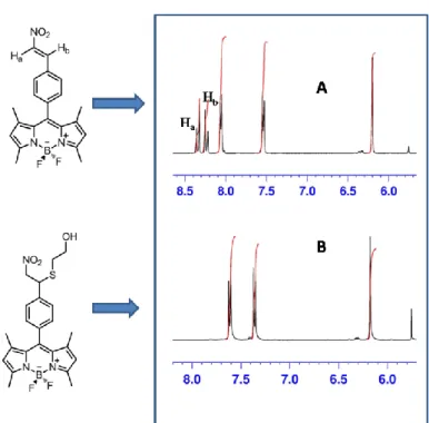
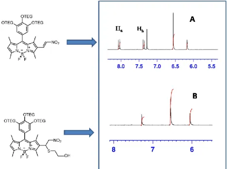
Figure 38: Comparison of aromatic regions of 1H-NMR of compound 4 and that of its conjugated form with mercaptoethanol ... 44
Figure 39: Comparison of aromatic regions of 1H-NMR of compound 11 and that of its conjugated form with mercaptoethanol ... 45
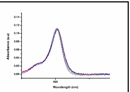
Figure 40: UV-VIS absorption spectra of compound 4 ... 46
Figure 41: Representation of the perpendicular stand of benzene ring on meso position of BODIPY ... 47
Figure 42: Fluorescence spectra of compound 4 upon increased L-Cys addition 47 Figure 43: Representation of blocking PeT process on compound 4 upon reaction with a thiol and turn-on fluorescence ... 48
Figure 44: Compound 4 and its L-cysteine conjugated form respectively under ambient light (on the left), under UV-light (on the right) ... 48
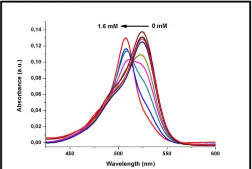
Figure 45: UV-VIS absorption spectra of compound 11 upon increased L-Cys concentrations ... 50
Figure 46: Emission spectra of compound 11 upon increased L-Cys addition .... 50
Figure 47: Compound 11 and its L-cysteine conjugated form respectively under ambient light (on the left), under UV-light (on the right) ... 51
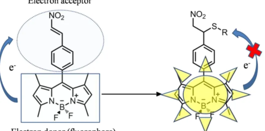
Figure 48: A nitroolefin functionalized BODIPY chemodosimeter ... 51
Figure 49: UV-VIS absorption spectra of compound 14 upon increased l-Cys concentrations ... 53
xi
Figure 50: Fluorescence spectra of compound 14 upon increased L-Cys
concentration ... 53
Figure 51: Compound 14 and its L-cysteine conjugated form respectively under ambient light (on the left), under UV-light (on the right) ... 54
Figure 52: Incubation for protein labeling (on top) & dialysis (on the bottom) .. 54
Figure 53: Absorbance spectra of dye 1 and BSA-dye 1 conjugate ... 56
Figure 54: Emission spectra of dye 1 and BSA-Dye 1 conjugate ... 57
Figure 55: Absorbance spectra of dye 2 and BSA-Dye 2 conjugate ... 57
Figure 56: Fluorescence spectra of dye 2 and BSA-dye 2 conjugate ... 58
Figure 57: Elimination of nitroolefin from reference 118 ... 58
Figure 58: 1H NMR spectrum of compound 2 ... 70
Figure 59: 1H NMR spectrum of compound 3 ... 71
Figure 60: 1H NMR spectrum of compound 4 ... 72
Figure 61: 1H NMR spectrum of compound 9 ... 73
Figure 62: 1H NMR spectrum of compound 10 ... 74
Figure 63: 1H NMR spectrum of compound 11 ... 75
Figure 64: 1H NMR spectrum of compound 12 ... 76
Figure 65: 1H NMR spectrum of compound 13 ... 77
Figure 66: 1H NMR spectrum of compound 14 ... 78
Figure 67: 13C NMR Spectrum of compound 2 ... 79
Figure 68: 13C NMR Spectrum of compound 3 ... 80
Figure 69: 13C NMR Spectrum of compound 4 ... 81
Figure 70: 13C NMR Spectrum of compound 9 ... 82
Figure 71: 13C NMR Spectrum of compound 10 ... 83
Figure 72: 13C NMR Spectrum of compound 11 ... 84
Figure 73: 13C NMR Spectrum of compound 12 ... 85
Figure 74: 13C NMR Spectrum of compound 13 ... 86
Figure 75: 13C NMR Spectrum of compound 14 ... 87
Figure 76: Mass spectrum of compound 2 ... 88
Figure 77: Mass spectrum of compound 3 ... 88
Figure 78: Mass spectrum of compound 4 ... 89
xii
Figure 80: Mass spectrum of compound 10 ... 89
Figure 81: Mass spectrum of compound 11 ... 90
Figure 82: Mass spectrum of compound 12 ... 90
Figure 83: Mass spectrum of compound 13 ... 90
xiii
LIST OF TABLES
1
CHAPTER 1
INTRODUCTION
1.1 Protein Labeling
Proteins belong to a class of major biomolecules existing in all living species. Their functions and interactions affect the whole organisation of the life. The term “protein labeling’’ stands for marking proteins to follow their mechanisms. Protein labeling has great biological importance in terms of visualizing cell-cell interactions, dynamics of proteins, mechanisms of life cycles of proteins, etc. The characterization of proteins in complex mixtures, as well as analysis of their biosynthesis, processing, intracellular mechanisms and degradation, generally requires the proteins to be labeled either in vivo or in vitro. Labeled protein is then isolated and analyzed by electrophoretic techniques1.
Labeling strategies include covalent or noncovalent attachment of the label to the target and since it is specific to the process, the strategy should be considered carefully in order not to disrupt the structure of the molecule. There are some factors affecting the choice of labeling strategy: Some proteins are affected negatively from fusion to another protein or peptide, some techniques are only suitable to surface proteins and others are for intracellular ones, different applications require different probes, techniques using disulfide bridges are influenced by the redox condition of the cell, etc.2.
The purposes of protein labeling includes lowering detection limits, obtaining turnover number, covalent modifications, analysis of 3D structures (Structure determination by NMR via radioactive labelling) and protein-protein interactions, immunological tests, affinity tests by nonradioactive labeling2.
2
1.1.1 In vivo labeling
Metabolic labeling is a strategy to label nucleic acids or proteins in a cell by culturing them with labeled nucleotides or amino acids. Cell culture in media containing labeled nucleic acids or amino acids results in all DNA, RNA or proteins becoming labeled via DNA replication, translation and protein turnover. The nucleic acid or protein of interest can later be purified for further experiments. The advantage of performing metabolic labeling is the consistent labeling of all nucleic acid or protein species. On the other hand, metabolic labeling can be toxic, depending on the type of label used, and the number of metabolic labeling reagents is not as broad as those for in vitro methods3.
Protein labeling with fluorescent probes or affinity reagents has facilitated in vitro studies of protein structure, dynamics and interactions of them with each other4. Since these traditional methods require purification of protein, chemical labeling, repurification and some invasive methods such as microinjection to reintroduce into cells, they are not adequate for in vivo studies. The general strategy of in vivo, site-specific protein labeling is summarized in figure 1.
3
a) Living cells are transfected with DNA encoding a protein of interest which is fused to a receptor domain b) a cell-permeable small molecule probe consisting of a ligand coupled to a detectable tag is added to cell growth medium c)Analysis of protein function in living cells via fluorescent microscopy or other detection methods.
There are both direct and indirect approaches to chemically modify proteins in
vivo5.
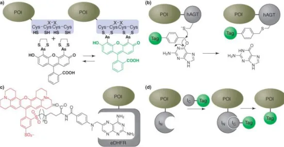
Most of the methods of in vivo labeling depends on ligand-receptor interactions. General ligand-receptor pairs include biotin-avidin system, hapten-antibody, various enzyme-antibody combinations, nitrilotriacetate(NTA)-oligohistidine sequence, biarsenical fluorophores that bind to cysteine-rich peptide sequences5. The biarsencial ligand/tetracysteine motif interaction studied by Roger Tsien's laboratory is the prototypical system for the specific chemical labeling of proteins
in vivo (Figure 2a)6-10. This strategy depends on the subnanomolar affinity between a short tetracysteine peptide (CCXXCC, where X is any amino acid except cysteine) and a biarsenical compound such as 4′,5′-bis(1,3,2-dithioarsolan-2-yl)fluorescein (FlAsH)5. In this method, the FlAsH dye is administered to the cells in the presence of an excess of 1,2-ethanedithiol (EDT) which outcompetes Figure 1: General strategy of in vivo, site-specific protein labeling. Picture adapted from ref 5 with permission. Copyright © 2012 Copyright Clearance Center, Inc.
4
endogenous proteins with closely spaced cysteine pairs. Therefore it minimizes non-specific binding and toxicity. The target protein of interest is expressed with the tetracysteine motif5. Besides the green FlAsH biarsenical, red (ReAsH)8, blue (ChoXAsH), and a biarsenical derivative of nile red have been synthesized11. In addition to direct chemical labeling, indirect approaches for modification of proteins also exist. Specific incorporation of unnatural amino acids based on suppressor tRNA technology12-14 is one of these. Protein splicing15-17 technique has also been adapted to protein labeling.
Figure 2: Some methods of labeling fusion proteins chemically in vivo
(a) FlAsH. Binding of a biarsenical fluorescein derivative to a short tetracysteine peptide fused to the protein of interest (POI) (b) Human O6-alkylguanine DNA alkyl transferase (hAGT) is labeled covalently with benzyl guanine derivatives. (c) Schematic representation of methotrexate-Texas Red™ dye bound to an E. coli dihydrofolate reductase (eDHFR) protein. (d) Split intein labeling5. Picture adapted from ref 5 with permission. Copyright © 2012 Copyright Clearance Center, Inc.
5
1.1.2 In vitro labeling
Chemical methods of protein labeling include the covalent attachment of labels to amino acids in laboratory environment. These groups, react with specific moieties on distinct amino acids. A few are also available that nonspecifically react with any amino acid at C-H and N-H bonds. Enzymatic methods are also used to label proteins and nucleic acids. These methods require the related polymerases, ATP and labeled amino acids or nucleotides. The expression of labeled proteins by in
vitro translation can be difficult due to the requirement for proper protein length,
folding and post-translational modifications that some commercial kits are unable to provide3.
1.1.2.1 Protein Labeling Procedure
Firstly, dye & protein solution is prepared. For lysine-sensitive labeling, pH should be greater than 8; for cysteine-directed labeling, pH should be around 7 and concentration greater than 50 µm is acceptable. Then conjugation reaction takes place by stirring, under protection from light and careful monitoring considering time and temperature. To remove excess dye; gel filtration, HPLC, dialysis or precipitation is done. For characterization of the conjugate, absorbance, fluorescence spectra could be obtained or mass spectroscopy could be useful in order to determine concentration, labeling ratio, label attachment site(s). Lastly, for biological testing, SDS, native gel denaturation experiments, etc. could be performed2.
1.1.3 Bioconjugation
Bioconjugation is the process of coupling two biomolecules via a covalent bond. Amine coupling of lysine amino acid residues, sulfhydyrl coupling of cysteine residues and photochemically initiated free radical reactions are common types of bioconjugation reactions. The product of bioconjugation is named as
6
“bioconjugate’’. Coupling of a small molecule such as biotin or a fluorescent dye to a protein or protein-protein conjugation such as antibody-enzyme conjugation are very common reactions of this type. Other less common molecules used in bioconjugation are oligosaccharides, nucleic acids, synthetic polymers such as polyethylene glycol18.
1.2
Protein Labeling Techniques and Probes
Various probes and techniques exist in the literature that are useful in a number of areas such as chemical, biochemical analysis, biotechnology and immunodiagnostics in terms of protein labeling19.
1.2.1 Chemical Labeling Techniques
There exists both covalent and noncovalent techniques to label proteins. In covalent techniques, chemical bonds are formed between the label and the protein whereas in noncovalent methods, there occurs a noncovalent interaction between the label and the target.
1.2.1.1 Covalent Labeling
One method to label cell-surface proteins is the ACP-PPTase system20. In this system, acyl carrier protein (ACP) undergoes post-translational modification by phosphopantetheine transferase (PPTase) as depicted in figure 3 which provides the transfer of 4′-phosphopantetheine from coenzyme A(CoA) to a serine residue of ACP21. This system is not commonly used to make labeling in living cells due to the activity of endogenous enzymes, limitation to cell-surface proteins and difficulty of cell permeability of the probes20 .
7
Figure 3: Labeling of ACP proteins on cell surface Picture adapted with permission from reference 22.Copyright © 2004, American Chemical Society Secondly, biotin ligase has been used to covalently label a short, acceptor peptide which can react with hydrazide or hydroxylamine fluorescent dyes23 and this technique could be used to biotinylate cell-surface proteins for labeling with streptavidin conjugates24
Another covalent labeling system depends on self alkylation reaction of human O6-alkylguanine transferase ( hAGT) to label hAGT fusion proteins with a type of fluorescent O6-benzylguanine substrates25,26 as depicted in figure 4.
Figure 4: Covalent labeling techniques. hAGT system uses the enzyme activity to conjugate molecules containing both the label and the enzyme’s substrate20
8
1.2.1.2 Noncovalent Labeling
There exists several noncovalent techniques which do not include endogenous enzymes or substrate problems faced in covalent ones. Tetracysteine-bioarsenical system that uses a nonendogenous, high affinity system is one of them. There is interaction between the enzyme dihydrofolate reductase (DHFR) and methotrexate to label DHFR fusion proteins by using a Texas-Red conjugate of methotrexate27. An improved system including bacterial DHFR and trimethoprim derivatives has also been published as shown in the figure 5.
Figure 5: Schematic representation of TMP-eDHFR labeling system
TMP is covalently attached to a fluorescent tag and bound by a chimeric fusion to eDHFR. Fluorescent TMP rapidly diffuses into mammalian cells when added into the culture medium. The orthogonal interaction of TMP–eDHFR creates specific fluorescent labeling of the protein. Picture adapted from reference 28 with permission. Copyright © 2007 WILEY-VCH Verlag GmbH & Co. KGaA, Weinheim
A single-chain antibody (scFV) that binds to a fluorescein conjugated hapten constitutes a specific receptor-ligand pair29 . In this process, the protein is labeled on the secretory pathway and provides the estimation of pH in the endoplasmic reticulum (ER) but as a disadvantage, the single-chain antibodies fold poorly in
9
the cytosol29. Nitriloacetate ligands that bind reversibly to short endogenous oligohistidine sequences (NTA-His system) have been applied more recently. These ligands, however, are very weakly fluorescent; they have very low quantum yield though they absorb photons efficiently. Consequently, they could be accepted as good FRET acceptors which is a property used to map the binding site of a fluorescent serotonin receptor antagonist30. Furthermore, other peptide-ligand pairs created by phage display selection of peptides that have the ability to bind to Texas red derivatives and can be used for labeling of fusion proteins have also been developed31,32. Since Texas-red based dyes can accumulate in mitochondria, it may cause nonspecific binding. Another approach useful to overcome nonspecificity has been proposed. In this approach, a mutant of FK-binding protein 12 (FKBP12 (F36V) ) binds to a fluorescent ligand which has a very high affinity towards it and this prevents binding of endogenous proteins endangering many chemical labeling techniques. The method has been used in many different cells by using different FKBP12 proteins and proved as efficient33.
Figure 6: Non-covalent labeling techniques. A ligand binds specifically to a moiety which is conjugated to the target protein20
10
1.2.2 Biological fluorophores
Phycobiliproteins are in this category. They are complexes purified from cyanobacteria and algae. They are highly soluble in water, have large Stokes shifts, very intense emission of light, broad and high absorption of light. R-Phyco-Erythrin and C-Phyco-Cyanine are some examples34.
Green Fluorescent Protein(GFP) is the major natural protein that is useful in protein labeling. It was first isolated from Aequorea Victoria jellyfish (Shimomura,1962)35 and Tsien’s research group has published many developments and they contributed to many other discoveries about GFP36-42. Shimomura, Tsien and Chalfie were Nobel prized for their study about GFP43. Though GFP has a highly efficiently emitting fluorophore with high-resolution crystals and it is useful as a marker of gene expression and protein targeting44 it is also claimed that since GFP and GFP-like molecules (FPs) are bulky, they are restricted due to their oligomeric structure and they may be harmful to the 3D structure of the protein to which they are linked45.
Figure 7: The Green Fluorescent Protein46
11
Luminescent probes have been widely used for protein labeling applications. Noncovalent fluorescent probes, near-IR fluorescent probes, Fluorescent derivatizing reagents reacting with protein at the N-terminus, rare earth ions together with their chealetes, chemiluminescence probes , resonance light scattering probes, nanoparticles and molecular beacons (MBs) are in this category19.
Fluorescent probes are very useful for labeling due to having the following features: High molar absorbance, high photostability, large Stokes shifts, excellent solubility and stability in water and/or organic solvents, choice to cover visible and far red spectrum, available as free acid form, amine- and thio-reactive derivatives. Free carboxy group could be conjugated to amine groups on proteins, maleimide groups are able to be conjugated to thiol groups and succinimidyl esters are available for amino groups on proteins, DNA, RNA34.
There are both covalent and noncovalent probes for protein detection. Dyes used as noncovalent probes are generally anionic dyes. These can bind to the residues of proteins carrying positive charges and pH is thus important19. Nile red47 and Sypro dyes48 are typical dyes in this category. Fluorescence quenching of dyes can also be used for protein detection19. For instance, Eosine Y gives strong fluorescence at pH 3.1 which is quenched after binding to proteins which could be used to detect proteins at a level of 1-100 µg49,50.
12
Fluorescence in near infrared region (600-1000 nm) is advantageous over ultraviolet and visible regions19. Low levels of background interference, reduced scatter, reduction in sample photodecomposition when longer wavelengths are used, excitation by cheap, stable, and compact diode lasers, ability to penetrate through the skin and tissues are some properties that could be observed in
NIR51,52. NIR dyes have good photopysical properties such as high quantum yield,
large Stokes shift, high molar absorptivity. There are three types of dyes in this category recorded in the literature19 which are namely, Cyanine dyes53-56, Squaraine dyes57,58, Thiazine and Oxazine derivatives59,60.
Proteins have both N-terminus and C-terminus. Since C-terminus is not very active, it is not used in labeling applications very often. Only proteins having Phe and Trp are known to exibit natural fluorescence and the others should be derivatized or labeled by some reagents. Since N-terminus is more reactive, there exists many fluorescent derivatizing reagents reacting with proteins at the N-terminus19. Ninhydrin48,49, fluorescein-5-isothiocyanate(FITC)63,64, naphthalene-2,3-dialdehyde/cyanide (NDA)65 are some of the reagents used in this category.
Figure 9: Structure of Ninhydrin
Rare earth ions, due to having luminescence characteristics such as narrow spectral width, large Stokes shift, long luminescence lifetime, etc. and comparable sizes to inorganic cations Ca2+ and Mg2+ are used as probes for proteins. Since they may have low sensitivity themselves, some rare earth ions (especially Eu3+ and Tb3+ ) are more useful when bound to chelates such as β-diketone19. Phenantroline derivatives66 and salicyclic acid derivatives67 are in this category. A phenantroline derivative, namely, 4,7-bis(chlorosulfophenyl)-1,10-phenantroline-2,9-dicarboxylic acid (BCPDA) was a good chelator of Eu3+ and it was used to label BSA68.
13
Figure 10: Structure of phenantroline derivatives
Resonance light scattering(RLS) is a phenomenon of elastic-light scattering19. RLS intensity of some organic compounds can be increased by biopolymers or inorganic ions due to supramolecular aggregations and some reactions and thus, the application of RLS could be used for detection of proteins19. Proteins are positively charged when pH is below their isoelectric point(PI) and they are more prone to aggregation on acidic dyes. Consequently, RLS intensity of dyes could be enhanced by the addition of proteins19. Porphyrins69-73 and acidic dyes are under the heading of organic dyes in this phenomena. Anion surfactants, dye-nonionic surfactants and resonance double scattering method are the other tools used for detection of proteins depending on RLS method19.
Figure 11: Structure of the simplest porphyrin
Chemiluminescence is another phenomenon which is made use of for detection of proteins. When a molecule relaxes to the ground state from an excited state, it
14
emits light which is called ‘’luminescence’’. If the energy to obtain the excited state is gained by a chemical reaction, it is called ‘’chemiluminescence’’74. Derivatization of proteins with a chemiluminogenic label could be used for labeling applications. Acylhydrazides, acridinium derivatives, dioxetanes, coelenterazines and peroxyoxalic derivatives are the five classes utilized as chemiluminescent probes19,75.
Molecular beacons76 and nanoparticle probes are the other types used as for protein detection. Nanoparticle probes could be subdivided as latex nanospheres77, luminescent quantum dots (Qdots) (semiconductor nanocrsytals)78,79 and optically active metal nanoparticles80. Though nanorparticles have unique physical and chemical properties especially useful for analytical chemistry, their application is still developing and will offer new oppurtuinites in the future19.
1.2.4 Biotin and Enzymes
When one of the groups or molecules of the two having high affinty towards each other is attached to a protein, the other affinity partner makes it possible to detect the protein. The best technique with this purpose is the biotinylation of proteins2. Biotin is an indirect label which is popular due to is versatility for detection, purification, and amplification systems34 and its extraordinarily strong binding to avidin, streptavidin or NeutrAvidin Protein3. It offers one of the strongest non-covalent interactions between a protein and a ligand and stands for one of the most compatible labels due to its relatively small size ( 244.3 Da). General formula of biotin derivatives and schematic representation of biotinylation are shown in the following figure.
15
Figure 12: Schematice representation of biotinylation reactive and general formula(on the left) and general formula of biotin derivatives(on the right)2
Labeling proteins or nucleotides, namely biotinylation process, could be done both by chemical and enzymatic ways of which chemical ways are the most preferred. There is a biotinyl group, a spacer arm and a reactive group which is responsible for the attachment to target functional groups on proteins. Spacer arms function to link the biotin molecule to a reactive group which interacts with certain functional groups on the amino acids of the target protein3.
There exists a wide range of biotinylation reagents with different reactive groups commercially available. Most common reactive groups with their respective targets on proteins can be listed as follows3:
N-hydroxysuccinimide(NHS) and sulfo-N-hydroxysuccinimide (Sulfo-NHS)- primary amines
Primary amines in combination with EDC-carboxyls
Hyrdrazines and alkoxyamines- glycoproteins
Maleimide, iodoacetyl gropus or pyridyl disulfides- sulfhydyrlsEnzyme labels are significantly larger than biotin and require the addition of a substrate to generate a chemiluminescent, chromogenic, or fluorescent signal that can be detected by different approaches. Due to multiple types of signal output, signal amplification and the wide selection of enzyme-labeled products(especially antibodies) enzymes are preferred. Horseradish peroxidase (HRP), alkaline phosphatase (AP), glucose oxidase and β-galactosidase are commonly used
16
enzymes for labeling. Enzyme probes can be used via conjugation to antibodies, streptavidin or other target proteins by multiple mechanisms, including glutaraldehyde, reductive amination following periodate oxidation of sugars to reactive aldehydes or by using heterobifunctional crosslinkers such as Sulfo-SMCC3.
1.2.5 Radioactive labeling
For radioactive labeling, the protein should be purified. Biological activity is affected from radiolysis due to effects in 3D structure and radicalic reactions. 3H,
125
I, 35S, 32P, 14C are some important radioactive isotopes. There is only isotope difference between natural amino acids and those labeled with 3H and 14C. Half– life of 32P is rather short. Labeling with this isotope is significant in metabolic regulation studies. Labeling with 35S is important in research of newly synthesized proteins2.
1.2.6 Novel methods
A recently developed ligand-directed tosyl (LDT) based technique allows attachment of several synthetic probes to proteins in living cells81.This is an advantageous method in terms of its simplicity and versatility82. During the labeling reaction, a nucleophile from the target surface attacks the probe’s electrophilic center to form a covalent adduct, with the release of the ligand that is attached to the other side of the tosyl leaving group. A series of benzenesulfonamide containing probes designed for three different detection modalities (fluorophore, biotin affinity tag or 19F NMR probe) were synthesized in order to demonstrate the efficiency of this technique. The fluorescent probes were efficient in labeling carbonic anhydrase II (CAII) in test tubes, in human red blood cells (RBCs) and in living animals83.
17
a)A disulfide linker cleaved by DTT and subsequently modified by probe-containing electrophiles such as iodoacetylated dansyl group b)Hydrazone linker converted to oxime with the restoration of enzyme activity c) LDT chemistry. P: probe, L: ligand, Nu: nucleophile. Picture adapted from reference 82 with permission. Copyright © 2012 Copyright Clearance Center, Inc.
HaloTag is a new protein tagging system that is applicable to be linked onto a single genetic fusion either in solutions or in living cells or in chemically fixed cells. HaloTag is a protein which is modified haloalkane dehalogenase designed to covalently bind to synthetic ligands (Halo-Tag ligands). The synthetic ligands are based on a chloroalkane linker attached to avariety of useful molecules, such as fluorescent dyes, solid surfaces. A highly specific covalent bond formation occurs between the protein tag and the chloroalkane linker. This system is utilized for cell imaging and protein immobilization84.
18
Development of affinity probes for protein labeling based on an epoxide reactive group has been achieved. Epoxide functionality possesses the special combination of stability and reactivity which makes it stable toward proteins in solution but reactive on the protein surface outside the active site (proximity-induced reactivity). Highly efficient and selective labeling of purified HCA II (human carbonic anhydrase II) was achieved by this way85.
Figure 14: A concept of an affinity labeling probe. Labeling may occur either at the active site (activity site affinity label) or outside the active site (general affinity label). FG: functional group. Picture adapted with permission from reference 85. Copyright © 2003, American Chemical Society
Based on the ‘’turn-on’’ fluorescence intensity phenomena, there exists a photoactive yellow protein labeling (PYP) system in the literature. Photoactive yellow protein is isolated from purple bacteria. In this method, fluorescent probes have been develeoped which begin to give emission when attached to PYP which makes a good probe for protein detection and applicable for research to determine biological functions86.
19
Figure 15: Principle of fluorogenic labeling system based on PYP.Picture adapted from reference 86 with permission. Copyright © 2009, American Chemical Society
There exists many other materials available to be used as probes for protein detection or startegies for protein labeling in the literature, each having its own advantages and disadvantages87-93.
1.3
BODIPY Dyes
Boradiazaindacene (BODIPY) dyes have gained popularity among the other known fluorescent dyes within the last decade. They have been the choice of many chemists, biochemists and other scientists. Kreuzer and Treibs were first to synthesize BODIPY dyes in 196894. These dyes have been developed and proposed as useful in many areas such as photodynamic therapy, light harvesting systems, energy transfer cassettes, molecular logic gates and sensitizers for solar cells.
BODIPY dyes offer many advantegous properties. Firstly, they have high molar extinction coefficients. Depending on the structure and environmental conditions, they offer strong absorption and fluorescence spectra in the visible region together with high quantum yields95. Secondly, they are relatively stable dyes, not sensitive to solvent polarity or pH of the solution and could be soluble in both organic and inorganic solvents depending on the functionalization of the available positions. Furthermore, BODIPY molecule has 8 positions available for functionalization which brings out new members together with different photophysical properties.
20
Substitution on 1th, 3th, 5th, 7th positions by condensation reactions gives out long wavelength absorbing and emitting derivatives96.97. Bare BODIPY core has an absorption value around 500 nm. Functionalization from meso does not offer significant and interesting change in photophysical properties of the dye98.
Position numbering of BODIPY core is given in the following figure. 8th position has a special name ‘’meso’’. α and β terms could also sometimes be useful.
Figure 16: Position numbering of BODIPY
2th and 6th positions have relatively less positive charge than the others and therefore more prone to electrophilic attacks. Halogenation on these positions is important in Suzuki and Sonogashira coupling reactions. On the other hand, halogenations, due to heavy atom effect, decreases the quantum yield. Both mono and di-halogenations are possible and the spectrum generally shifts to red region when the dyes are halogenated98. 1th, 3th, 5th, 7th positions can also be functionalized by Knoavanagel reactions. Since these methlys are acidic, condensation is possible with different aldehydes. Derivatization of BODIPY dyes still continues all over the world by many research groups such as Akkaya, Ziessel and Burgess.
21
1.3.1 Applications of BODIPY dyes
BODIPY dyes have found many areas of application due to offering many chemical and photophysical properties. Photosensitizers for solar energy conversion99 , through-bond (Dexter type) and through-space (Förster type) energy transfer cassettes100,101 are some of them.
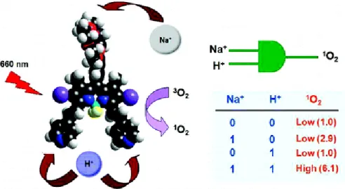
Photodynamic therapy(PDT) is a significant application area of BODIPY dyes which is a novel alternative treatment way for cancer. In PDT, e photosensitizer and a near-IR light source is used. These cause the production of singlet oxygen from molecular oxygen and since singlet oxygen is fatal to cancer tissue, the treatment is achieved. Since the skin tolerates between 650-800 nm, wavelength of light used is in this region. There is a study of Akkaya et al. about this subject in the literature102. A PDT agent were synthesized which can produce singlet oxygen when treated with 660 nm light. The molecule could also detect aciditiy and concentration of Na+ inside the cell and is able to target the cancer tissue. The study is represented in figure 17:
Figure 17: Photodynamic therapy agent and AND logic gate by Akkaya et al. Copyright © 2009 American Chemical Society. Adapted with permission
Light scattering is reduced at longer wavelengths; therefore fluorophores emitting beyond 650 nm are good agents for biological sensing. Two examples of
red-22
emitting BODIPY dyes developped by Akkaya et al. are given in the following figure, namely bis(2-pyridyl)-substituted boratriazaindacene (on the left)103 and a fluorescent chemosensor for anions (on the right)104.
Figure 18: Examples of red-emitting BODIPY dyes
Protein labeling was one of the first applications of BODIPY dyes due to high quantum yields and photostabilities105.
Figure 19 shows two examples from the literature. First one was published in
Org.Biomol.Chem106 (on the left), the other was published in
Angew.Chem.Int.Ed.107 (on the right) which are BODIPY dyes for protein labeling.
23
Figure 19: Literature examples of BODIPY dyes for protein labeling
1.4
Fluorescence Phenomenon
Electrons move to a higher electronic level (Sn, n>0) which is also known as
‘’excited state’’ from their ground state (S0) when molecules are irradiated with
light. The excited electron returns to its ground state via some mechanisms. The Perrin-Jablonski Diagram ( Figure ) is useful for visualizing possibilities of the processes that can occur after excitation. S0 is the lowest electronic state, S1, S2,..
are singlet electronic states. T1, T2.. are triplet excited states. Lateral lines
denoted as thinner than those standing for electronic states are vibrational levels. The excited electron firstly goes to S1 (the lowest electronic state) by scattering
some of its energy which is called as ‘’internal conversion’’. For movement to S0
from S1, there are some possibilities108. Turning back to ground state with
emission of photon is ‘’fluorescence’’ which is the most significant of all the mechanisms. Non-radiative dissipation of energy as heat is another way. Collisional quenching is another case in which excited molecules transfer their energy to nearby moelcules. Transition from a singlet state to a triplet state T1 is
the last possibility called as ‘’internal conversion’’. The electron is then de-excited to the ground state from the de-excited triplet state which is namely ‘’phosphorescence’’ 108
24
Figure 20: Jablonski diagram
As it is seen on the Jablonski Diagram, emitted light has lower energy than the absorbed light and hence it has higher wavelength than that of the absorbed light. In other words, the emission spectrum of the fluorophore is red-shifted to a higher wavelength. Fluorophore is a chromophore emitting light.
25
The red-shift which was firstly observed in 1852 by Sir George Stokes is known as ‘’Stokes’ shift’’ 109
. The main reason for Stokes’ shift is the rapid decay of electron to the lowest vibrational level of S1. Solvent effects, complex formation,
reactions at the excited state and energy transfer are the other causes of Stokes’ shift110.
1.4.1 Photoinduced Electron Transfer (PeT)
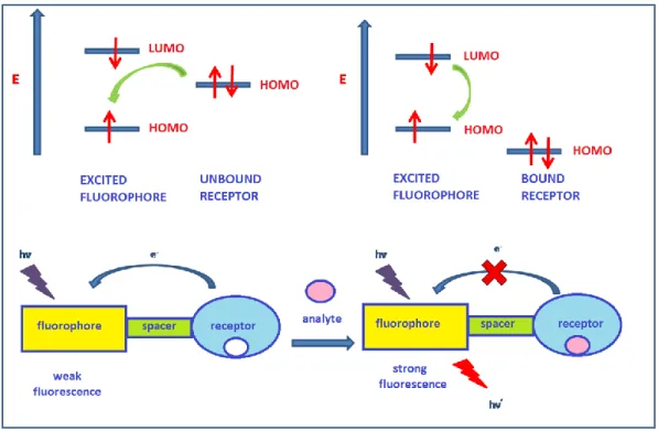
Photoinduced electron transfer (PeT) is a signalling case in which emission is either quenched or enhanced, encountered in fluorophore-spacer-receptor systems111. In figure 22, mechanism of PeT is given. The highest occupied molecular orbital (HOMO) of the receptor is between the the lowest unoccupied molecular orbital (LUMO) and the highest occupied molecular orbital (HOMO) of the fluorophore in the figure. The receptor also contains an electron rich group. One of the electrons of the fluorophore is in the excited state due to irradiation of light. An electron from HOMO of the receptor transfers to the HOMO of the fluorophore and pairs with the single electron in HOMO of it. This situation prevents returning of the excited electron of flurophore from LUMO to HOMO and as a result, the fluorescence is ‘quenched’. On the other hand, if an analyte is present in the medium to stabilize the receptor, the energy level of its HOMO is lowered and it can not transfer its electron to HOMO of the fluorophore. Consequently, strong fluorescence is observed.
26
Figure 22: Reprentation of PeT working principle
27
There also exists reverse PeT (oxidative PeT) mechanism. This event occurs if the receptor includes electron withdrawing group112. If there is no analyte in the medium, the electron in LUMO returns to HOMO and strong fluorescence is observed. However, in the presence of an analyte binding, HOMO of the receptor decreases and the electron in LUMO of the fluorophore is transferred to LUMO of the receptor and the result is weak fluorescence. In other words, fluorescence is quenched as a result of the process known as ‘’reverse PeT’’ or ‘’oxidative PeT’’ which is summarized in the figure 23.
1.4.2 Internal Charge Transfer (ICT)
In this phenomenon, there exist a fluorophore and a receptor without a spacer between them. The fluorophore has an acceptor part which is electron poor and a donor part which is electron rich. Excitation of such a fluorophore ends up with electron redistribution and creation of a dipole which result in electron donation from donor to acceptor112.
The receptor, existing as a part of the Π-electron system of the fluorophore can be either an electron donating or electron accepting group. In the first case, consider an electron donating receptor such as an amino acid. When a cation is added to the medium, the excited state will be more destabilized as compared to the ground state and this will cause an increase in the energy gap between the excited state and the ground state. This scenerio means that the electron donating ability of the receptor decreases as a cation is present in the environment and the overall result is a ‘’blue shift’’ in the absorbance spectrum. In the second case, the receptor is an electron acceptor such as a carbonyl and the presence of a cation will enhance the electron withdrawing capacity of it. The excited state will be more stabilized as compared to the ground state and energygap will decrease causing a ‘’red shift’’ in the absorbance spectrum. The IcT phenomenon is explained in figure 24.
28
Figure 24: Schematic representation of ICT working principle
29
CHAPTER 2
EXPERIMENTAL
2.1 General
Chemical compounds and solvents were obtained from Sigma-Aldrich and were used without further purification. Thin layer chromatography to monitor reactions were performed by using Merck TLC Silica gel 60 F254. In addition, Merck Silica
gel 60 (particle size: 0.040-0.063 mm, 230-400 mesh ASTM) was used as the silica gel for column chromotography.
1
H NMR and 13C NMR spectra were recorded on the instrument Bruker DPX-400 (operating at 400 MHz for 1H NMR and 100 MHz for 13C-NMR) at room temperature in CDCl3 and DMSO-d6 with tetramethylsilane (TMS) as internal
standard. Coupling constants (J values) are reported in Hz and chemical shifts are given in parts per million (ppm). Splitting patterns are designated as s(singlet), d (doublet), t(triplet), q(quartet), m(multiplet), and p(pentet).
Agilent Technologies 6530 Accurate-Mass Q-TOF LC/MS instrument was used to collect mass spectra data at Bilkent University, UNAM. A Varian Cary-100 spectrophotometer was used to record absorption spectra. Furthermore, to obtain emission spectra, a Varian Eclipse Spectrofluoremeter instrument was used at Bilkent University, UNAM.
30
2.2 Synthesis Scheme
31
2.3 Syntheses
Three nitroolefin functionalized BODIPY dyes have been synthesized as potential protein labeling agents. Compound 1113 and compound 8114 were synthesized according to the literature.
2.3.1. Synthesis of Compound 2
4-(hydroxymethyl) benzaldehyde (0.5 g, 3.68mmol, 1 equiv.) was dissolved in 400 mL Ar-degassed DCM in a 1 L round bottom flask. 2,4-dimethylpyrrole (0.83 mL, 769.5 mg, 2.2 equiv.) was added. This was followed by the addition of 1-2 drops of TFA. The mixture was stirred about 3 hr at room temperature. After TLC showed no starting material, p-chloroanil (903.93 mg, 1.1 equiv.) was poured into the reaction vessel. After 1 hr stirring, 6 mL TEA was added dropwise to the solution over a period of 5 min. The color turned out to be brown and it was stirred for an additional 30 min. Lastly, 6 mL BF3.OEt2 was added to the reaction
in a dropwise manner over a period of 5 min, as well. The mixture was left to stir overnight at room temperature. Extraction was made with water (3x300 mL) and the organic layer was dried over anhydrous Na2SO4. After concentrating the
organic layer in vacuo, it was purified by flash column chromotography with the eluant DCM. The product was obtained as red solid (380 mg, 29.2% yield).
1 H NMR (400 MHz, CDCl3) δ 7.51 (d, J = 7.8 Hz, 2H), 7.38 (d, 2H), 6.04 (s, J = 31.5 Hz, 2H), 4.83 (s, 2H), 2.60 (s, J = 17.9 Hz, 6H), 1.46 (s, J = 51.7 Hz, 6H). 13C NMR (100 MHz, CDCl 3) δC: 155.49, 143.08, 141.87, 141.54, 134.20, 128.18, 127.38, 121.23, 64.78, 14.47.
MS (TOF-ESI): m/z: Calcd:353.17 [M-H]-, Found:353.1673 [M-H]-, ∆=-7.64 ppm.
32
1 2
Figure 26: Synthesis of compound 2
2.3.2 Synthesis of Compound 3
Compound 2 (150 mg, 0.42 mmol) was dissolved in a minimum amount of Ar-degassed DCM. Dess-Martin periodinane (359.2 mg, 0.84 mmol) was also dissolved in min. amount of Ar-degassed DCM and this was added dropwise to the previous one at 0oC. When the addition is completed, the reaction left to stir at room temperature for about 2 hrs. When TLC showed no starting material, the mixture was quenched with 20 mL sat’d Na2S2O3 solution. The organic layer was
then washed with sat’d NaHCO3 solution (2x20 mL). It was lastly washed with
water (2x20 mL) and dried over anhydrous Na2SO4. Flash column
chromotography was performed to purify the organic layer by using 95:5/ DCM:MeOH as the eluant. The product was obtained as orange solid (90 mg, 60% yield). 1 H NMR (400 MHz, CDCl3) δH: 10.14 (s, J = 4.6 Hz, 1H), 8.04 (d, J = 6.2 Hz, 2H), 7.53 (d, J = 8.0 Hz, 2H), 6.019 (s, 2H), 2.58 (s, 6H), 1.37 (s, 6H). 13C NMR (100 MHz, CDCl 3) δC:191.43, 156.25, 142.75, 141.40, 139.68, 136.68, 130.32, 129.15, 121.63, 121.60, 14.61, 14.50.
MS (TOF-ESI): m/z: Calcd: 351.16 [M-H]-, Found:351.1548 [M-H]-, ∆=-14.8 ppm.
33
2 3
Figure 27: Synthesis of compound 3
2.3.3. Synthesis of Compound 4
Compound 3 (80 mg, 0.23 mmol) was dissolved in 4 mL nitromethane. Oil bath was stabilized at 90oC. Solution was placed in oil bath and waited for 30 min. After that, a pinch of NH4OAc was added to the reaction vessel. The reaction was
completed as monitored by TLC after 24 hr stirring. Nitromethane was evaporated
in vacuo and flash column chromotography was performed to purify the product
by using 95:5/DCM:MeOH as the eluant. The nitroolefin functionalized protein labeling agent was obtained as red crystals ( 45 mg, 49.5% yield).
1 H NMR (400 MHz, d6-DMSO) δH: 8.35(d, J=18 Hz, 1H), 8.25 (d, J=14 Hz, 1H), 8.1(d, J=8 Hz, 2H), 7.6(d, J=8 Hz, 2H), 6.2(s, 2H), 2.45(s, 6H), 1.4(s,6H) 13C NMR (100 MHz, d6-DMSO) δ C: 156.23, 142.69, 139.66, 139.04, 138.02, 137.86, 130.95, 130.83, 129.73, 129.45, 121.60, 14.62, 14.57.
34
3 4
Figure 28: Synthesis of compound 4
2.3.4. Synthesis of Compound 9
3,4,5-tris(2-(2-(2-methoxyethoxy)ethoxy)benzaldehyde (500 mg, 0.85mmol) was mixed with 2,4-dimethylpyrrole (176 mg, 1.85 mmol) in 350 ml argon degassed dichloromethane (DCM). 1-2 drops of TFA was added followingly. The mixture was stirred at room temperature for about 3 hours. When TLC showed no starting material, p-chloranil (229.8 mg, 0.93 mmol) was added to the reaction and it was stirred for an additional hour. Then, 7 ml Et3N was added to the mixture dropwise
over a period of 5 min and the resulting brown solution was stirred for 30 min. After that, 7 ml BF3•OEt2 was added dropwise over another period of 5 min to
the reaction and the resulting mixture was allowed to stir overnight. The reaction mixture was washed with water (3×300 mL) and extracted into DCM. The organic layer was dried over anhydrous Na2SO4, concentrated in vacuo and the crude
product was purified by flash column chromotography packed with silica gel by using 95:5/ DCM:MeOH as the eluant. The product was obtained as a waxy dark orange solid (370 mg, 54% yield).
1H NMR (400 MHz, CDCl 3) δH: 6.55 (s, 2H), 5.99 (s, 2H), 4.22 (t, J= 4.97 Hz, 2H), 4.12 (t, J= 4.72 Hz, 4H), 3.84 (t, J= 5.29 Hz, 6H), 3.51-3.76 (m, 25H), 3.38 (s, 2H), 3.36 (s, 6H), 2.54 (s, 6H), 1.52 (s, 6H). 13C NMR (100 MHz, CDCl 3) δC: 155.8, 153.7, 142.9, 141.1, 139.0, 131.1, 129.9,
35
121.1, 107.7, 2.7, 71.9, 71.9, 70.8, 70.7, 70.6, 70.5, 69.7, 69.2, 58.8, 29.6 14.4, 14.2.
MS (TOF-ESI): m/z: Calcd: 809.4286 [M-H]-, Found: 809.4306 [M-H]-, ∆= 2.47 ppm.
9
Figure 29: Synthesis of compound 9
2.3.5. Synthesis of Compound 10
0.5 mL POCl3 was added into anhydrous DMF (0.5 mL) dropwise in ice bath.
Addition was done under N2. This mixture (pale yellow viscous liquid) was left to
stir for 30 min at room temperature. Compound 9 (405.215mg, 0.5 mmol) was dissolved in 25 mL DCE and then slowly introduced to the previous mixture. The resulting brown solution was stirred at 60oC for 3 hr. 60oC is the critical temperature to get the mono-formylated product 10, side products could be obtained otherwise at higher temperatures. Then the reaction was cooled to room temperature and poured into ice-cold saturated NaHCO3 solution. Extraction was
performed with DCM (2x50 mL) and the organic layer was dried over Na2SO4.
The organic layer was concentrated in vacuo and purified by flash column chromotography with 98:2/DCM:MeOH as the eluant. Target compound 10 was obtained as orange crytals(185 mg, 93% yield).
36 1H NMR (400 MHz, CDCl 3) δH: 9.98 (s,1H), 6.53 (s, 2H), 6.15 (s, 1H), 4.23 (d, J= 4.34 Hz, 2H), 4.12 (d, J= 4.29 Hz, 4H), 3.83 ( d, J= 4.81 Hz, 6H), 3.75-3.49 (m, 25H), 3.37 (s, 2H), 3.34 (s, 6H), 2.80 (s, 3H), 2.59 (s, 3H), 1.78 (s, 3H), 1.57 (s, 3H). 13C NMR (100 MHz, CDCl 3) δC: 185.81, 161.64, 156.47, 154.02, 147.17, 143.07, 142.68, 139.54, 133.98, 129.65, 128.89, 126.25, 123.92, 107.04, 72.76, 71.93, 71.88, 70.84, 70.68, 70.62, 70.55, 70.53, 69.69, 69.32, 58.96, 14.76, 11.55.
MS (TOF-ESI): m/z: Calcd: 837.4235 [M-H]-, Found: 837.4094 [M-H]-, ∆=-16,83ppm
9 10
Figure 30: Synthesis of compound 10
2.3.6. Synthesis of Compound 11
Compound 10 (140 mg, 0.17 mmol) was dissolved in 2 mL nitromethane. This solution was stabilized at 90 oC in oil bath in 30 min. Then NH4OAc (1.28 mg,
0.017 mmol) was added to the mixture. The reaction was stirred for 5 days. When TLC showed no starting material, nitromethane was removed in vacuo. Flash column chromotography packed with silica gel was used to purify the crude product by using 95:5/ DCM:MeOH as the eluant. Compound 11 was obtained as a wine-red waxy solid (43 mg, 28.6% yield).
37 1H NMR (400 MHz, CDCl 3) δH: 8.05 (d, J= 13.8 Hz, 1H), 7.37 (d, J= 13.63 Hz, 1H9, 6.56 (s, 2H), 6.18 (s, 1H), 4.26 (t, J= 4.64 Hz, 2H), 4.14 (t, J= 4.52 Hz, 4H), 3.86 (t, J=5.15Hz, 6H), 3.52- 3.76 (m, 25H), 3.40 (s, 2H), 3.37 (s, 6H), 2.72 (s, 3H), 2.63 (s, 3H), 1.64 (s, 3H), 1.59 (s, 3H). 13C NMR (100 MHz, CDCl 3) δC: 161.54, 154.23, 154.03, 147.18, 147.22, 140.16, 139.65, 134.60, 133.71, 130.71, 130.62, 128.89, 128.84, 123.93, 120.03, 107.41, 72.81,71.96, 71.90, 70.86, 70.71, 70.68, 70.58, 70.56, 69.71, 69.36, 65.30, 59.01, 29.67, 14.72, 12.85,11.09.
MS (TOF-ESI): m/z: Calcd: 880.7612 [M-H]-, Found: 880.4134 [M-H]-, ∆= -2.09 ppm.
10 11
Figure 31: Synthesis of compound 11
2.3.7. Synthesis of Compound 12
3,4,5-tris(2-(2-(2-methoxyethoxy)ethoxy)benzaldehyde (1500 mg, 2,55mmol) was dissolved in 450 ml argon degassed dichloromethane (DCM). 3-ethyl-2,4-dimethyl-pyrrole (528 mg, 5.61 mmol) was added. 1 drop of TFA was added to mixture followingly. It was stirred for about 3 hr. Reaction was monitored by TLC and when it showed no starting material, p-chloranil (698,4 mg, 2.79 mmol) was added in one portion. After an additional 1 hr stirring, 8 ml Et3N was added to
38
stirred for 30 min. Thereafter, 8 ml BF3•OEt2 was added dropwise over another
period of 5 min to the reaction and the resulting mixture was left to stir overnight at room temperature. The resultant mixture was washed with water (3×400 mL) and extracted into DCM. The organic layer was dried over anhydrous Na2SO4,
concentrated in vacuo and the crude product was purified by flash column chromotography packed with silica gel by using 95:5/ DCM:MeOH as the eluant. The product was obtained as a waxy dark orange solid (1034 mg , 46.7% yield).
1 H NMR (400 MHz, CDCl3) δH: 6.55 (s, J = 1.2 Hz, 2H), 4.24 (t, J = 7.4 Hz, 2H), 4.12 (t, J = 4.5 Hz, 4H), 3.85 (t, J = 4.7 Hz, 6H), 3.78 – 3.47 (m, 24H), 3.40 – 3.31 (m, 8H), 2.53 (s, 6H), 2.32 (q, J = 7.4 Hz, 4H), 1.42 (s, 6H), 1.00 (t, J = 7.35 Hz, 6H). 13C NMR (100 MHz, CDCl 3) δC: 153.79, 153.61, 139.66, 136.25, 132.74, 132.72, 72.70, 71.91, 70.86, 70.71, 70.64, 70.55, 69.73, 69.18, 59.01, 58.98, 17.06, 14.59, 12.47, 11.61
MS (TOF-ESI): m/z: Calcd:865.49 [M-H]-, Found: 865.4904 [M-H]-, ∆= -4.05 ppm.
12 Figure 32: Synthesis of compound 12
2.3.8. Synthesis of Compound 13
2,8-diethyl-5,5-difluoro-1,3,7,9-tetramethyl-10-(3,4,5-tris(2-(2-(2-
methoxyethoxy)ethoxy)ethoxy)phenyl)-5H-dipyrrolo[1,2-c:2',1'-f][1,3,2]diazaborinin-4-ium-5-uide (Compound 12) (400 mg, 0.46 mmol) was dissolved in 18 mL, Ar-degassed THF in 100 mL round bottom flask. DDQ(420
39
mg, 0.54 mmol) was dissolved in 6 mL, Ar-degassed THF and 0.16 mL distilled water was added. This mixture was added dropwise to the previous one at 0oC under Ar. When the addition is completed, the reaction was left to stir for 3 days. When TLC showed no starting material, the resultant mixture was purified by flash column chromotography filled with silica gel. 95:5/ EtOAc:MeOH was used as eluant. Product was obtained as red solid(350 mg, 86% yield).
1 H NMR (400 MHz, CDCl3) δH: 10.38 (s, 1H), 6.56 (s, 2H), 4.26 (s, 2H), 4.14 (s, 4H), 3.80-3.51 (m, 30H), 3.44 – 3.30 (m, 9H), 2.72 (s, 2H), 2.61(s, 3H), 2.36 (s,2H), 1.55 (s,3H), 1.41 (s, 3H), 1.05 (s, 6H). 13 C NMR (100 MHz, CDCl3) δC: 213.74, 185.97, 166.16, 153.90, 143.72, 141.50, 139.87, 139.23, 137.71, 137.03, 135.60, 134.33, 132.05, 129.58, 107.47, 77.37, 77.05, 76.73, 72.73, 71.90, 71.86, 70.80, 70.61, 70.59, 70.50, 70.46, 69.66, 69.23, 59.02, 59.01, 58.98, 17.59, 17.12, 14.34, 14.02, 13.68, 12.26, 10.44.
MS (TOF-ESI): m/z: Calcd: 879.47 [M-H]-, Found:879.468 [M-H]-, ∆= -2.52 ppm.
12 13
Figure 33: Synthesis of compound 13
2.3.9 Synthesis of Compound 14
2,8-diethyl-5,5-difluoro-3-formyl-1,7,9-trimethyl-10-(3,4,5-tris(2-(2-(2-
methoxyethoxy)ethoxy)ethoxy)phenyl)-5H-dipyrrolo[1,2-c:2',1'-40
f][1,3,2]diazaborinin-4-ium-5-uide (Compound 13) (340 mg, 0.39 mmol) was dissolved in 5 ml nitromethane. It was placed in an oil bath at 90oC. A pinch of NH4OAc was added. The reaction was stirred at this temperature for 3 hr. Product
was obtained as violet-purple solid (90 mg, 25.2% yield).
1 H NMR (400 MHz, CDCl3) δH: 8.36 (d, J = 13.7 Hz, 1H), 7.84 (d, J = 13.8 Hz, 1H), 6.55 (s, 2H), 4.29 – 4.17 (m, 2H), 4.17 – 4.01 (m, 4H), 3.90 – 3.82 (m, 6H), 3.82 – 3.33 (m, 34H), 2.72 – 2.32 (m, 7H), 2.08 – 1.68 (m, 1H), 1.50 (s, 3H), 1.46 (d, J = 26.5 Hz, 6H), 1.07 (m, J = 19.8, 7.5 Hz, 6H). 13 C NMR (100 MHz, CDCl3) δC: 165.16, 153.95, 143.06, 140.18, 139.36, 137.86, 137.81, 137.76, 137.51, 137.47, 136.35, 135.43, 135.09, 133.94, 129.50, 127.51, 107.56, 77.37, 77.06, 76.74, 72.76, 71.95, 71.89, 70.85, 70.70, 70.65, 70.56, 69.70, 69.27, 59.03, 59.00.
MS (TOF-ESI): m/z: Calcd: 922.48 [M-Na]+, Found: 946.47 [M-Na]+, ∆= -8.73 ppm.
13 14