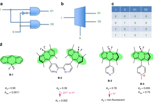
Molecular demultiplexer as a terminator
automaton
Ilke S. Turan
1
, Gurcan Gunaydin
2
, Seylan Ayan
3
& Engin U. Akkaya
1,3
Molecular logic gates are expected to play an important role on the way to information
processing therapeutic agents, especially considering the wide variety of physical and
che-mical responses that they can elicit in response to the inputs applied. Here, we show that a
1:2 demultiplexer based on a Zn
2+-terpyridine-Bodipy conjugate with a quenched
fluorescent
emission, is efficient in photosensitized singlet oxygen generation as inferred from trap
compound experiments and cell culture data. However, once the singlet oxygen generated by
photosensitization triggers apoptotic response, the Zn
2+complex then interacts with the
exposed phosphatidylserine lipids in the external lea
flet of the membrane bilayer,
autono-mously switching off singlet oxygen generation, and simultaneously switching on a bright
emission response. This is the con
firmatory signal of the cancer cell death by the action of
molecular automaton and the con
finement of unintended damage by excessive singlet
oxy-gen production.
DOI: 10.1038/s41467-018-03259-z
OPEN
1UNAM-National Nanotechnology Research Center, Bilkent University, 06800 Ankara, Turkey.2Department of Basic Oncology, Hacettepe University,
06100 Ankara, Turkey.3Department of Chemistry, Bilkent University, 06800 Ankara, Turkey. Ilke S. Turan and Gurcan Gunaydin contributed equally to this
work. Correspondence and requests for materials should be addressed to E.U.A. (email:eua@fen.bilkent.edu.tr)
123456789
M
ore than two decades after the seminal work by de Silva
et al.
1, molecule-based logic gates have reached a level
of considerable sophistication
2–4. Many examples of
basic Boolean logic gates and various implementations of both
combinatorial and sequential logic were reported
5–10. While it is
clear that more advanced digital designs may require novel
integration mechanisms of chemical logic gates, functional
equivalents of more complex information processing is still
possible with simple molecules or molecular assemblies as a result
of their unique characteristics. However, despite this impressive
progress, the power of chemical and molecular logic gates beyond
the exploratory phase is yet to be convincingly demonstrated,
11with what is colloquially referred to as a killer app
12.
The most likely
field where molecule-based information
pro-cessing agents would
find a niche for true utility is therapeutic
medicine. We previously described various protocols to combine
molecular logic gate notions with photosensitized generation of
short-lived cytotoxic species, singlet oxygen
3,13,14. Photosensitized
generation of singlet oxygen in tumors, in or around cancer cells,
is the essence of photodynamic therapy of cancer
15–18. The fate of
the singlet excited state (S
1) of the photosensitizer is strongly
related to the relative efficiencies and rates of photophysical
processes involved. Among these, the most relevant are radiative
transition from S
1to S
0(fluorescence) and intersystem crossing to
T
1triplet state (isc). Efficiency of access to the triplet manifold is
directly linked to the singlet oxygen quantum yield. Thus,
con-sidering the fact that in the photosensitization process,
fluores-cence and intersystem crossing (hence, singlet oxygen generation)
are mutually antagonistic, and at least in principle, it should be
possible to switch between these two processes. The digital
equivalent is that of a demultiplexer (DEMUX) circuit
19, which
takes single data input, and uses n select/address inputs to switch
between 2
npossible outputs (Fig.
1
).
Chemical implementation of this idea relies on the
photo-physics of the meso-pyridyl (or oligopyridyl) substituted Bodipy
dyes (Fig.
1
d). Bodipy dyes are exceptionally versatile and
che-mically malleable chromophores. The reference Bodipy
chro-mophore (B-1) has high
fluorescence quantum yield with a small
Stokes’ shift and, on excitation, would have negligible access to
the triplet manifold. This is also in part the reason for higher
chemical stability of this class of dyes under ambient conditions.
In our earlier investigations
20aiming cation-responsive
fluor-ophores (chemosensors), we observed a sharp decrease in the
emission of bipyridyl-Bodipy compound (B-2) when an acid
(trifluoroacetic acid (TFA)) or zinc perchlorate was added.
Apparently, protonation of the ligand, or complexation with a
+2
charged cation, transforms the ligand into a more easily reducible
species, and photoinduced electron transfer from the excited
Bodipy dye to the bipyridyl ligand becomes more efficient.
Resulting charge transfer state (CTS) is responsible for the overall
non-radiative relaxation (hence quenching). The orthogonal
arrangement of the ligand and the chromophore is expected to
facilitate intersystem crossing to the triplet state, as the charge
recombination is typically accompanied with the population of
the triplet excited state
21. Considering recent data on the
pho-tophysics of the similar Bodipy-pyridyl ligand systems
22, we
concluded that such protonated pyridinium, or quaternized
pyridinium substituted Bodipy dyes, and the Zn
2+complexes are
likely to be more efficient generators of singlet oxygen compared
to the parent compounds from which they are derived due to
enhanced yield of the triplet state. Another useful aspect of this
change is that it is chemically reversible; i.e, if the protonation is
reversed, or charge on the Zn
2+ion is neutralized (even
par-tially), a return to the original state of affairs should be expected.
Thus, the addition of phosphate to the B-2/Zn
2+complex
destabilizes the CT state and restores the
fluorescence emission
intensity.
a
b
c
d
I S O1 O2 I S O1 O2 I S O1 O2 0 0 0 0 0 1 0 0 1 0 1 0 1 1 0 1 N B N F F N B N F F N B N F F N N N B N F F N N B N F F N+ B-1 B-2 B-3 B-4 + Zn2+ or H+ + H+ fl = 0.90 isc = 0.0011 fl = 0.39 fl < 0.002 fl = 0.78 fl = 0.005 isc = 0.75 fl = non-fluorescentFig. 1 DEMUX combinatorial circuit and structures of relevant Bodipy compounds. a DEMUX circuit with standard logic gate symbols. b Isosceles trapezoid is a common symbol for the MUX/DEMUX circuits. I is the data input, S is the select or address input, and O1 and O2 are two different outputs. c The truth table for a 1:2 DEMUX circuit is on the right.d Structures of the parent Bodipy compound B-131, and meso-pyridyl22/bipyridyl20substituted derivatives, together with their respectivefluorescence and intersystem crossing quantum efficiencies are demonstrated. Protonation of both B-2 and B-3 result in the pyridinium cation, which switches on the intramolecular charge transfer process. Coordination of Zn2+ions or quaternization at the pyridine nitrogen (B-4) results in the same photophysical consequences
In a demultiplexer, the select input would determine the choice
between the possible outputs. Searching for a candidate as the
select input, we looked into the possibility of making use of
structural changes taking place in the cell membranes during
apoptosis. Fluorescence imaging of apoptosis relies on the loss of
membrane asymmetry (adenosine triphosphate (ATP)-dependent
enzyme called
flippase normally keeps phosphatidylserine inside
the cell)
23,24as the negatively charged lipids and particularly
phosphatidylserine is
flipped to the extracellular side of the
bilayer. The change is mediated by enzymes such as scramblase,
which exposes phosphatidylserine on the cell’s surface without
consuming ATP
23,25. A selective probe for this event is the
pro-tein Annexin V, which selectively binds to phosphatidylserine.
Since Annexin V can be conveniently labeled with
fluorescent
dyes of different emission colors, it provides a set of useful tools
for detecting apoptotic cells. There is also an interest in
finding
simpler, non-protein reporters of apoptosis, and in most of the
reported examples
26, the part which interacts with the
phos-phatidylserine unit is a Zn
2+complex, in which the metal ion is
held in place by pyridine-derived ligands, presenting coordination
sites to the anionic groups of the phosphatidylserine
27.
In this work, we present a unique molecular device based on
above considerations. The molecular automaton described here
initially generates singlet oxygen to trigger apoptosis in cancer
cells, and then, in response to apoptotic changes in the membrane
structure, shuts off singlet oxygen generation and produces
emission as a result of its interaction with the exposed
phos-phatidylserines in the outer leaflet of the apoptotic cell
mem-branes. This could be interpreted as the response of a molecular
demultiplexer which takes light as an input and
phosphati-dylserines as the switch. The two alternative outputs are singlet
oxygen and light.
Results
Operation of the automaton. The structures of the targeted
compounds T-1 and T-2 for synthesis are shown in Fig.
2
a.
Terpyridyl-Bodipy compounds T-1 and T-2 differ only in the
groups attached to the boron bridge. While compound T-2 is
appropriate for chemical characterization, compound T-1 is
better for biological media due to additional solubilizing
oli-goethyleneglycol units. The use of terpyridine instead of
bipyridine ligand is favored due to the stronger affinity of the
former ligand to Zn
2+ions in aqueous media. The changes in
emission were studied in acetonitrile. A 2.0
μM solution of the
model DEMUX T-2 has an intense emission band (ϕ
F= 0.34)
with a maximum at 517 nm. Titration with Zn
2+ions in the
form of perchlorate salt results in sharp loss of emission
intensity, with emission band moving to longer wavelengths, as
the CT component in the emission becomes more prominent.
However, the addition of tetrabutylammonium phosphate
completely reverses this change at 60
μM concentration in
acetonitrile. We then studied singlet oxygen generation rates
under the same conditions for
+/– phosphate: the singlet
oxygen trap used is 1,3-diphenylisobenzofuran, its absorbance
at 410 nm decreases due to a [4+2] cycloaddition with singlet
oxygen, which is followed by decomposition. In accordance
with our design expectations, when irradiated with a
λ = 522
nm light-emitting diode array (at 98.0 µmol m
−2s
−1photon
flux), the Zinc
2+complex is quite an effective photosensitizer.
The singlet oxygen quantum yield (ϕ
Δ) was determined in
reference to Eosin Y, and found to be 0.11. However, the
addition of 2.0 mM phosphate in the form of
tetrabutylammo-nium phosphate essentially stops singlet oxygen generation
(Fig.
3
f). Thus, it is clear that
fluorescence emission intensity
and the singlet oxygen generation efficiency are inversely
coupled, and the switch is the phosphate ions in solution.
Operation of the molecular DEMUX circuit based on
compound T-2 was confirmed. We also determined the singlet
oxygen quantum yield of T-1 as 0.10.
Once the chemical validation was obtained, we ventured into
cell culture experiments. Cell culture assays were performed
with a human cancer suspension cell line-chronic myelogenous
a
b
S1 (2.29 eV) Ground state CTS’ +Phosphate or phosphatidylidylserine CTS (2.17 eV) T1 (1.58 eV) 3O 2 1O 2 L N N N N B B F F T-2 O O O O O O O O T-1 N N N N N N L Zn2+ Zn2+ L LFig. 2 The structures of the molecular automata T-1 and T-2 and the Jablonski diagram depicting processes involved. a L indicates solvent molecules as ligands, water, or acetonitrile. Bodipy and the terpyridyl planes are orthogonal to each other due to the presence of 1,7-dimethyl substitution of the Bodipy chromophore. Double mTEG (methoxytriethyleneglycol) substitution at the boron center enhances water solubility.b Thicker green arrow is absorption, the other green arrows indicate radiative relaxations. Black-dashed arrows are non-radiative relaxation processes. Blue arrows indicate transitions between various excited states. CTS to T1transition is particularly enhanced because of the orthogonal geometry of the terpyridyl-Bodipy diad. Energy levels were experimentally estimated using the spectral data (S1, CTS) in analogy to previous literature22, or based on the phosphorescence data32for similar Bodipy
leukemia (K562). The cells were incubated with Dulbecco’s
modified Eagle's medium (DMEM) supplemented with 20%
fetal bovine serum at the environmental conditions of 37° C, 5%
CO
2, and 60% humidity. Cells were treated with varying
concentrations of the molecular automaton T-1 (187.5 nM–1.5
µM) and illuminated with a green light source (λ = 522 nm
light-emitting diode array, 98.0 µmol m
−2s
−1photon
flux) for
a continuous duration of 12 h. This 12 h period of illumination
was followed by 12 h of incubation in the dark (total 24 h). The
control group of the cells were incubated in the dark, for the
exact duration of 24 h under identical environmental
condi-tions. The MTT (3-(4,
5-dimethylthiazolyl-2)−2,5-diphenylte-trazolium bromide) assay was used in order to assess cell
viability and cytotoxicity. Even the low doses of the automaton
T-1
seem to have resulted in a significant decrease of the cell
viability (Fig.
3
a, points and error bars designate means and
a
b
c
PS 0 0 0 0 0 1 0 0 1 0 1 0 1 1 0 1Input Switch Out1 Out2
h @520 nm Input h (exc) h (em) 1O 2 Switch (PS=0) 1O 2 100
f
100 T2+Zn T2+Zn+P Eosin Y L L N N FBF On 1O2 1O2 Off N N FBF N N N O O O Zn2+ Zn2+ O P – N N N 80 Normalized absorbance @411 nm (%) 60 40 20 0 0 500 1000 1500 Secondse
d
1000 800 h Off h 600 400 200 0 500 550 600 0 µM 20 µM 650 Wavelength (nm) Emission intensity 1000 800 On 600 400 200 0 500 550 600 0 µM 60 µM HO P O –O –O 650 Wavelength (nm) Emission intensity 80 Cell death (%) 60 40 20 0 0 250 500 750 1000 T-1 concentration (nM) 1250 1500 Zn2+Fig. 3 Operation of the molecular automata as evidenced by spectroscopic and cell culture data. a MTT assay data: Green open circles correspond to percent death of K562 cells under irradiation at 522 nm for 12 h followed by continued incubation in dark for another 12 h. Solid black circles correspond to cells kept under identical conditions of incubation with the agent, but in dark. Positive control (dashes at 100% line) corresponds to cells incubated in DMSO-growth medium mixture (50/50, v/v). b The truth table with the data and switch inputs, and the corresponding outputs clearly identified, and the particular set of conditions valid under the light irradiation conditions on power-up were highlighted. c Switch input 0 (no added phosphate in the model system or lack of phosphatidylserine (PS) in the external leaflet of the cell membrane in the cell cultures) selects singlet oxygen as the primary output.d The model compound T-2, which is the Zn2+complex of the meso-terpyridyl-bodipy compound, has a very lowfluorescence emission intensity in acetonitrile. e The addition of phosphate ions results in a very sharp increase in emission intensity. The low emission intensity is due to the availability of a charge transfer state (CTS) resulting in enhanced intersystem crossing, which in turn leads to efficient generation of singlet oxygen. f The decrease in the absorbance at 411 nm, in an oxygen saturated ethanol solution of selective singlet oxygen trap DPBF (50µM) in the presence of 2.0 µM T-2 and under irradiation with 522 nm green LED light source (solid blue squares). The singlet oxygen quantum yield (ϕΔ) ofT-2 is 0.11. The addition of phosphate (red open circles) destabilizes the CTS state, blocking access to the triplet manifold. The absorbance data presented is the net absorbance values obtained by subtracting any background decrease in the probe absorbance due to light alone
standard deviations, respectively). The CC
50(50% cytotoxic
concentration) value of the T-1 subjected to green light was
estimated by
fitting a model with non-linear regression
(approximately 365 nM; the CC
50value of the compound in
the dark condition cannot be estimated since its projection is
clearly out of the scale of the model
fit).
Flow cytometry and microscopy. In order to confirm the MTT
assay results, and the switch from singlet oxygen generation to the
emission/signaling mode; Annexin V detection protocol for
apoptosis was performed using
flow cytometry. The percentage of
fluorescent-labeled Annexin V (phycoerythrin (PE))-stained cells
was much higher in the illuminated cell population compared
with those incubated in the dark (73.7 vs. 18.0%; Fig.
4
a, blue
shaded areas represent the cells in the dark, green shaded areas
represent the irradiated cells). In addition, 71.7% of irradiated
K562 cells (Fig.
4
b) were positive for T-1 in contrast to 18.0% of
the cells incubated in the dark (Fig.
4
c), demonstrating that
Annexin V and the automaton targets the same kind of cell
membranes with outer leaflet enriched in phosphatidylserine,
which are undergoing apoptosis. Cancer cells, which are not
killed, or not undergoing apoptosis, are not marked by the agent,
but subjected to cytotoxic singlet oxygen attack, when the
auto-maton is powered up by irradiation. Photocytotoxicity was
fur-ther revealed in a 2-color analysis with PE-Annexin V in order to
specifically target and identify apoptotic cells in conjunction with
the T-1 (Fig.
4
d). Approximately 70% of the illuminated cells
were positive for both PE-Annexin V and T-1 and about 16% of
the cells were negative for both (area A++ and A−− in Fig.
4
d,
respectively), confirming that the terminator automaton turns on
the emission signal only when the cells are undergoing apoptotic
death process.
Microscopy. Confocal microscopy provided further evidence
corroborating the cytotoxicity results from MTT assays and
flow
cytometry, as well as the co-staining of PE-Annexin V and T-1 in
a
b
c
d
e
f
g
h
i
h (exc) PS 0 0 0 0 0 1 0 0 1 0 1 0 1 1 0 1Input Switch Out1 Out2
h @488 nm Input h @520 nm 100 80 60 Count Count 40 20 0 100 80 60 Count 40 20 0 60 40 20 0 100 103 102 101 100 A–– 100 101 102 103 PE-Annexin V A+– A++ A–+ T-1 100 101 102 103 T-1 100 101 102 103 T-1 T-1 (+) T-1 (+) 101 102 103 PE-Annexin V Annexin V (+) 1O 2 h (em) Switch (PS=1)
Fig. 4 T-1 signals apoptosis by switching to diagnostic mode. a PE-Annexin labels most of the cells incubated with T-1 under light irradiation (Annexin V (+) region of the green area). b Green channel: cells incubated with T-1 under irradiation. c Green channel: cells incubated with T-1 in the dark. d The 2D plot for both green and red channels:T-1 and PE-Annexin V (a specific apoptosis marker) stain the same kind of cells with a large (86%) agreement: 70% of the irradiated cells were co-stained with bothT-1 and PE-Annexin V, indicating only apoptotic cells arefluorescently labeled with T-1. e The truth table with input, the switch and the outputs clearly identified, and the particular set of conditions were highlighted. f Switch input (appearance of
phosphatidylserine in the external leaflet of the cell membrane) selects fluorescence emission as the primary output. g Cells treated with PE-Annexin V and theT-1, and kept in dark, show no signs of morphological change, and the cellular membranes are not stained with either one of the agents. h, i Cells were incubated withT-1, irradiated with the LED light source, then treated with PE-Annexin V. The two agents (T-1 and PE-Annexin V, green and red, respectively) label the same regions in the cells undergoing apoptosis. (scale bar, 10μm)
flow cytometry analyses. The data demonstrate that just like
PE-Annexin V, the automaton shows
fluorescence signal only when
attached to apoptotic cell membranes. Cellular membranes of the
illuminated cells incubated with T-1 were shown to be stained
with both Annexin V and T-1 (1.5 µM); in contrast to their
counterparts incubated in the dark, which were not stained by
either T-1 (1.5 µM) or PE-Annexin V. Cell membranes of the
illuminated cells in the presence of T-1 appear bright green when
excited at 488 nm and bright red when excited at 543 nm.
However, their counterparts, which had been incubated in the
dark, were negative for either green or red emission. Figure
4
g–i
shows the differential interference contrast (DIC) image, T-1,
Annexin V and merged images, respectively.
A graphical representation of the operation of the automaton
T-1
is shown in Fig.
5
.
Discussion
It appears that judiciously designed molecular logic devices can
carry out critical information processing in or around the cells.
Widely speculated nanorobots are not expected to be
miniatur-ized versions of their macroscopic counterparts, but as illustrated
here, most likely to be developed by careful control of
photo-physics and chemical reactivity at the individual molecule level,
resulting in molecular entities with intelligent responses and
actions. The zinc complex of the terpyridyl-bodipy, T-1, is
approximately 3 nm at the longest dimension, yet it is capable of
killing cancer cells when powered up by light, and then
autono-mously switching to a signaling mode in response to changing
membrane characteristics, thus confirming apoptosis by a strong
emission signal. As evidenced by the spectroscopic experiments
with T-2, and
flow cytometry data obtained with T-1, emission
intensity and the singlet oxygen production is inversely coupled,
and the fact that singlet oxygen generation is turned off once the
cells show signs of apoptosis is very valuable in containing
unintended damage by singlet oxygen. This clearly qualifies T-1
as a molecular terminator automaton, or an example of a
func-tional molecular robot. While most nano-sized robotics work is at
present focussed on mobility, it is interesting to note that mobility
is far from being the number one issue for a functional,
molecule-sized robot. Most infectious microorganisms, cells
fighting against
these agents, and drug molecules are efficiently carried passively
via the circulatory system to any part of the body. It is the
intelligent autonomous operation that provides the most crucial
challenge, and the terminator automaton T-1 demonstrates one
way of moving ahead to meet that challenge.
Methods
Cell culture and MTT assay. K562 human chronic myelogenous leukemia sus-pension cells were cultured in 25 cm2cultureflasks containing DMEM (Gibco, 11971-025) supplemented with 20% fetal bovine serum in a cell culture incubator at 37 °C, 5% CO2, and 60% humidity. The main functional goals of the current study are to study the cytotoxicity of T-1 under illumination and its staining ability of the apoptotic cells. Since phosphate in the growth media can interfere with experimental results, we used DMEM w/o phosphate supplemented with 20% fetal bovine serum that has been dialyzed extensively at 4 °C against isotonic saline (0.15 M NaCl) using dialysis tubing (Sigma-Aldrich, USA (D7884)), since the viability of the cells cultured in such conditions is known to be not hampered for periods even more than 24 h28. This cell culture medium was also utilized for
analyses withflow cytometry and confocal laser scanning microscopy to study cytotoxicity and cellular staining.
The compound was diluted in cell culture medium and assay concentrations were freshly prepared. Cell viability/death was evaluated by MTT assay. Briefly, 50 µl cell suspensions in culture medium containing 3 × 10⁴K562 cells were plated in 96-wellflat-bottom culture plates (Corning, MA, USA) and incubated for 12 h to recover from handling. Varying concentrations of the chemical compound in cell culture medium were added into each well (thefinal concentrations were 187.5 nM–1.5 µM) in quadruplicate. The experimental group of the cells were illuminated with a green light source (λ = 522 nm light-emitting diode array, 98 µmol m−2s−1photonflux, distance between light source and cells: 10 cm) for a continuous duration of exactly 12 h in a culture incubator (37 °C, 5% CO2, 60% humidity). This 12 h period of illumination was followed by 12 h of incubation solely in the dark (total 24 h) also in the incubator. The control group of the cells were incubated in the dark, for the exact duration of 24 h under identical environmental conditions except illumination. According to the assay protocol, 25 µl of the MTT reagent (Sigma-Aldrich, MO, USA) was added to each well in order to assess cell viability (final concentration: 1 mg ml−1) at the end of the 24 h of incubation period. Following 4 h of incubation of the cells with the MTT reagent, the generated formazan precipitates were solubilized by the addition of the lysing buffer (80 µl, pH: 4.7), which is composed of 23% sodium dodecyl sulfate (SDS) dissolved in a solution of 45% N,N-Dimethylformamide (DMF). After an overnight incubation at 37 °C, the absorbance values (of each well) were measured at 570 nm in a microtiter plate reader (Spectramax Plus, Molecular Devices, CA, USA) at 25 °C. Cells incubated in culture medium only (without the compound) served as the control for cell viability both for the illuminated plates and for the ones kept in the dark; whereas dimethyl sulfoxide (DMSO; 50%, v/v) was used to observe 3O 2 1O 2 O O H2O OH Apoptosis N B N N O O O O O O O O N Zn N 2+ N B N N O O O O O O O O N N Zn2+
Fig. 5 T-1 induces apoptosis and then switches to diagnostic mode andfluorescently tags apoptotic cells. Blue polar heads represent phosphatidylcholine and sphingomyelins, whereas yellow, pink and purple heads represent phosphatidylserine, phosphatidylinositol and other negatively charged lipids. Apoptosis is accompanied by a loss of membrane asymmetry
maximum cell death (positive control). Cell death (%) was assessed with the normalization of the values calculated by the formula 'optical density (OD) of control cells− OD of treated cells'. The CC50values of the compound under illumination conditions were estimated byfitting a model with non-linear regression.
Flow cytometry. Illuminated and control K562 cells (in the dark) were stained with PE-Annexin V, as described in the technical data sheet (BioLegend, USA), and were analyzed by FACS Aria II (equipped with 488 nm and 635 nm lasers) using FACS Diva software.
Confocal laser scanning microscopy. Illuminated and control (dark) K562 cells were analyzed under a confocal laser scanning microscope (Zeiss LSM 510 META, Germany) at the excitation wavelengths of 488 or 543 nm to view the green (T-1) and red (PE-Annexin V)fluorescence, respectively, in order to investigate the correlation of staining of the cells with T-1 and Annexin V; operating in the sequential (multitrack) excitation/recording mode to eliminate a possible cross-talk between the channels with recording thefluorescence signal in the green (BP 505–530 nm) or red (LP 560) channel. Annexin V serves as a sensitive marker for detection of cells that are undergoing apoptosis. Images were captured at a magnification of ×63, 1.4 numerical aperture objective and a scan speed of 400 Hz.
Synthesis of T-2. TFA (0.22 ml, 2.87 mmol) was added dropwise to a vigorously stirring solution of 4′-formyl-2,2′:6′,2”-Terpyridine29,30(0.5 g, 1.91 mmol) and
2,4-dimethylpyrrole (0.473 ml, 4.58 mmol) in 500.0 ml argon deaerated dichloromethane (DCM). The resulting solution was left to stir at room tem-perature in the dark for 1 day. p-Chloranil (0.47 g, 1.91 mmol) was added in one portion and reaction was left to stir for 2 h. Diisopropylethylamine (8.0 ml) was added dropwise to this mixture over a period of 15 min, and the resulting dark brown solution was allowed to stir for an additional 30 min. BF3.OEt2(8.0 ml) was then added dropwise over a period of 15 min and the resulting dark red solution was allowed to stir at room temperature in the dark for 1 day. The slurry reaction mixture was washed with water (3 × 300 ml) and dried over anhydrous Na2SO4. The solvent was evaporated and the residue was purified by using neutral Al2O3using DCM: Hexane (1:1, v/v) as the eluent to afford compound T-2(0.37 g, 40.4%).1H nuclear magnetic resonance (NMR) (400 MHz, CDCl3): δH8.70–8.73 (m, 4H), 8.57 (s, 2H), 7.92 (td, J = 1.9, 7.6 Hz, 2H), 7.37–7.40 (m, 2H), 6.01 (s, 2H), 2.60 (s, 6H), 1.56 (s,6H).13C NMR (100 MHz, CDCl3)δ 156.4, 156.1, 155.3, 149.4, 145.3, 142.8, 138.8, 136.9, 130.5, 124.3, 121.6, 121.1, 120.6, 30.9, 15.2 ppm. High-resolution mass spectrometry (HRMS) (time-of-flight electrospray-ionization (TOF-ESI)): m/z: calculated: 479.22019, Found: 479.22179 [M+ H]+,Δ = −3.21 ppm.
Synthesis of T-1. A sample of T-2 (0.05 g, 0.10 mmol) was dissolved in 3.0 ml DCM. Triethyleneglycol monomethyl ether (0.168 g, 1.0 mmol) was added to the reaction mixture which was stirred at 45 °C. The reaction was started with the addition of AlCl3(0.031 g, 0.23 mmol). The progress of the reaction was followed by thin-layer chromatography (neutral Al2O3, DCM:MeOH [98:2, v/v]). When all the starting material was consumed, reaction medium was cooled down to room temperature andfiltered. The filtrate was concentrated under vacuum and the residue was purified by using neutral Al2O3using DCM: MeOH (98:2, v/v) as the eluent to afford compound T-1 (0.068 g, 88.3%).1H NMR (400 MHz, CDCl3):δH 8.75–8.70 (m, 4H), 8.54 (s, 2H), 7.95–7.90 (m, 2H), 7.41–7.37 (m, 2H), 5.95 (s, 2H), 3.66–3.54 (m, 2H), 3.39 (s, 6H), 2.59 (s, 6H), 1.52 (s, 6H).13C NMR (100 MHz, CDCl3)δ 156.6, 156.2, 155.4, 149.3, 146.1, 144.5, 141.1, 140.7, 138.3, 137.0, 131.2, 124.3, 121.5, 121.2, 120.8, 96.4, 73.1, 72.3, 71.9, 70.7, 70.5, 70.4, 60.7, 59.0, 15.3, 14.9. HRMS (TOF- ESI): m/z: calculated: 789.39940, Found: 789.40056 [M+ H]+, Δ = −1.47 ppm.
Data availability. The authors declare that the data supporting thefindings of this study are available within the article and its Supplementary Informationfiles.
Received: 13 June 2017 Accepted: 31 January 2018
References
1. de Silva, P. A., Gunaratne, N. H. Q. & McCoy, C. P. A molecular photoionic AND gate based onfluorescent signalling. Nature 364, 42–44 (1993). 2. de Silva, A. P. & Uchiyama, S. Molecular logic and computing. Nat.
Nanotechnol. 2, 399–410 (2007).
3. Ozlem, S. & Akkaya, E. U. Thinking outside the silicon box: molecular AND logic as an additional layer of selectivity in singlet oxygen generation for photodynamic therapy. J. Am. Chem. Soc. 131, 48–49 (2009).
4. Pischel, U., Andréasson, J., Gust, D. & Pais, V. F. Information processing with molecules—quo vadis? Chemphyschem 14, 28–46 (2013).
5. Andreasson, J. & Pischel, U. Molecules with a sense of logic: a progress report. Chem. Soc. Rev. 44, 1053–1069 (2015).
6. de Silva, A. P. Molecular logic gate arrays. Chem. Asian J. 6, 750–766 (2011). 7. Margulies, D., Melman, G. & Shanzer, A. A molecular adder and
full-subtractor, an additional step toward a moleculator. J. Am. Chem. Soc. 128, 4865–4871 (2006).
8. de Ruiter, G., Tartakovsky, E., Oded, N. & van der Boom, M. E. Sequential logic operations with surface-confined polypyridyl complexes displaying molecular random access memory features. Angew. Chem. Int. Ed. 49, 169–172 (2010).
9. Credi, A., Balzani, V., Langford, S. J. & Stoddart, J. F. Logic operations at the molecular level. An XOR gate based on a molecular machine. J. Am. Chem. Soc. 119, 2679–2681 (1997).
10. Baytekin, H. T. & Akkaya, E. U. A molecular NAND gate based on Watson −Crick base pairing. Org. Lett. 2, 1725–1727 (2000).
11. Ball, P. Chemistry meets computing. Nature 406, 118–120 (2000). 12. Merriam-webster.com. Definition of KILLER APP. Available at:https://www.
merriam-webster.com/dictionary/killer%20app(2017).
13. Erbas-Cakmak, S. & Akkaya, E. U. Cascading of molecular logic gates for advanced functions: a self-reporting, activatable photosensitizer. Angew. Chem. Int. Ed. 52, 11364–11368 (2013).
14. Erbas-Cakmak, S., Cakmak, F. P., Topel, S. D., Uyar, T. B. & Akkaya, E. U. Selective photosensitization through an AND logic response: optimization of the pH and glutathione response of activatable photosensitizers. Chem. Comm. 51, 12258–12261 (2015).
15. Agostinis, P. et al. Photodynamic therapy of cancer: an update. CA Cancer J. Clin. 61, 250–281 (2011).
16. Brown, S. B., Brown, E. A. & Walker, I. The present and future role of photodynamic therapy in cancer treatment. Lancet Oncol. 5, 497–508 (2004). 17. Kochevar, I. E., Lynch, M. C., Zhuang, S. & Lambert, C. R. Singlet oxygen, but not oxidizing radicals, induces apoptosis in HL-60 cells. Photochem. Photobiol. 72, 548–553 (2000).
18. Agarwal, R. et al. Apoptosis is an early event during phthalocyanine photodynamic therapy-induced ablation of chemically induced squamous papillomas in mouse skin. Photochem. Photobiol. 63, 547–552 (1996). 19. Andreasson, J. et al. A molecule-based 1: 2 digital demultiplexer. J. Phys.
Chem. C 111, 14274–14278 (2007).
20. Turfan, B. & Akkaya, E. U. Modulation of boradiazaindacene emission by cation-mediated oxidative PET. Org. Lett. 4, 2857–2859 (2002). 21. Okada, T. et al. Ultrafast intersystem crossing in some intramolecular
heteroexcimers. J. Phys. Chem. 85, 3957–3960 (1981).
22. Harriman, A., Mallon, L. J., Ulrich, G. & Ziessel, R. Rapid intersystem crossing in closely-spaced but orthogonal molecular dyads. Chemphyschem 8, 1207–1214 (2007).
23. Nagata, S., Suzuki, J., Segawa, K. & Fujii, T. Exposure of phosphatidylserine on the cell surface. Cell Death Differ. 23, 952–961 (2016).
24. Daleke, D. L. Phospholipidflippases. J. Biol. Chem. 282, 821–825 (2007). 25. van Meer, G., Voelker, D. R. & Feigenson, G. W. Membrane lipids: where they
are and how they behave. Nat. Rev. Mol. Cell Biol. 9, 112–124 (2008). 26. Hanshaw, R. G., Lakshmi, C., Lambert, T. N., Johnson, J. R. & Smith, B. D.
Fluorescent detection of apoptotic cells by using zinc coordination complexes with a selective affinity for membrane surfaces enriched with
phosphatidylserine. Chembiochem 6, 2214–2220 (2005).
27. Lakshmi, C., Hanshaw, R. G. & Smith, B. D. Fluorophore-linked zinc(II) dipicolylamine coordination complexes as sensors for phosphatidylserine-containing membranes. Tetrahedron 60, 11307–11315 (2004).
28. Ermolli, M., Menne, C., Pozzi, G., Serra, M. A. & Clerici, L. A. Nickel, cobalt and chromium-induced cytotoxicity and intracellular accumulation in human hacat keratinocytes. Toxicology 159, 23–31 (2001).
29. Husson, J., Dehaudt, J. & Guyard, L. Preparation of carboxylate derivatives of terpyridine via the furan pathway. Nat. Protoc. 9, 21–26 (2014).
30. Boechat, N. et al. Simple reduction of heteroaromatic esters to alcohols using a sodium borohydride–methanol system. Syn. Commun. 35, 3187–3190 (2005). 31. Schmitt, A., Hinkeldey, B., Wild, M. & Jung, G. Synthesis of the core
compound of the BODIPY dye class: 4,4′-Difluoro-4-bora-(3a,4a)-diaza-s-indacene. J. Fluoresc. 19, 755–758 (2009).
32. Wu, W. et al. Long-lived room-temperature near-IR phosphorescence of BODIPY in a visible-light-harvesting N^C^N PtII–acetylide complex with a directly metalated BODIPY chromophore. Chem. Eur. J. 18, 1961–1968 (2012).
Acknowledgements
Author contributions
I.S.T. and S.A. synthesized and characterized (chemically and spectroscopically) the target compounds; G.G. carried out the cell culture experiments; G.G. and S.A. per-formed microscopy experiments; E.U.A. conceived the concept and wrote the manuscript with contributions from all co-authors.
Additional information
Supplementary Informationaccompanies this paper at https://doi.org/10.1038/s41467-018-03259-z.
Competing interests:The authors declare no competing interests.
Reprints and permissioninformation is available online athttp://npg.nature.com/ reprintsandpermissions/
Publisher's note:Springer Nature remains neutral with regard to jurisdictional claims in published maps and institutional affiliations.
Open Access This article is licensed under a Creative Commons Attribution 4.0 International License, which permits use, sharing, adaptation, distribution and reproduction in any medium or format, as long as you give appropriate credit to the original author(s) and the source, provide a link to the Creative Commons license, and indicate if changes were made. The images or other third party material in this article are included in the article’s Creative Commons license, unless indicated otherwise in a credit line to the material. If material is not included in the article’s Creative Commons license and your intended use is not permitted by statutory regulation or exceeds the permitted use, you will need to obtain permission directly from the copyright holder. To view a copy of this license, visithttp://creativecommons.org/ licenses/by/4.0/.