Therapeutic Nanomaterials, First Edition. Edited by Mustafa O. Guler and Ayse B. Tekinay.
© 2016 John Wiley & Sons, Inc. Published 2016 by John Wiley & Sons, Inc.
NANOMATERIALS FOR BONE
TISSUE REGENERATION AND
ORTHOPEDIC IMPLANTS
Gulcihan Gulseren, Melis Goktas, Hakan Ceylan,
Ayse B. Tekinay, and Mustafa O. Guler
Institute of Materials Science and Nanotechnology, National Nanotechnology Research Center (UNAM), Bilkent University, Ankara, Turkey
6.1 INTRODUCTION
Bone is a highly mineralized connective tissue with complex biochemical and biophysical cues including structural, compositional, and cellular properties that cooperate to direct bone function and remodeling. Hier archical organization and specialized composition of bone extracellular matrix (ECM) control the cellular processes including proliferation, migration, and differentiation for continuous modulation and maintenance of structure (Fernandez‐Yague et al., 2014). In general, bone tissue consists of inorganic components such as hydroxyapatite and a wide variety of organic components, most of which is type I collagen. Inorganic mineral phase constitutes 65% of the wet weight of the bone tissue, while the organic components contribute to the rest with water (Triffit, 1980). Organic extracellular components found in bone matrix can be classified as insol uble (e.g., collagen) and soluble (e.g., growth factors (GFs), transcription
factors) factors and play a critical role in bone organization and remodeling (Athanasiou et al., 2000). Organic components, primarily collagen, provide resistance to tension, while the inorganic mineral phase contributes to resistance against compression. When the bone is demineralized, remaining organic phase provides flexibility and resistance to fracture, while removal of organic matrix makes the bone rigid and brittle. As a composite struc ture consisting of these organic and inorganic components, hierarchically organized bone matrix not only serves as a structural and mechanical support to cells but also provides biochemical cues that regulate cell and tissue functions.
6.2 BONE MATRIX
6.2.1 Organic Matrix and Bioactivity
6.2.1.1 Insoluble Factors The organic matrix of bone is predominantly
constituted by collagens. Collagens, mostly type I along with small amounts of type V and type XII, form 90% of the organic bone matrix and the rest 10% is composed of noncollagenous matrix proteins and proteoglycans (Triffit, 1980). Generally, nonmineralized part of the bone is composed of a variety of network organizations including random network of coarse collagen bundles as well as hierarchically organized parallel fibrils and bundles. Type I collagen fibrils are bundled together to form parallel col lagen fibers with a large scale in diameter ranging from 0.2 to 12 µm (Kielty and Grant, 2002). This specific organization into large diameter parallel fibrils plays the primary role in supporting bone tissue to withstand tensile stress. However, collagen not only provides mechanical strength to bone but also contains short peptide sequences that exhibit bioactive signals such as aspartic acid–glycine–glutamic acid–alanine (DGEA) peptide motif, which can activate adhesion and differentiation of osteoblastic cells and mesenchymal stem cells through interaction with cell surface receptors called integrins (Hennessy et al., 2009; Mizuno et al., 2000).
Bone also contains a wide variety of noncollagenous proteins pro duced by osteoblasts including osteonectin, osteocalcin, osteopontin, bone sialoprotein (BSP), and small proteoglycans, which play important roles in matrix organization, mineralization, and cellular functions. Bone cells contain a variety of transmembrane receptors such as integrins, cadherins, and selectins. Among these, integrins are the primary receptors regulating the cell–matrix interactions (Hynes, 1992). Through the interaction with ligand motifs such as arginine–glycine–aspartic acid (RGD) adhesion
BONE MATRIX 121 sequence present in the structure of many noncollagenous bone matrix proteins, integrin signaling pathways regulate cellular functions including osteoblast survival, gene expression, and matrix mineralization (Garcia and Reyes, 2005).
The most abundant noncollagenous bone matrix protein in mineralized bone is osteonectin. It has been proposed that osteonectin contains calcium and collagen binding domains that act as nucleation sites for hydroxyapatite formation and mineralization (Engel et al., 1987; Maurer et al., 1996; Young et al., 1992). Another calcium‐binding matrix protein is osteocal cin, which is also called as bone Gla protein. Due to the acidic residues (γ‐carboxyglutamic acid) found in its structure, this protein has a Ca2+/
hydroxyapatite affinity (Ducy et al., 1996). Osteocalcin also acts as a regu lator of bone remodeling through suppression of bone formation by osteo blasts and promotion of osteoclast migration (ButLer, 2000). Osteopontin is another acidic glycoprotein found in bone tissue, which is involved in the regulation of mineral deposition and cell migration (de Oliveira and Nanci, 2004). It has been proposed that hydroxyapatite formation is inhib ited by the suppression of crystallite growth through high‐level phosphor ylation and negative charge density of osteopontin preventing ionic growth via electrostatic repulsion of ions (Pampena et al., 2004). Osteopontin also contains cell‐adhesive RGD amino acid sequence that provides cell attach ment and regulates osteoclast motility and migration during bone resorp tion (ButLer, 2000; Oldberg et al., 1986). BSP constitutes 15% of the noncollagenous bone matrix proteins and contributes to bone mineraliza tion and remodeling through facilitation of HA nucleation (Hunter et al., 1996; Wuttke et al., 2001). Also, due to the RGD peptide sequence found in its protein structure, BSP promotes osteoblast adhesion and differentiation (Oldberg et al., 1988).
6.2.1.2 Soluble Factors Bone matrix also contains soluble and mobile
biochemical cues synthesized by local osteoblasts or by cells outside of the bone and then delivered to bone matrix via the bloodstream. GFs are a group of these soluble factors, which take place in the regulation of cel lular functions such as cell growth, motility, proliferation, differentiation, and bone formation (Mackie, 2003; Urist, 1965). Bone matrix contains a variety of GFs including bone morphogenic proteins (BMPs), transform ing growth factor‐β (TGF‐β) family, insulin‐like growth factor I and II (IGF‐I, IGF‐II), fibroblast growth factors (FGFs), and vascular endothe lial growth factor (VEGF) (Canalis et al., 1988; Gowen et al., 1983; Hauschka et al., 1986; Mohan and Baylink, 1991; Sporn et al., 1986; Urist et al., 1983).
BMPs are the only GFs that can stimulate differentiation of mesen chymal stem cells into both chondroblasts and osteoblasts. Therefore, they play a critical role in endochondral bone formation where cartilage forms first and then it is replaced by bone (Wozney and Rosen, 1998). Other GFs such as TGF‐β, IGF, and FGFs take place in the proliferation of already differentiated bone‐forming cells and matrix deposition. Also, FGFs and VEGF act in bone formation and remodeling by inducing vascularization (angiogenesis).
In addition to GFs, osteoblasts carry other soluble factors in their matrix in vesicles built from phospholipid membranes, which play an important role in nucleation of hydroxyapatite and mineralization (Bosetti et al., 2005; Eanes, 1989; Raggio et al., 1986). Matrix vesicles contain calcium, phosphorus, and alkaline phosphatase (ALP). ALP functions by hydro lyzing organic phosphate esters and producing free inorganic phosphate, which initiates biomineralization and hydroxyapatite deposition on ECM proteins (Beertsen and van den Bos, 1992; Storrie and Stupp, 2005). 6.3 INORGANIC MATRIX, MINERALIZATION,
AND BONE ORGANIZATION
The inorganic matrix of bone consists of a mineral phase in the form of carbonated hydroxyapatite crystals (Reznikov et al., 2014). Mineral phase functions as an ion reservoir to keep the extracellular fluid concen trations within the proper ranges for physiological functions and also provides stiffness and strength to bone tissue to withstand the mechanical forces. Bone mineral crystals contain 99% of the total body calcium, 85% of the phosphorus, and more than 40% of the sodium and magnesium (Glimcher, 1992).
Basically, mineralization of bone occurs as a phase transformation of soluble calcium and phosphate found in the organic matrix into solid calcium phosphate crystals. Solid calcium phosphate first forms as a poor crystalline apatite and the crystallinity of bone apatite increases in time (Roberts et al., 1992). Calcification of bone first starts in specific hole regions of the collagen fibrils formed by type I collagen. Hierarchical array structure built by the triple helical molecules of type I collagen results in formation of holes within the fibrils that are further aligned to construct grooves (1.5 nm thick) in which the mineral crystals form (Traub et al., 1989). Crystals of carbonated hydroxyapatite nucleate in these grooves as a precursor phase and the growth of crystals extends to include the zone of collagen fibrils between the grooves. Eventually,
INORGANIC MATRIX, MINERALIZATION, AND BONE ORGANIZATION 123 mineral phase deposits all over the space within fibrils and mineralized collagen fibrils in the form of plate‐shaped crystal layers are formed (Traub et al., 1992; Weiner and Traub, 1986). Mineralization usually takes place very rapidly so that 60% of the total mineral capacity is achieved within hours. After the initial phase, mineral concentration, density, and stiffness of bone increase, while the water content and the noncollagenous protein concentration decrease, but the collagen organization and concentration remain the same (Triffit, 1980).
6.3.1 Mechanical Properties and Structural Hierarchy of Bone Tissue
Bone tissue is a hierarchically organized connective tissue with specialized structural features, material properties, and cellular organization that direct bone function in an interactive manner. According to its structural organiza tion, porosity, and mechanical properties, bone tissue can be classified as either cortical or cancellous bone. Cortical bone has less than 20% porosity and a specialized compartmentalized organization that consists of closely packed osteons and cylindrical systems called Haversian canals with a central channel enclosing a blood vessel surrounded by concentric rings (lamellae) of bone matrix (Fernandez‐Yague et al., 2014). Macroscale organization of this compartmentalized system provides a char acteristic mechanical anisot ropy to long bones. According to the direction of applied force, mechanical stiffness of the cortical bones differs. When a force is applied along the Haversian canals, the elastic modulus of the system can be measured as approximately E = 20 GPa, while it can be altered to 8 GPa along the transverse axis (Katsamenis et al., 2013). Cancellous bone (spongy bone) is more porous (>90% porosity), softer, and flexible than cortical bone. However, mechanical stiffness of cancellous bone is more isotropic depending on the density and porosity of the trabeculae and can change depending on the loading rate. Elastic modulus of cancellous bone can vary between 50 and 100 MPa (Beddoe et al., 1976; Goldstein et al., 1983). Bone cells including osteocytes, osteoblasts, osteoclasts, and osteoprogenitor cells are able to sense the mechanical properties of their environment and the mechanical stress arising from physical loading during skeletal movement, and they produce cellular biochemical responses via integrin‐mediated mechanotransduction to main tain bone remodeling (Sikavitsas et al., 2001). Therefore, in order to properly simulate the mechanical physiological environment in natural bone tissue, design of a scaffold should comprise similar mechanical properties as well as the hierarchical organization of native bone since the factors such as porosity could compromise the mechanical competence of the material.
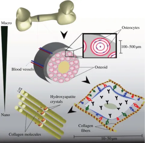
Macro‐ to nanoscale hierarchical structure and porosity of the bone are also important at cell–material interface in terms of regulating miner alization, vascularization, and cellular functions such as proliferation, migration, and ECM production (Fig. 6.1). Macroscale porosity (>400 µm) and interconnectivity of pores promote osteogenesis by enabling cell migration, vascularization, nutrient transportation, and waste disposal. Micro‐ and nanoscale architectures also serve as a cell and mineral binding template. Organization of type I collagens into fibers with diameters of 3–10 nm and lengths of 300 µm supports the binding of nanoscale hydroxyapatite crystals (10–50 nm long) and proteins. Cell adhesion and mineral nucleation directed by microscale collagen template contribute to the resilience and strength of bone tissue (Mistry and Mikos, 2005; Nair et al., 2013). Macro Blood vessels Hydroxyapatite crystals Collagen molecules Collagen fibers 1 nM Nano Osteoid Osteocytes 100–500 µm 10–50 µm
REGULATION OF BONE MATRIX IN ADULT TISSUE 125 6.4 REGULATION OF BONE MATRIX IN ADULT TISSUE
Bone is a rigid but dynamic living tissue that continuously undergoes remodeling and regeneration to maintain its structural integrity and function. Angiogenesis plays an important role in bone remodeling as well as in fracture healing. Vasculature supplies bone tissue with oxygen, nutrients, GFs, hormones, and cytokines as well as osteoblast and osteo clast precursor cells, which are important for bone remodeling processes, and also provides communication between bone and neighboring tissues (Chim et al., 2013; Kanczler and Oreffo, 2008).
Bone remodeling occurs in two important steps accomplished by osteo clasts and osteoblasts. Osteoclasts perform bone resorption by removing old and damaged bone through degradation of bone matrix. Then, mineral ization and deposition of new bone matrix are carried out by osteoblasts in the resorption area. This process takes place in specialized vascularized structures called bone remodeling compartments (BRCs) (Hauge et al., 2001). BRC is a narrow sinus formed between the bone marrow and the remodeling surface, which contains osteoclasts, osteoblasts, osteocytes, bone lining cells, and the capillary blood supply. BRC serves as an anatom ical structure that couples the blood supply with bone remodeling process. Osteoblast and osteoclast precursor cells as well as cytokines and systemic hormones are recruited to the remodeling site via circulating blood supply. BRC also facilitates the direct cell‐to‐cell contact between osteoblast and osteoclast precursors since the secretion of membrane‐bound receptor activator of NF‐κB ligand by osteoblasts is required for differentiation and activation of osteoclast precursors, which enter the BRC through the blood supply to start the remodeling process (Yasuda et al., 1998).
The initial stage of bone remodeling after a stimulus such as an injury is the formation of new blood vessels connecting the BRC to the existing vasculature. Early vascularization and blood flow to the injured site are essential for a proper healing and osteogenic repair since vessels provide nutrients and other factors required for the regeneration process (Fang et al., 2005). Previous studies demonstrated that angiogenesis takes place before the initiation of osteogenesis and the formation of new blood ves sels is crucial for the transport of osteoblast and osteoclast precursors to remodeling sites (Sojo et al., 2005). Therefore, the intercellular communi cation between bone‐forming cells and vessel‐forming endothelial cells plays an important role in remodeling and regeneration of bone tissue. Communication between these cell types is conducted by direct or indirect cell contact and by the secretion of diffusible factors. It has been indicated that the diffusible factors produced by osteoblasts and osteoclast affect
endothelial cell functions such as proliferation, survival, migration, and angiogenesis (Brandi and Collin‐Osdoby, 2006). Recent studies demon strated that osteocytes also play an important role in the initiation of remodeling process by secreting angiogenic factors including RANKL, VEGF, and BMPs (Chim et al., 2013).
6.4.1 Angiogenic Factors in Bone Remodeling
A large variety of angiogenic factors such as VEGF, FGF, BMPs, TGF‐β, and platelet‐derived growth factor (PDGF) are involved in bone remod eling process. Among these, VEGF is a potent mitogen and angiogenic factor for endothelial cells and it serves as a central regulator for the other diffusible factors. At the initial stage of bone remodeling, VEGF plays an important role in the formation of new blood vessels since it regulates the mobilization of endothelial progenitor cells, their differ entiation into endothelial cells, and proliferation (Ferrara et al., 2003). VEGF also acts as a strong chemoattractant for osteoblasts and osteoclasts and supports their survival (Henriksen et al., 2003; Mayr‐Wohlfart et al., 2002). It has been reported that inhibition of VEGF blocks angiogenic activity of FGF and BMP‐2. Blocking of BMP‐2 activity in turn results in inhibition of osteoblast differentiation and bone formation (Deckers et al., 2002; Peng et al., 2005). It has been reported that the inhibition of VEGF pre vents in vitro osteoblast differentiation, while its in vivo inhibition results in decreased blood vessel formation and osteoclastic activity during bone remodeling, as well as reduced mineralization and trabec ular bone healing (Gerber et al., 1999; Maes et al., 2002, 2004; Street et al., 2002).
FGF is another angiogenic factor produced by various cell types inclu ding fibroblasts, endothelial cells, and osteoblasts (Globus et al., 1989; Montero et al., 2000; Sato et al., 1991). FGF stimulates angiogenesis via inducing endothelial cell proliferation and migration and increases expres sion of GFs and integrins involved in angiogenesis (Klein et al., 1993; Seghezzi et al., 1998). It also induces osteogenesis through promoting proliferation of osteoblast precursor cells and their differentiation into mature osteoblasts (Hanada et al., 1997; Pitaru et al., 1993).
BMPs including BMP‐2, BMP‐4, and BMP‐7 also play important roles in promotion of angiogenesis and osteogenesis. BMPs stimulate the prolif eration and differentiation of MSCs and osteoprogenitor cells to promote bone regeneration (Bax et al., 1999; Fiedler et al., 2002; Lee et al., 2006; Li et al., 2005). They also induce endothelial cell proliferation, migration, and angiogenesis (David et al., 2009).
STRATEGIES FOR BONE TISSUE REGENERATION 127 Both TGF‐β and PDGF are known to be potent chemoattractants produced by degranulating platelets at the injury site. TGF‐β serves as a chemoattractant for MSCs and it promotes differentiation of osteoblasts (Erlebacher et al., 1998; Tang et al., 2009), while PDGF is known to be a chemoattractant and mitogenic factor for osteoblasts at the bone remod eling sites (Mehrotra et al., 2004). TGF‐β also induces VEGF expression in endothelial cells within the BRC to increase blood supply during remod eling process (He and Chen, 2005; Shao et al., 2009).
6.5 STRATEGIES FOR BONE TISSUE REGENERATION 6.5.1 Hard Grafts for Bone Regeneration
Bone implants are medical devices used to replace or fix injured bone parts for a temporary period of time or permanently. They are used exten sively in dental, maxillofacial, and orthopedic surgeries (Dohan Ehrenfest et al., 2010; Smalley et al., 1988; Yeo, 2014). Bone anchoring is also an integral part of cochlear and spinal implants and artificial limbs (Gittens et al., 2014; Khan et al., 2012; Wazen et al., 2007). Global estimates suggest that more than 12 million patients receive oral implants and around half a million patients undergo hip replacement surgery per annum (Albrektsson et al., 2014; Hoption Cann et al., 2003; Mundi et al., 2014). As these devices are designed to bear and transfer mechanical loads, tight and stable bone bonding is essential for long‐term functioning. In this regard, under standing and directing complex interactions occurring at the bone–implant interface has critical implications for developing effective healing strategies. When an implant comes into contact with the native bone, there are two possible outcomes. If the bone–implant interface is properly filled with a bone‐like mineral, biological bone bonding, that is, osseointegration, is established. Coined first by Brånemark, osseointegration is the clinically desired state of healing where no progressive relative movement between the implant and bone tissue is observed under mechanical loading (Adell, 1985; Branemark et al., 2001; Khan et al., 2012). If this process is delayed or permanently fails, the interface is instead filled with a collagenous, nonmineralizable soft matrix, which isolates implant from the rest of the body, leading to implant failure (Le Guéhennec et al., 2007; Southam and Selwyn, 1970). In this case, a revision surgery becomes inevitable, which is usually more expensive and invasive, with increased risk factors compared to the primary intervention (Vanhegan et al., 2012). On the one hand, remarkable progress has been made over the last four decades in the
understanding of short‐ and long‐term contributors to bone bonding and functioning. Recent clinical surveys suggest that the long‐term success rates are above 80% for various types of implants (Melo et al., 2006; Penarrocha‐Diago et al., 2012; Simonis et al., 2010). On the other hand, there remain a number of major challenges associated with unsuccessful osseointegration, or failure in the longer period. Among the failed implants, the majority is early term failures caused by softening (Penarrocha‐Diago et al., 2012). Particularly in patients with impaired osteoblastogenesis, such as osteoporosis, mineralization process takes much longer time and is more prone to failure (Alghamdi et al., 2013). This and other factors that impair the osseointegration process and long‐term functioning of bone‐anchored prosthetic devices are discussed in detail in the following. Current accomplishments to overcome such challenges are highlighted along with prospective technologies that would have profound impact on the recovery time and the overall life quality of patients.
After implantation of a bone‐anchored prosthesis, formation of a tight, mineralized interface is vital for stable osseointegration. This process is under competitive pressure from fibrosis progression, and hence softening, so accelerated osteoblast activity is needed for proper mineralization (Daculsi et al., 1990; Lavenus et al., 2010; LeGeros, 2008; Yuan et al., 2010). Bone implants are made mainly from titanium and cobalt–chromium (molybdenum) alloys due to their suitable mechanical properties and corrosion resistance (Le Guéhennec et al., 2007; Linder, 1989; Mavrogenis et al., 2011). In terms or bioactivity, however, these materials are inert, that is, not osteoinductive, thereby requiring an additional bioinstructive support to promote osteoblast activity. As a result, the main challenge in the field is to define the toolbox of engineering implant surface so as to impart bioactivity for promoted osteoblast activity and osseointegration. However, the irreproducible complexity of dynamic interactions regu lating osteoblast formation and subsequent biomineralization has limited the existing efforts to reductionist approaches where one or few bioactive components are displayed on the implant surface, endowing with a com petitive advantage for the desired regenerative response. To understand mechanistic relations of surface parameters with osteoblast behavior and bone formation, various in vitro, that is, cell culture, and in vivo, that is, animal, models have been developed. Primary osteoblasts, preosteoblastic cell lines, and progenitor stem cells obtained from mice, rats, and humans have provided invaluable insight into the molecular biology of the biomin eralization process (Ceylan et al., 2012, 2014; Fratzl‐Zelman et al., 1998; Kalajzic et al., 2005; Kocabey et al., 2013; Marinucci et al., 2006; Olivares‐ Navarrete et al., 2008; Qi et al., 2003; Wang et al., 2014). Early studies
STRATEGIES FOR BONE TISSUE REGENERATION 129 toward this purpose concentrated on exploiting topography and wettability of the implant surface to modulate surface free energy at local sites and thereby altering the adsorbed matrix protein profile, that is, conformation, orientation, and composition, from the biological fluids (Arima and Iwata, 2007; Chug et al., 2013; Marinucci et al., 2006; Ruardy et al., 1995; Song and Mano, 2013; Sousa et al., 2008). Interaction of cells with the implant surface through this adsorbed layer of proteins strongly influences early adhesion, spreading, viability, proliferation, and differentiation (Chug et al., 2013; Wilson et al., 2005). For example, the presence of micrometer‐ scale surface roughness promotes higher osteoblast activity compared to the smooth surface (Gittens et al., 2014; Puleo and Nanci, 1999; von Wilmowsky et al., 2014). Then again, the actual method of choice for cre ating roughness has varying impacts on certain osteoblast behaviors. Both micro‐ and macrosand blasted surfaces promote cell proliferation, whereas osteoblast differentiation is induced only by macrosand blasting (Marinucci et al., 2006). Osteoblasts can also discriminate micro‐ and nanoscale topo graphic features (Olivares‐Navarrete et al., 2014). Nanoscale roughness can induce differentiation of mesenchymal stem cells into mature osteo blasts and promotes osseointegration (Aboushelib et al., 2013; Bjursten et al., 2010; Mendonca et al., 2009). In addition to its direct impact on osteoblast functionality, roughening also provides greater surface area for primary bone bonding. For this reason, the majority of commercial bone implants are manufactured to have a roughness of about 1–2 µm, which leads to an approximately sixfold increase in the total surface area (Albrektsson and Wennerberg, 2004; von Wilmowsky et al., 2014). It is also noteworthy that the minimum bone ingrowth pore size on a material surface is approx imately 50 µm, which implies that osseointegration is established over a much less part of the actual implant surface area (Bobyn et al., 1980). The impact of surface wettability on osteoblast behavior and osseointegration is rather vague. Several reports have linked increase in implant hydrophi licity to enhanced osteoblast functioning and osseointegration (Ceylan et al., 2012; Schwarz et al., 2009). However, depending on the actual surface chemistry, hydrophilicity might not be a striking factor for establishment of osseointegration or it can even be associated with the decrease in oste oblast adhesion in vitro (Unal Gulsuner et al., 2015; Vasak et al., 2014). Hydroxyapatite mineral coating is another strategy to accelerate minerali zation at the bone–implant interface. Depending on the micro‐ and nanoscale geometry and porosity, hydroxyapatite can exhibit osteoinductivity in addition to its osteoconductive properties (Chien and Tsai, 2013; LeGeros, 2008). In this regard, precipitation of carbonated biological apatite on the implanted material would be a useful platform for promoting adhesion,
survival, and osteogenic differentiation of the progenitor cells (Ceylan et al., 2014; Layrolle, 2011; LeGeros, 2008). Altogether, the interplay between different topographic features with varying surface chemistry and wetta bility represents a complex state of understanding, which requires more detailed investigation to develop optimal surface parameters for osteoblast behavior and stable osseointegration. Nevertheless, indirect modulation of surface bioactivity through altering the surface free energy is ultimately limited by the lack of selectivity for promoting adhesion of particular cell types, such as osteoblasts with respect to fibroblasts. Reconstitution of artificial microenvironments by means of ECM‐mimetic, synthetic cues to direct cellular activities in a controlled way can provide effective guidance for osseointegration through adhesion, growth, and differentiation of oste oblasts and their progenitor cells. Targeting molecular level interactions through cell surface receptors has recently drawn broad attention. For example, a simple tetrapeptide, Asp–Gly–Glu–Ala (DGEA), derived from collagen type I α1 chain, can induce osteogenic differentiation of human mesenchymal stem cells and mouse preosteoblastic cells via binding to integrin receptor α2β1. Presentation of this molecule on a solid support is critical in differentiation, adhesion, spreading, migration, and proliferation of human mesenchymal stem cells (Anderson et al., 2009b; Ceylan et al., 2014; Hennessy et al., 2009; Mizuno et al., 2000; Mizuno and Kuboki, 2001; Popov et al., 2011; Staatz et al., 1991; Yoo et al., 2011). In this approach, selecting proper cues is the crucial step to modulate desired biological response. For example, a well‐established biomimetic tripeptide, Arg–Gly– Asp (RGD), of fibronectin interacts with integrin α5β1, which is critical for adhesion and proliferation, while not having any impact on osteogenic differentiation (Hennessy et al., 2009; Olivares‐Navarrete et al., 2008; Yoo et al., 2011). Furthermore, being a rather “universal” cell‐adhesive molecule, RGD can also promote adhesion and growth of fibroblasts, which potentially promote fibrosis (Anselme, 2000; Gailit et al., 1997; Shu et al., 2004). In addition to the biochemical identity, surface density, spatial availability, and patterning are the other principal parameters determining the overall cellular response. For example, a threshold density of RGD ligand is necessary on a polymer substrate to induce focal adhesion of osteoblasts (Chollet et al., 2009). Nanostructuring of bioactive cues has been sought as an appealing method for increasing their display density and optimal presentation geometry to the cell surface receptors (Anderson et al., 2009a and 2009b; Englund et al., 2012; Lim et al., 2009; Niece et al., 2003; Silva et al., 2004; Yoo et al., 2011). Self‐assembled peptide nanofibers have attracted special attention for this purpose because of their inherent biocompatibility and chemical versatility (Cui et al., 2010;
SOFT GRAFTS FOR BONE REGENERATION 131 Hartgerink et al., 2001; Zhang, 2003). Controlled assembly of chemically well‐defined building blocks further allows combinatorial display of mul tiple cues. Nevertheless, one major drawback of the strategy relying on the modification of implant surfaces with biomimetic cues is the adhesion chemistry that is applied in the form of a robust coating. Water molecules, dissolved ions, and polyionic biomolecules in the biological fluid as well as mechanical abrasiveness during implant placement create a challenging environment for adhesives to operate efficiently (Ceylan et al., 2013). To overcome this challenge, natural adhesives adapted to living underwater pro vide a plethora of inspirations toward developing biologically safe and reliable synthetic adhesives for medical applications. Mussel‐inspired surface functionalization has been proposed a viable alternative for bio functionalization of implant surfaces (Black et al., 2012; Ceylan et al., 2011, 2012; Kang et al., 2012; Lee et al., 2007). As discussed in detail in Chapter 8, development of a biomedical adhesive is of paramount impor tance in developing robust implant coatings. Combining the aforemen tioned strategies, recapitulation of a synthetic matrix coating comprised from a bioactive Lys–Arg–Ser–Arg (KRSR) sequence known to selectively bind and promote osteoblastic cell behaviors over fibroblasts, as well as from mussel‐mimetic adhesive moiety, Dopa, represents one of the state‐ of‐the‐art approaches (Nelson et al., 2006).
In summary, a complex variety of parameters create an array of interac tions at the bone–implant interface, the outcome of which has an integral role on the establishment of osseointegration. The aforementioned factors constitute a useful toolbox in the form of source of inspiration for guiding cellular behavior toward efficient bone bonding as well as aspiration for the prospective strategies.
6.6 SOFT GRAFTS FOR BONE REGENERATION
Native bone tissue is mainly a nanocomposite structure with physical and biological features guiding osteoinductive and osteoconductive properties. For bone tissue regeneration, peptide‐ or polymer‐based biomaterials have offered a framework to design interactive molecules displaying bone com posite properties to mimic living bone tissue. Biomimetic peptide and polymer materials can enable the appropriate matrix environment for bone regeneration, integrate desirable biological properties, and provide con trolled release of multiple soluble factors or GFs during regeneration pro cess. In the following, structure and properties of peptide‐ and polymer‐based soft grafts for bone tissue regeneration will be reviewed with a summary
of upcoming goals and challenges in the future of these versatile materials. This section basically covers types and applications of soft bone grafts, directed bone regeneration from biocompatible and bioactive biomaterials, and nanocomposite scaffolds for bone tissue regeneration.
6.6.1 Peptide‐Based Bone Grafts
Degradable small molecules emerged as an alternative approach for devel oping implantable materials for tissue regeneration. Bone regeneration studies have been primarily focused on polymers and synthetic proteins. However, these structures have several shortcomings like toxic preparation components (monomers, cross‐linking agents, etc.), shrinkage after appli cation, and low biodegradability. Various types of supramolecular nano structures and hydrogels attained great interest in regenerative medicine because of their significant features, such as simple production, complete biodegradability, and biocompatibility. These inert nanostructures can be applied to deformed bone tissue as implantable or injectable materials with simple procedures and minimal invasiveness. Investigation of self‐ assembled peptide nanostructures is a growing field with great potential to generate new, facile, and effective bone regenerative applications. Here, we describe some of the significant contributions to the field of bone regeneration with self‐assembled peptide structures.
Bone formation is a well‐orchestrated and complex biological process; therefore, bone regeneration strategies should meet rigid requirements to constitute proper remodeling of deformed site. During bone regeneration, osteoinduction and osteoconduction are governed by different factors (mechanical, biological, and chemical) that interplay each other, and these components should be considered carefully while constructing materials for bone regeneration. While a diverse set of materials have been utilized to build such scaffolds, inert and mechanically supportive metals and alloys have so far been used as permanent bone implants. These metallic implants and surface modification techniques lack osteoinductive prop erties, despite their success in osteoconductive features to accelerate the bone healing process. To improve cell attachment and to induce bone differentiation process, bioactive molecule (ECM proteins, GFs) attach ments to implant surfaces are critical to obtain adequate bone healing and controlled mineralization. Proteins and GFs are large molecules bearing short peptide sequences, which can trigger downstream processes, espe cially cell adhesion and differentiation among many other roles. Accordingly, peptide structures with short bioactive units draw significant attention for bone remodeling studies.
SOFT GRAFTS FOR BONE REGENERATION 133 In general, bioactive short peptides are cell‐binding epitopes including RGDS, IKVAV, and YIGSR. The RGDS sequence has been frequently used to direct cell attachment. The RGD is found in fibronectin, osteopontin, and sia loprotein, and fibrous materials that display RGD can mimic protein function (Puleo and Bizios, 1991; Pytela et al., 1987). In early studies, adhesion of bone marrow‐derived stem cells was investigated on RGDS‐containing peptide amphiphile (PA) surfaces, and activity of scaffold was compared with epitope‐ free peptides. Stem cells were encapsulated into PAs and the coassembled system was injected in vivo. RGDS‐containing PA gels promoted cell viability notably better than epitope‐free control group (Guler et al., 2006).
The primary component of bone ECM is collagen I fibers, and bioactive short sequences derived from this protein are preferential targets for induction of bone tissue remodeling. The most studied collagen I epitope, DGEA, is found in its α1 helix, and its osteoinductive characteristics were utilized intensively for bone regeneration studies. For example, DGEA peptide‐coated hydroxyapatite surfaces enhanced differentiation of mesenchymal stem cell into osteogenic fate (Harbers and Healy, 2005). However, in some cases, bio activity is not enough for adequate regeneration, and more than one compo nent may be required to enhance activity. For example, another study using CGGDGEAG sequence reported lack of adhesion by rat calvarial osteoblasts onto peptide surfaces (Anderson et al., 2011). To improve adhesion and osteo inductive potential of DGEA‐PA, RGDS‐PA, and S‐PA peptides was utilized with different combinations. According to histochemical staining and PCR results, the RGDS‐PA and DGEA‐PA combination upregulated osteogenic differentiation (Anderson et al., 2009b). Another short peptide sequence adopted from collagen is GFOGER peptide, which binds to osteogenesis regulating inte grin α2β1. Differentiation of osteoprogenitor cells into osteoblasts can be triggered by using this sequence, and GFOGER‐coated polycaprolactone scaffolds were shown to support remodeling of critical‐sized defects in rat bone. GFOGER sequence induced significant regeneration in nonhealing femoral defects compared to peptide‐free control groups (Dee et al., 1998).
In addition to cell attachment and proliferation, peptide sequences were also used as GF binding units. Binding units located onto gel scaffold can attract GFs to the desired location to sustain improvement during healing process. A well‐known bone differentiation‐regulating factor, bone mor phogenetic protein‐2 (BMP‐2), which plays significant roles during osteo genesis, was targeted for this purpose. BMP receptor‐binding peptides with osteopromotive domains, DWIVA and A4G3EDWIVA, were adequate for maintaining bone regeneration process (Gelain et al., 2006). In the ECM, glycosaminoglycans also interact with BMP‐2 and sulfated GAG‐mimetic peptide–BMP interaction was used to promote osteoblast maturation and
mineralization of osteogenic cells. The GAG‐mimicking ability of the pep tide nanofibers and their interaction with BMP‐2 promoted osteogenic activity and mineralization of osteoblastic cells (Webber et al., 2010).
RADA16‐I is another scaffold that shows stable beta sheet formation, and these peptides can be used with different active groups to form nano structures to enhance tissue regeneration. In one study, RADA16‐I was linked to variable bioactive signal inducing short peptides like ALK (ALKRQGRTLYGF) osteogenic growth motif, DGR (DGRGDSVAYG) osteopontin‐based cell adhesion sequence, and PGR (PRGDSGYRGDS) two‐repeat RGD adhesion sequence to enhance osteogenic differentiation and support (Horii et al., 2007).
Mussel‐inspired Dopa‐mediated surface adhesion strategy was applied to direct efficient osteoblast maturation on implant surfaces. For this purpose, E3‐PA/Dopa‐PA and DGEA‐PA/Dopa‐PA combination was used to induce osteoblast formation and subsequent bone‐like mineralization. Hydroxyapatite‐boosting glutamic acid and Dopa residues on fiber sur faces induced significant osteogenic activity as a consequence of significant mineral deposition, since inorganic materials like hydroxyapatite minerals can also induce osteoblast maturation during bone formation process (Tashiro et al., 1989). Therefore, mineral‐inducing peptide scaffolds have important roles for bone remodeling studies.
Peptide‐based soft bone grafts can also serve as structural frameworks. These materials can be designed in order to have structural components presenting a complementary effect to signaling epitope to enhance bioac tivity. For example, micropatterned RGDS‐modified scaffolds bearing 20–40 nm holes upregulated osteogenic marker expression compared to nonpatterned surfaces. The hole‐patterned surfaces presented high‐density epitopes that were able to enclose cells and stimulated differentiation (Sur et al., 2012). In another example, phosphoserine‐bearing peptide amphi phile nanofibers were used to induce mineralization. This matrix presented convenient template for hydroxyapatite crystal (HA)—basic bone mineral— deposition as a result of its suitable structure for crystal packing (Anderson et al., 2009b). By the virtue of allowing nanopatterned structure design, peptides are versatile building blocks for generating templates that can induce inorganic material deposition.
6.6.2 Polymer Nanocomposites as Bone Grafts
Bone autografts and allografts are already widely used in clinical practice for bone regeneration studies. However, these grafts are not ideal scaffolds for osteogenesis, especially because of biocompatibility and handling
SOFT GRAFTS FOR BONE REGENERATION 135 issues. Producing demineralized bone scaffolds to regenerate native bone tissue requires tedious work, and mineralization of these grafts can be less controllable compared to synthetic scaffolds. These shortcomings of auto grafts and allografts boosted the interest in utilization of synthetic polymer scaffolds as another useful strategy for bone regeneration. Polymers can be constructed from various building blocks depending on the desired outcome and can be connected or decorated through covalent/noncovalent linkages to create ordered composite.
Biodegradability and biocompatibility are important parameters for bone engineering applications. Natural polymer scaffolds are basically derived from collagen, gelatin‐like proteins, or alginate‐, hyaluronate‐, and chitin‐like polysaccharides. However, despite their biocompatibility or biodegradability, these molecules have several shortcomings. These natural constructs were shown to exhibit variable physical characteristics for each batch, such as molecular weight, crystallinity, polydispersity, degradation kinetics, structure, etc., which causes different scaffold properties. On the other hand, synthetic polymeric materials (poly(glycolic acid) (PGA), poly(lactic acid) (PLA)) and their copolymers offer facile and more con trollable methods for clinical bone regeneration applications.
Aliphatic polymers were the earliest examples of polymers that were used in the field of bone tissue engineering. PLA, PGA, and poly(lactic‐ co‐glycolic acid) (PLGA) and their copolymers are well‐known aliphatic polymer types. The most widely utilized subtypes of these polymers are D‐PLA PDLA, L‐PLA (PLLA) forms of PLA, and blend of D, L‐PLA (PDLLA), PLA, PGA, and PLGA, especially used in bone fixation devices, sutures, drug carriers, and bioregeneration scaffolds. In addition to aliphatic polymers, high‐molecular‐weight aliphatic polyesters, poly(l‐lactide), PLA, and PCL, are also used for similar regeneration applications. There are also amorphous polymers, poly(orthoesters), which are hydrophobic and pH‐sensitive structures. Poly(orthoesters), like pH‐sensitive polymers, are especially used as drug delivery systems, because of enabled pH‐responsive degradation that can lead to controlled drug release for medical studies.
Mineralization is a crucial part of bone formation and CaP minerals are the building blocks of bone composite; hence, mineral‐inducing matrices are important for osteogenesis process. During natural bone formation, col lagen I fibrils serve as a template upon which HA deposition occurs to form crystalline bone material. Collagen matrix comprises anionic proteins that are nucleator and inhibitor of mineralization, and density of these proteins changes according to bone type or age for controlling deposition (Kocabey et al., 2013). Accordingly, anionic polymers are used to mimic mineraliz able collagen matrices for bone mineral nucleation (Palmer et al., 2008).
For polymer‐based mineralization studies, simulated body fluid (SBF), which mimics human plasma in terms of ion concentrations, is a widely applied solution to evaluate ex vivo mineral deposition. Synthetic PLGA and poly‐l‐lactic acid (PLLA) are early examples of scaffolds mineralized in SBF. Mineralized PLGA polymer construct implanted in a mouse cal varial defect and after 4 weeks showed bone mineral growth, whereas non mineralized scaffolds did not induce any mineralization (Ceylan et al., 2014). Polymers can also be utilized by mixing with biomolecules; in one example, PLLA mixed with collagen I and SBF closely mimicked natural bone mineral nucleating surface (Mata et al., 2009).
Different monolayers such as dihydrogen phosphate (PO4H2), carboxylic acid (COOH), and methyl (CH3) were also investigated for mineralization, and it was found that negatively charged groups are potent inducers of mineralization, where noncharged methyl monolayer did not induce min eralization (Chen et al., 2006; Cowan et al., 2004). Moreover, different monolayers showed different Ca : P ratios and different crystal morphol ogies, meaning that mineral phase is highly dependent charged groups on templates. Charge‐functionalized surface moieties induce interaction between fibronectin and functional groups, and this cooperation attracts integrin binding by osteoblasts and probably simulates integrin binding to induce cell‐dependent mineralization. In one example of anionic group‐ functionalized polymer, carboxymethyl attached to poly(2‐hydroxyethyl methacrylate) (PHEMA) hydrogels displayed better mineralization and cell spreading compared to only PHEMA surfaces after incubation with SBF (Tanahashi and Matsuda, 1997).
Phosphorus is one of the most effective mineralization‐inducing chemical units, among the other effective groups. The main role of phosphate ion is stabilization of calcium ions on the polymer surfaces. Phosphate‐ modified polymers mineralize faster both in in vivo and in vitro conditions (Filmon et al., 2002). Addition of these chemical units also enhances prop erties of polymers such as vinylphosphonic acid (VPA), which showed better swelling, protein uptake, and adhesion and proliferation of osteo blast (Tan et al., 2005). In another study, PLGA films stabilized and used for enhanced mineralization by the attachment of phosphate ion (Cowan et al., 2004).
Poly(ethylene glycol) (PEG) polymer is one of the most widely used polymers for regenerative studies. Phosphoester‐containing PEG hydro gels demonstrate unique characteristics, where polymer backbone allows cleavage of phosphate group by ALP enzyme (Stancu et al., 2004). This material has shown osteoinductive and osteoconductive features by both its phosphate source and phosphate‐modified surface characteristics.
SOFT GRAFTS FOR BONE REGENERATION 137 However, some of phosphorus‐deficient polymer materials are also capable of surface mineralization like phosphorus‐containing analogs. Poly(phosphazene)‐based composites interacted with hydroxyapatite due to surface hydrolysis, hydrolyzed carboxylate groups on polymer chain, and nucleated hydroxyapatite layer by interacting with Ca ions in the surrounding environment (Li et al., 2006).
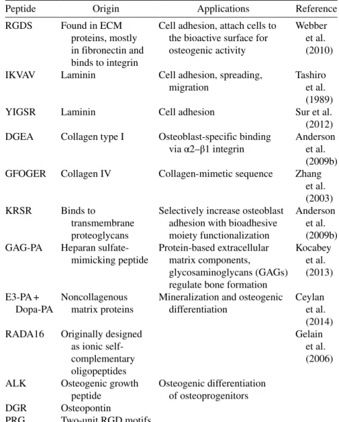
TABLE 6.1 Short Peptides for Bone Regeneration
Peptide Origin Applications Reference
RGDS Found in ECM proteins, mostly in fibronectin and binds to integrin
Cell adhesion, attach cells to the bioactive surface for osteogenic activity
Webber et al. (2010) IKVAV Laminin Cell adhesion, spreading,
migration
Tashiro et al. (1989)
YIGSR Laminin Cell adhesion Sur et al.
(2012) DGEA Collagen type I Osteoblast‐specific binding
via α2–β1 integrin Anderson et al. (2009b) GFOGER Collagen IV Collagen‐mimetic sequence Zhang
et al. (2003) KRSR Binds to
transmembrane proteoglycans
Selectively increase osteoblast adhesion with bioadhesive moiety functionalization
Anderson et al. (2009b) GAG‐PA Heparan sulfate‐
mimicking peptide
Protein‐based extracellular matrix components,
glycosaminoglycans (GAGs) regulate bone formation
Kocabey et al. (2013) E3‐PA + Dopa‐PA Noncollagenous matrix proteins
Mineralization and osteogenic differentiation
Ceylan et al. (2014) RADA16 Originally designed
as ionic self‐ complementary oligopeptides Gelain et al. (2006) ALK Osteogenic growth
peptide
Osteogenic differentiation of osteoprogenitors DGR Osteopontin
6.7 FUTURE PERSPECTIVES
Bone grafts can offer promising platforms for bone regeneration studies and therapeutic applications by integrating dynamics and functions of different materials to reach high responsivity to stimuli, healing potential, and envi ronmental adaptation. The expansion of knowledge on bone structure and reconstruction at molecular level is continuing to lead to improvement of bone regeneration methods. Yet, further information is required to fill remaining gaps in this area. Particularly, short peptide‐mediated regen eration requires more detailed analysis in terms of investigation of new sequences and their cellular activities during skeletal repair to elicit cellular– molecular interactions for developing novel and effective regenerative materials. Further understanding in this area, with the integration of a wide variety of material‐based strategies, could be the key to develop integrated and efficient bone regeneration methods. Hence, integration and mani pulation of bone regeneration strategies that approximate natural bone cascade can lead to successful treatment of many bone diseases including aging‐ or genetic‐related illnesses and bone traumas (Table 6.1).
REFERENCES
Aboushelib, M.N., Salem, N.A., Taleb, A.L., and El Moniem, N.M. (2013). Influence of surface nano‐roughness on osseointegration of zirconia implants in rabbit femur heads using selective infiltration etching technique. The Journal of Oral Implantology 39, 583–590.
Adell, R. (1985). Tissue integrated prostheses in clinical dentistry. International Dental Journal 35, 259–265.
Albrektsson, T., Dahlin, C., Jemt, T., Sennerby, L., Turri, A., and Wennerberg, A. (2014). Is marginal bone loss around oral implants the result of a provoked foreign body reaction? Clinical Implant Dentistry and Related Research 16, 155–165.
Albrektsson, T. and Wennerberg, A. (2004). Oral implant surfaces: part 1—review focusing on topographic and chemical properties of different surfaces and in vivo responses to them. The International Journal of Prosthodontics 17, 536–543. Alghamdi, H.S., Bosco, R., van den Beucken, J.J.J.P., Walboomers, X.F., and
Jansen, J.A. (2013). Osteogenicity of titanium implants coated with calcium phosphate or collagen type‐I in osteoporotic rats. Biomaterials 34, 3747–3757. Anderson, J.M., Andukuri, A., Lim, D.J., and Jun, H.W. (2009a). Modulating
the gelation properties of self‐assembling peptide amphiphiles. ACS Nano 3, 3447–3454.
REFERENCES 139 Anderson, J.M., Kushwaha, M., Tambralli, A., Bellis, S.L., Camata, R.P., and
Jun, H.W. (2009b). Osteogenic differentiation of human mesenchymal stem cells directed by extracellular matrix‐mimicking ligands in a biomimetic self‐ assembled peptide amphiphile nanomatrix. Biomacromolecules 10, 2935–2944. Anderson, J.M., Vines, J.B., Patterson, J.L., Chen, H., Javed, A., and Jun, H.W.
(2011). Osteogenic differentiation of human mesenchymal stem cells synergis tically enhanced by biomimetic peptide amphiphiles combined with conditioned medium. Acta Biomaterialia 7, 675–682.
Anselme, K. (2000). Osteoblast adhesion on biomaterials. Biomaterials 21, 667–681.
Arima, Y. and Iwata, H. (2007). Effect of wettability and surface functional groups on protein adsorption and cell adhesion using well‐defined mixed self‐assembled monolayers. Biomaterials 28, 3074–3082.
Athanasiou, K.A., Zhu, C., Lanctot, D.R., Agrawal, C.M., and Wang, X. (2000). Fundamentals of biomechanics in tissue engineering of bone. Tissue Engineering 6, 361–381.
Bax, B.E., Wozney, J.M., and Ashhurst, D.E. (1999). Bone morphogenetic protein‐2 increases the rate of callus formation after fracture of the rabbit tibia. Calcified Tissue International 65, 83–89.
Beddoe, A.H., Darley, P.J., and Spiers, F.W. (1976). Measurements of trabecular bone structure in man. Physics in Medicine and Biology 21, 589–607.
Beertsen, W. and van den Bos, T. (1992). Alkaline phosphatase induces the mineralization of sheets of collagen implanted subcutaneously in the rat. The Journal of Clinical Investigation 89, 1974–1980.
Bjursten, L.M., Rasmusson, L., Oh, S., Smith, G.C., Brammer, K.S., and Jin, S. (2010). Titanium dioxide nanotubes enhance bone bonding in vivo. Journal of Biomedical Materials Research, Part A 92, 1218–1224.
Black, K.C.L., Yi, J., Rivera, J.G., Zelasko‐Leon, D.C., and Messersmith, P.B. (2012). Polydopamine‐enabled surface functionalization of gold nanorods for cancer cell‐targeted imaging and photothermal therapy. Nanomedicine 8, 17–28. Bobyn, J.D., Pilliar, R.M., Cameron, H.U., and Weatherly, G.C. (1980). The
optimum pore size for the fixation of porous‐surfaced metal implants by the ingrowth of bone. Clinical Orthopaedics and Related Research, 263–270. Bosetti, M., Lloyd, A.W., Santin, M., Denyer, S.P., and Cannas, M. (2005). Effects
of phosphatidylserine coatings on titanium on inflammatory cells and cell‐ induced mineralisation in vitro. Biomaterials 26, 7572–7578.
Brandi, M.L. and Collin‐Osdoby, P. (2006). Vascular biology and the skeleton. Journal of Bone and Mineral Research 21, 183–192.
Branemark, R., Branemark, P.I., Rydevik, B., and Myers, R.R. (2001). Osseointegration in skeletal reconstruction and rehabilitation: a review. Journal of Rehabilitation Research and Development 38, 175–181.
ButLer, W.T., ed. (2000). Noncollagenous proteins of bone and dentin: a brief overview (Rosemont IL:AAOS).
Canalis, E., McCarthy, T., and Centrella, M. (1988). Growth factors and the regu lation of bone remodeling. The Journal of Clinical Investigation 81, 277–281. Ceylan, H., Kocabey, S., Tekinay, A.B., and Guler, M.O. (2012). Surface‐adhesive
and osteogenic self‐assembled peptide nanofibers for bioinspired functional ization of titanium surfaces. Soft Matter 8, 3929–3937.
Ceylan, H., Kocabey, S., Unal Gulsuner, H., Balcik, O.S., Guler, M.O., and Tekinay, A.B. (2014). Bone‐like mineral nucleating peptide nanofibers induce differentiation of human mesenchymal stem cells into mature osteoblasts. Biomacromolecules 15, 2407–2418.
Ceylan, H., Tekinay, A.B., and Guler, M.O. (2011). Selective adhesion and growth of vascular endothelial cells on bioactive peptide nanofiber functionalized stainless steel surface. Biomaterials 32, 8797–8805.
Ceylan, H., Tekinay, A.B., and Guler, M.O. (2013). Mussel‐inspired adhesive inter faces for biomedical applications. In Biological and biomimetic adhesives: challenges and opportunities, R. Santos, N. Aldred, S. Gorb, and P. Flammang, eds. (Cambridge: The Royal Society of Chemistry), pp. 103–116.
Chen, Y., Mak, A.F., Wang, M., and Li, J. (2006). Composite coating of bonelike apatite particles and collagen fibers on poly‐l‐lactic acid formed through an accelerated biomimetic coprecipitation process. Journal of Biomedical Materials Research Part B: Applied Biomaterials 77, 315–322.
Chien, C.‐Y. and Tsai, W.‐B. (2013). Poly(dopamine)‐assisted immobilization of Arg‐Gly‐Asp peptides, hydroxyapatite, and bone morphogenic protein‐2 on titanium to improve the osteogenesis of bone marrow stem cells. ACS Applied Materials & Interfaces 5, 6975–6983.
Chim, S.M., Tickner, J., Chow, S.T., Kuek, V., Guo, B., Zhang, G., Rosen, V., Erber, W., and Xu, J. (2013). Angiogenic factors in bone local environment. Cytokine & Growth Factor Reviews 24, 297–310.
Chollet, C., Chanseau, C., Remy, M., Guignandon, A., Bareille, R., Labrugere, C., Bordenave, L., and Durrieu, M.C. (2009). The effect of RGD density on oste oblast and endothelial cell behavior on RGD‐grafted polyethylene terephthalate surfaces. Biomaterials 30, 711–720.
Chug, A., Shukla, S., Mahesh, L., and Jadwani, S. (2013). Osseointegration— molecular events at the bone–implant interface: a review. Journal of Oral and Maxillofacial Surgery, Medicine, and Pathology 25, 1–4.
Cowan, C.M., Shi, Y.Y., Aalami, O.O., Chou, Y.F., Mari, C., Thomas, R., Quarto, N., Contag, C.H., Wu, B., and Longaker, M.T. (2004). Adipose‐derived adult stromal cells heal critical‐size mouse calvarial defects. Nature Biotechnology
22, 560–567.
Cui, H., Webber, M.J., and Stupp, S.I. (2010). Self‐assembly of peptide amphiphiles: from molecules to nanostructures to biomaterials. Biopolymers 94, 1–18.
REFERENCES 141 Daculsi, G., Passuti, N., Martin, S., Deudon, C., Legeros, R.Z., and Raher, S.
(1990). Macroporous calcium phosphate ceramic for long bone surgery in humans and dogs. Clinical and histological study. Journal of Biomedical Materials Research 24, 379–396.
David, L., Feige, J.J., and Bailly, S. (2009). Emerging role of bone morpho genetic proteins in angiogenesis. Cytokine & Growth Factor Reviews 20, 203–212.
de Oliveira, P.T. and Nanci, A. (2004). Nanotexturing of titanium‐based surfaces upregulates expression of bone sialoprotein and osteopontin by cultured osteogenic cells. Biomaterials 25, 403–413.
Deckers, M.M., van Bezooijen, R.L., van der Horst, G., Hoogendam, J., van Der Bent, C., Papapoulos, S.E., and Lowik, C.W. (2002). Bone morphogenetic pro teins stimulate angiogenesis through osteoblast‐derived vascular endothelial growth factor A. Endocrinology 143, 1545–1553.
Dee, K.C., Andersen, T.T., and Bizios, R. (1998). Design and function of novel osteoblast‐adhesive peptides for chemical modification of biomaterials. Journal of Biomedical Materials Research 40, 371–377.
Dohan Ehrenfest, D.M., Coelho, P.G., Kang, B.‐S., Sul, Y.‐T., and Albrektsson, T. (2010). Classification of osseointegrated implant surfaces: materials, chem istry and topography. Trends in Biotechnology 28, 198–206.
Ducy, P., Geoffroy, V., and Karsenty, G. (1996). Study of osteoblast‐specific expression of one mouse osteocalcin gene: characterization of the factor binding to OSE2. Connective Tissue Research 35, 7–14.
Eanes, E.D. (1989). Biophysical aspects of lipid interaction with mineral: lipo some model studies. The Anatomical Record 224, 220–225.
Engel, J., Taylor, W., Paulsson, M., Sage, H., and Hogan, B. (1987). Calcium binding domains and calcium‐induced conformational transition of SPARC/ BM‐40/osteonectin, an extracellular glycoprotein expressed in mineralized and nonmineralized tissues. Biochemistry 26, 6958–6965.
Englund, E.A., Wang, D., Fujigaki, H., Sakai, H., Micklitsch, C.M., Ghirlando, R., Martin‐Manso, G., Pendrak, M.L., Roberts, D.D., Durell, S.R., et al. (2012). Programmable multivalent display of receptor ligands using peptide nucleic acid nanoscaffolds. Nature Communications 3, 614.
Erlebacher, A., Filvaroff, E.H., Ye, J.Q., and Derynck, R. (1998). Osteoblastic responses to TGF‐beta during bone remodeling. Molecular Biology of the Cell
9, 1903–1918.
Fang, T.D., Salim, A., Xia, W., Nacamuli, R.P., Guccione, S., Song, H.M., Carano, R.A., Filvaroff, E.H., Bednarski, M.D., Giaccia, A.J., et al. (2005). Angiogenesis is required for successful bone induction during distraction osteogenesis. Journal of Bone and Mineral Research 20, 1114–1124.
Fernandez‐Yague, M.A., Abbah, S.A., McNamara, L., Zeugolis, D.I., Pandit, A., and Biggs, M.J. (2014). Biomimetic approaches in bone tissue
engineering: integrating biological and physicomechanical strategies. Advanced Drug Delivery Reviews 84, 1–29.
Ferrara, N., Gerber, H.P., and LeCouter, J. (2003). The biology of VEGF and its receptors. Nature Medicine 9, 669–676.
Fiedler, J., Roderer, G., Gunther, K.P., and Brenner, R.E. (2002). BMP‐2, BMP‐4, and PDGF‐bb stimulate chemotactic migration of primary human mesenchymal progenitor cells. Journal of Cellular Biochemistry 87, 305–312.
Filmon, R., Grizon, F., Basle, M.F., and Chappaard, D. (2002). Effects of negatively charged groups (carboxymethyl) on the calcification of poly(2‐hydroxyethyl methacrylate). Biomaterials 23, 3053–3059.
Fratzl‐Zelman, N., Fratzl, P., Horandner, H., Grabner, B., Varga, F., Ellinger, A., and Klaushofer, K. (1998). Matrix mineralization in MC3T3‐E1 cell cultures initiated by beta‐glycerophosphate pulse. Bone 23, 511–520.
Gailit, J., Clarke, C., Newman, D., Tonnesen, M.G., Mosesson, M.W., and Clark, R.A. (1997). Human fibroblasts bind directly to fibrinogen at RGD sites through integrin alpha(v)beta3. Experimental Cell Research 232, 118–126. Garcia, A.J. and Reyes, C.D. (2005). Bio‐adhesive surfaces to promote osteoblast
differentiation and bone formation. Journal of Dental Research 84, 407–413. Gelain, F., Bottai, D., Vescovi, A., and Zhang, S. (2006). Designer self‐assembling
peptide nanofiber scaffolds for adult mouse neural stem cell 3‐dimensional cultures. PLoS One 1, e119.
Gerber, H.P., Vu, T.H., Ryan, A.M., Kowalski, J., Werb, Z., and Ferrara, N. (1999). VEGF couples hypertrophic cartilage remodeling, ossification and angiogenesis during endochondral bone formation. Nature Medicine 5, 623–628.
Gittens, R.A., Olivares‐Navarrete, R., Schwartz, Z., and Boyan, B.D. (2014). Implant osseointegration and the role of microroughness and nanostructures: lessons for spine implants. Acta Biomaterialia 10, 3363–3371.
Glimcher, M.J. (1992). The nature of the mineral component of bone and the mechanisms of calcification (New York: Raven Press).
Globus, R.K., Plouet, J., and Gospodarowicz, D. (1989). Cultured bovine bone cells synthesize basic fibroblast growth factor and store it in their extracellular matrix. Endocrinology 124, 1539–1547.
Goldstein, S.A., Wilson, D.L., Sonstegard, D.A., and Matthews, L.S. (1983). The mechanical properties of human tibial trabecular bone as a function of metaph yseal location. Journal of Biomechanics 16, 965–969.
Gowen, M., Wood, D.D., Ihrie, E.J., Mcguire, M.K.B., and Russell, R.G.G. (1983). An interleukin‐1 like factor stimulates bone‐resorption in vitro. Nature
306, 378–380.
Guler, M.O., Hsu, L., Soukasene, S., Harrington, D.A., Hulvat, J.F., and Stupp, S.I. (2006). Presentation of RGDS epitopes on self‐assembled nanofibers of branched peptide amphiphiles. Biomacromolecules 7, 1855–1863.
REFERENCES 143 Hanada, K., Dennis, J.E., and Caplan, A.I. (1997). Stimulatory effects of basic
fibroblast growth factor and bone morphogenetic protein‐2 on osteogenic differentiation of rat bone marrow‐derived mesenchymal stem cells. Journal of Bone and Mineral Research 12, 1606–1614.
Harbers, G.M. and Healy, K.E. (2005). The effect of ligand type and density on osteoblast adhesion, proliferation, and matrix mineralization. Journal of Biomedical Materials Research, Part A 75, 855–869.
Hartgerink, J.D., Beniash, E., and Stupp, S.I. (2001). Self‐assembly and mineral ization of peptide‐amphiphile nanofibers. Science 294, 1684–1688.
Hauge, E.M., Qvesel, D., Eriksen, E.F., Mosekilde, L., and Melsen, F. (2001). Cancellous bone remodeling occurs in specialized compartments lined by cells expressing osteoblastic markers. Journal of Bone and Mineral Research 16, 1575–1582.
Hauschka, P.V., Mavrakos, A.E., Iafrati, M.D., Doleman, S.E., and Klagsbrun, M. (1986). Growth‐factors in bone‐matrix—isolation of multiple types by affinity‐ chromatography on heparin‐Sepharose. Journal of Biological Chemistry 261, 2665–2674.
He, C. and Chen, X. (2005). Transcription regulation of the vegf gene by the BMP/Smad pathway in the angioblast of zebrafish embryos. Biochemical and Biophysical Research Communications 329, 324–330.
Hennessy, K.M., Pollot, B.E., Clem, W.C., Phipps, M.C., Sawyer, A.A., Culpepper, B.K., and Bellis, S.L. (2009). The effect of collagen I mimetic peptides on mesenchymal stem cell adhesion and differentiation, and on bone formation at hydroxyapatite surfaces. Biomaterials 30, 1898–1909.
Henriksen, K., Karsdal, M., Delaisse, J.M., and Engsig, M.T. (2003). RANKL and vascular endothelial growth factor (VEGF) induce osteoclast chemotaxis through an ERK1/2‐dependent mechanism. Journal of Biological Chemistry
278, 48745–48753.
Hoption Cann, S., van Netten, J., and van Netten, C. (2003). Dr. William Coley and tumour regression: a place in history or in the future. Postgraduate Medical Journal 79, 672–680.
Horii, A., Wang, X., Gelain, F., and Zhang, S. (2007). Biological designer self‐ assembling peptide nanofiber scaffolds significantly enhance osteoblast prolif eration, differentiation and 3‐D migration. PLoS One 2, e190.
Hunter, G.K., Hauschka, P.V., Poole, A.R., Rosenberg, L.C., and Goldberg, H.A. (1996). Nucleation and inhibition of hydroxyapatite formation by mineralized tissue proteins. The Biochemical Journal 317 (Pt 1), 59–64.
Hynes, R.O. (1992). Integrins: versatility, modulation, and signaling in cell adhesion. Cell 69, 11–25.
Kalajzic, I., Staal, A., Yang, W.‐P., Wu, Y., Johnson, S.E., Feyen, J.H.M., Krueger, W., Maye, P., Yu, F., Zhao, Y., et al. (2005). Expression profile of
osteoblast lineage at defined stages of differentiation. Journal of Biological Chemistry 280, 24618–24626.
Kanczler, J.M. and Oreffo, R.O. (2008). Osteogenesis and angiogenesis: the potential for engineering bone. European Cells & Materials 15, 100–114. Kang, S.M., Hwang, N.S., Yeom, J., Park, S.Y., Messersmith, P.B., Choi, I.S.,
Langer, R., Anderson, D.G., and Lee, H. (2012). One‐step multipurpose surface functionalization by adhesive catecholamine. Advanced Functional Materials 22, 2949–2955.
Katsamenis, O.L., Chong, H.M.H., Andriotis, O.G., and Thurner, P.J. (2013). Load‐bearing in cortical bone microstructure: selective stiffening and hetero geneous strain distribution at the lamellar level. Journal of the Mechanical Behavior of Biomedical Materials 17, 152–165.
Khan, S.N., Ramachandran, M., Senthil Kumar, S., Krishnan, V., and Sundaram, R. (2012). Osseointegration and more—a review of literature. Indian Journal of Dentistry 3, 72–76.
Kielty, C.M. and Grant, M. E. (2002). The collagen family: structure, assembly, and organization in the extracellular matrix. In Connective tissue and its heri table disorders: molecular, genetic, and medical aspects, 2nd edn. (eds P. M. Royce and B. Steinmann, John Wiley & Sons, Inc., Hoboken).
Klein, S., Giancotti, F.G., Presta, M., Albelda, S.M., Buck, C.A., and Rifkin, D.B. (1993). Basic fibroblast growth factor modulates integrin expression in micro vascular endothelial cells. Molecular Biology of the Cell 4, 973–982.
Kocabey, S., Ceylan, H., Tekinay, A.B., and Guler, M.O. (2013). Glycosaminoglycan mimetic peptide nanofibers promote mineralization by osteogenic cells. Acta Biomaterialia 9, 9075–9085.
Lavenus, S., Louarn, G., and Layrolle, P. (2010). Nanotechnology and dental implants. International Journal of Biomaterials 2010, 9.
Layrolle, P. (2011). 1.112—Calcium phosphate coatings. In Comprehensive biomaterials, D. Paul, ed. (Oxford: Elsevier), pp. 223–229.
Le Guéhennec, L., Soueidan, A., Layrolle, P., and Amouriq, Y. (2007). Surface treat ments of titanium dental implants for rapid osseointegration. Dental Materials
23, 844–854.
Lee, D.H., Park, B.J., Lee, M.S., Lee, J.W., Kim, J.K., Yang, H.C., and Park, J.C. (2006). Chemotactic migration of human mesenchymal stem cells and MC3T3‐ E1 osteoblast‐like cells induced by COS‐7 cell line expressing rhBMP‐7. Tissue Engineering 12, 1577–1586.
Lee, H., Dellatore, S.M., Miller, W.M., and Messersmith, P.B. (2007). Mussel‐ inspired surface chemistry for multifunctional coatings. Science 318, 426–430. LeGeros, R.Z. (2008). Calcium phosphate‐based osteoinductive materials.
REFERENCES 145 Li, G., Cui, Y., McIlmurray, L., Allen, W.E., and Wang, H. (2005). rhBMP‐2,
rhVEGF(165), rhPTN and thrombin‐related peptide, TP508 induce chemo taxis of human osteoblasts and microvascular endothelial cells. Journal of Orthopaedic Research 23, 680–685.
Li, Q., Wang, J., Shahani, S., Sun, D.D., Sharma, B., Elisseeff, J.H., and Leong, K.W. (2006). Biodegradable and photocrosslinkable polyphosphoester hydro gel. Biomaterials 27, 1027–1034.
Lim, Y.‐b., Moon, K.‐S., and Lee, M. (2009). Recent advances in functional supra molecular nanostructures assembled from bioactive building blocks. Chemical Society Reviews 38, 925–934.
Linder, L. (1989). Osseointegration of metallic implants. I. Light microscopy in the rabbit. Acta Orthopaedica Scandinavica 60, 129–134.
Mackie, E.J. (2003). Osteoblasts: novel roles in orchestration of skeletal architecture. The International Journal of Biochemistry & Cell Biology 35, 1301–1305. Maes, C., Carmeliet, P., Moermans, K., Stockmans, I., Smets, N., Collen, D.,
Bouillon, R., and Carmeliet, G. (2002). Impaired angiogenesis and endochon dral bone formation in mice lacking the vascular endothelial growth factor isoforms VEGF164 and VEGF188. Mechanisms of Development 111, 61–73. Maes, C., Stockmans, I., Moermans, K., Van Looveren, R., Smets, N., Carmeliet,
P., Bouillon, R., and Carmeliet, G. (2004). Soluble VEGF isoforms are essen tial for establishing epiphyseal vascularization and regulating chondrocyte development and survival. The Journal of Clinical Investigation 113, 188–199. Marinucci, L., Balloni, S., Becchetti, E., Belcastro, S., Guerra, M., Calvitti, M.,
Lilli, C., Calvi, E.M., and Locci, P. (2006). Effect of titanium surface rough ness on human osteoblast proliferation and gene expression in vitro. The International Journal of Oral & Maxillofacial Implants 21, 719–725.
Mata, A., Hsu, L., Capito, R., Aparicio, C., Henrikson, K., and Stupp, S.I. (2009). Micropatterning of bioactive self‐assembling gels. Soft Matter 5, 1228–1236. Maurer, P., Hohenester, E., and Engel, J. (1996). Extracellular calcium‐binding
proteins. Current Opinion in Cell Biology 8, 609–617.
Mavrogenis, A.F., Papagelopoulos, P.J., and Babis, G.C. (2011). Osseointegration of cobalt‐chrome alloy implants. Journal of Long‐Term Effects of Medical Implants 21, 349–358.
Mayr‐Wohlfart, U., Waltenberger, J., Hausser, H., Kessler, S., Gunther, K.P., Dehio, C., Puhl, W., and Brenner, R.E. (2002). Vascular endothelial growth factor stimulates chemotactic migration of primary human osteoblasts. Bone
30, 472–477.
Mehrotra, M., Krane, S.M., Walters, K., and Pilbeam, C. (2004). Differential reg ulation of platelet‐derived growth factor stimulated migration and proliferation in osteoblastic cells. Journal of Cellular Biochemistry 93, 741–752.