Copyright © 2016 The Korean Association of Internal Medicine
This is an Open Access article distributed under the terms of the Creative Commons Attribution Non-Commercial License (http://creativecommons.org/licenses/ by-nc/3.0/) which permits unrestricted noncommercial use, distribution, and reproduction in any medium, provided the original work is properly cited.
pISSN 1226-3303 eISSN 2005-6648 http://www.kjim.org
IMAGE OF INTEREST
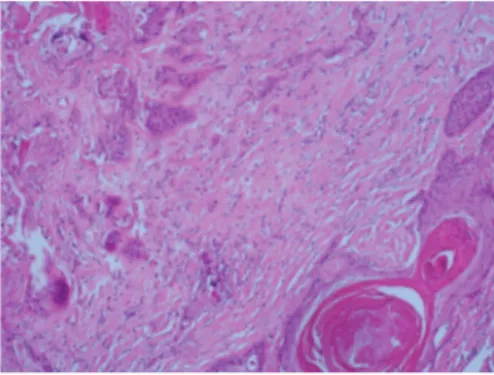
Korean J Intern Med 2016;31:411-412 http://dx.doi.org/10.3904/kjim.2015.081The patient was a 26-years-old female with a history of lumbosacral menin-gomyelocele with paraplegia. She was admitted to the hospital due to the lumbosacral wound having become infecting and producing a discharge for 6-months. The patient underwent excisional biopsy of the lumbosacral region. The pathological diagnosis was consistent with metastatic moderate-ly-differentiated squamous cell carci-noma (SCC) (Fig. 1). Fluorodeoxyglu-cose-positron emission tomography/ computed tomography (FDG-PET/CT) imaging was performed for the detec-tion and localisadetec-tion of the primary tu-mor and metastatic evaluation (Fig. 2).
The development of carcinoma asso-ciated with meningomyelocele is rare and only limited sporadic cases have been reported in the literature.
Report-ed cases were usually of SCC, as in our case. It has been proposed that malig-nancy arises at these sites as a result of chronic mechanical irritation and chronic infection, again as in our case. The term Marjolin’s ulcer is often used to describe the formation of neoplastic changes in the scar tissue of chronic ulcers. The most common cell type is SCC, and those SCCs resulting from Marjolin’s ulcers have a much great-er tendency to metastasize than those SCCs arising from other causes, which makes early diagnosis imperative.
Imaging studies are useful for the characterization of the tumor, the eval-uation of its extent, and are essential for treatment planning. FDG-PET/CT whole-body imaging is a widely used technique for the evaluation of many types of malignancies including SCC and is particularly useful for the detec-tion of the primary tumor of unknown origin. FDG-PET/CT can also identify additional sites of metastases that can alter the patient’s management. In a recent study, PET/CT was shown to be useful in differentiating Marjolin ulcer from benign inflammatory con-ditions of chronic nonhealing ulcer in burn scars and in the evaluation of the depth of invasion in Marjolin’s ulcer cases.
Fluorodeoxyglucose-positron emission
tomogra-phy/computed tomography imaging of squamous
cell carcinoma arising in a meningomyelocele
Seval Erhamamcı1, Mehmet Reyhan1, and Nebil Bal2Departments of 1Nuclear Medicine
and 2Pathology, Baskent University
Faculty of Medicine, Ankara, Turkey
Received : March 24, 2015 Revised : May 5, 2015 Accepted : May 12, 2015 Correspondence to Seval Erhamamcı, M.D. Tel: +90-332-257-0606 Fax: +90-332-257-0637 E-mail: [email protected]
Figure 1. Atypical squamous cells have ke-ratinization that infiltrating desmoblastic stroma (H&E, ×100).
412 www.kjim.org
The Korean Journal of Internal Medicine Vol. 31, No. 2, March 2016
http://dx.doi.org/10.3904/kjim.2015.081
Conflict of interest
No potential conflict of interest relevant to this article was reported.
Figure 2. (A) Maximum intensity projection, (B) sagittal fusion positron emission tomography/computed tomography (PET/CT), and (C) CT images show heterogeneously increased high fluorodeoxyglucose (FDG) uptake (maximum standardized uptake value [SUVmax], 12.7) including hypometabolic area within the lesion in the lumbosacral region due to squamous cell carcinoma, and mild FDG uptake within right axillary (SUVmax, 2.6) and bilateral inguinal (SUVmax, 3.4) lymph nodes.
![Figure 2. (A) Maximum intensity projection, (B) sagittal fusion positron emission tomography/computed tomography (PET/CT), and (C) CT images show heterogeneously increased high fluorodeoxyglucose (FDG) uptake (maximum standardized uptake value [SUVmax],](https://thumb-eu.123doks.com/thumbv2/9libnet/3955490.51474/2.892.90.816.102.516/intensity-projection-tomography-tomography-heterogeneously-increased-fluorodeoxyglucose-standardized.webp)
