Ankara Üniv Vet Fak Derg, 58, 291-294, 2011
Short Communication / Kısa Bilimsel Çalışma
Cutaneous candidiasis in two guinea pigs
Mehmet ŞAHAL1, Serkal GAZYAĞCI2, Kerem URAL3, Hakan YARDIMCI4
1Ankara University Faculty of Veterinary Medicine, Department of Internal Medicine, 4Department of Microbiology, Ankara, 2Kirikkale University Faculty of Veterinary Medicine, Department of Internal Medicine, Kirikkale, 3Adnan Menderes University
Faculty of Veterinary Medicine, Department of Internal Medicine, Aydin, Turkey.
Summary: Two approximately 6-month-old guinea pigs were admitted to the Department of Internal Medicine, Faculty of Veterinary (DIMFV), University of Ankara because of lethargy of several weeks’ duration, anorexia, bilateral allopecia, and itching.
Candida albicans, was isolated from the skin lesions. Both patients were treated with ketoconazole (10 mg/kg PO every 12 h) and
topical clotrimazole. The female guinea pig died during the seventh day of the treatment period. Key words: Candida albicans, guinea pig, clotrimazole, ketoconazole
İki kobayda Kutanöz Kandidiyazis
Özet: Yaklaşık 6 aylık iki kobay haftalardır süren letarji, iştahsızlık, çift taraflı tüy dökülmesi ve kaşıntı şikayetiyle Ankara Üniversitesi Veteriner Fakültesi İç Hastalıkları Anabilim dalına getirildi. Derideki yaralardan yapılan izolasyonda Candida albicans tespit edildi. Her iki kobaya tedavi olarak oral ketokonazol (10mg/kg, 12 saat arayla) ve topikal klotrimazol verildi. Dişi kobay tedavinin 7. gününde öldü.
Anahtar sözcükler: Candida albicans, kobay, ketokonazol, klotrimazol.
Candida albicans, an opportunistic fungal pathogen
of humans, has been described to cause systemic infections in immuno-compromised patients (2).
The presence of C. albicans, is widespread in healthy person’s mucous membranes. Adhesion, penetration and morphologic transition are the mechanisms that relates with the pathogenesis of Candida spp. Human body protects itself from Candida invasion with the nonspesific defence mechanism and additionally spesific immune response (17). The present article indicates that cutaneous candidiasis in two guinea pigs and also in the
owner of the guinea pigs.To the authors’ knowledge, this
is the first report in Ankara, Turkey and exotic animals may be one source of the Candidiasis because of the close interactions they have with the owners.
Two approximately 6-month-old guinea pigs were admitted to the Department of Internal Medicine, Faculty of Veterinary (DIMFV), University of Ankara because of lethargy of several weeks’ duration, anorexia, bilateral allopecia, and itching. The owner had initially noticed that, prior to the onset of clinical singns the owner had direct contact with the female guinea pig (guinea pig 1) and slept together at nights for the preceding weeks. According to the owner clinical signs were first noticed 3 weeks previously for 1 of the guinea pigs (female guinea
pig 1) and 2 weeks previously for the other guinea pig (male guinea pig 2) and also admitted that the owner had a complaint of itching and bilateral alopecia in her ear margins. These guinea pigs had always been fed a commercially available diet of cereals and pellets formulated for this species. The guinea pigs were not given fresh food or a mixture of fresh vegetables and there were no history of dietary indiscretion. On admission to the DIMFV, guinea pig 1 had skin lesions involving the ear margins, dorsal lumbosacral area, nose, head and neck, and vaginal discharge with exudate, licking of the vulva and perianal region. There was marked alopecia of the ears and nose with extensive and generalized skin eruptions with erythema, vesicles and scaling which were strongly suggestive of Candidiasis (Fig. 1).
Fungal cultures were made from lesions occurring in the ear margins of the guineapigs; The samples were cultured on Sabouraud’s dextrose agar containing antibacterial antibiotics (20 I.U.penicillin and 40 μg/ml streptomycin) at 25 °C and 37 °C for two weeks. The identification of the yeast species was made considering the assimilation and fermentation properties with different biochemical sources. Germ-test tube was also performed (8). Candida albicans (C.albicans) was
Mehmet Şahal - Serkal Gazyağcı - Kerem Ural - Hakan Yardımcı 292
isolated from the skin lesions of both guinea pigs and the owner (Fig. 1 and 2). In an attempt to identify the microscopic evidence of candidiasis, biopsy from the lesional parts was discussed with the owner, who declined further evaluation. Examination of the oral cavity revealed no abnormality. Fecal analysis were unremarkable. Radiography revealed increased bronchial and a miliary intersitial markings. For guinea pig1, routine haematology revealed neutrophilic-leucocytosis, neutrophils (71%) and lymphopenia (25%). Serum
biochemistry testing were unremarkable except for hyperglycemia (blood glucose levels; 184 mg/dl and 193 mg/dl, for guinea pig 1 and 2).
The 6-month old male guinea pig (guinea pig 2) had skin lesions around the ear margins similar to guinea pig 1 (Fig 1 c-d). The guinea pig was afebrile and there was a moderate discomfort at respiration. Radiography was unremarkable.
Both guinea pigs of this report had cutaneous Candidiasis and guinea pig 1 had also pneumomycosis
Fig.1. The guinea pig 1:Extensive and generalized skin eruptions with erythema, vesicles and scaling on the dorsolumbosacral area (a), nose (b) and The guinea pig 2:ear margins (c-d).
Şekil 1. Kobay 1 de: dorso lumbal bölgede (a) ve burunda(b) Kobay 2 de: kulak kenarlarında yoğun ve yaygın eritem, vezikül oluşumu ve pullanma şeklinde görülen deri yıkımlanması.
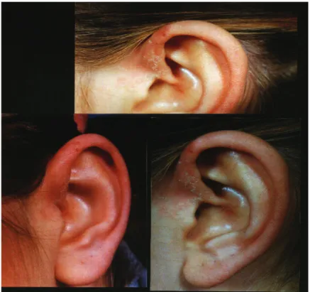
Fig.2. The dermatitic changes on the ear margin in the owner of the guinea pigs: Squamatosus desquamation on the erythematosus base.
Şekil 2: Kobay sahibinde kulak kenarlarında görülen dermatik değişiklikler: skuamatöz deskuamasyon ve bazal eritamatoz.
Ankara Üniv Vet Fak Derg, 58, 2011 293
due to C. albicans. Both patients were treated with ketoconazole (10 mg/kg PO every 12 h for five weeks) and topical clotrimazole solution and vitamin C 30 mg/kg i.m daily. The owner had noticed that the guinea pig 1 was dead 1 week after initial examination. This may be due to the fact that there was severe pneumomycosis in the guinea pig1. The guinea pig 2 was still alive 2 months after diagnosis and is still well at the time of writing.
Contact sensitivity to Candida antigen has an important role in the formation of skin lesions in humans who are predisposed to C. albicans. It has been reported that in humans contact sensitivity to Candidiasis occurs
during later life (19).However the owner of the guinea
pigs of this report, who also had Candidiasis, was 18-year-old and had no history of a particular infection in the past or any other systemic disease. In a previous study in guinea pigs and in humans dermatitic changes has been induced even on normal skin with a highly concentrated antigen solution (19) As in the present report, to the author’s knowledge the actual cutaneous Candidiasis in the guinea pigs has been generated by this procedure and the owner of the guinea pigs began to show contact sensitivity to C. albicans antigen with the development of hypersensitivity reactions. Although the underlying inflammatory process that facilitates the contact sensitivity interacted between the owner and the guinea pigs of this report was not certain and fortuitous, it seems more likely that the guinea pigs may be the source of the infection.
In guinea pigs described in the present report blood parameters are in accordance with those published by Laird (7) and Quillec et al. (15), except for neutrophilic-leucocytosis (71%) and lymphopenia (25%) on guinea pig 1. To the authors’ knowledge severe stress and/or a secondary bacterial infection may be associated with the latter changes. Considering the serum biochemical changes, serum biochemical profile was unremarkable except for hyperglycemia in both of the present guinea pigs. However diabetes mellitus was ruled out in the present cases as the causes of hyperglycemia. This may be due to the fact that diabetes mellitus is a rare condition in guinea pigs with the exception of colonies prone to genetic conditions or unidentified infectious agent (4).
In a previous report with albino guinea pigs experimentally infected with C. albicans, one of the animals presented extensive pneumonia due to C.
albicans and B. bronchiseptica (23).For the guinea pigs report, vaginitis evident on initial examination and the radiological changes were unusual in cutaneous candidiasis. These radiological changes in the guinea pig 1 of this report, although non-spesific, are crude means of pneumomycosis in aggreement with the latter report.
In guinea pigs, vitamin C deficiency has been reported to be the most commonly disorder affecting
bone metabolism (3,14). Low levels of dietary ascorbic acid increase susceptibility to Candidiasis (16). Therefore in the present article although the guinea pigs had always been fed a commercially available diet with enough vitamin C levels, it was considered that treatment should include vitamin C supplementation as signs of conjunctivitis or upper respiratory disease should evoke deficiency (4).
Experimental cutaneous C. albicans infection in guinea pigs have been reported previously (9-11,18,20,24). It has been reported that the rate of translocation of C. albicans increases with the thermal injury in guinea pigs (5). However in guinea pigs of our report, thermal injury was ruled out as there was no
history of burn injury. Kitabatake et al.(6) described that
exposure to NO2 or SO2 increases bronchopulmonary
reactions in guinea pigs induced by Candida albicans. However the guinea pigs described in the present report
had no history of exposure to NO2 or SO2. Experimental
cutaneous Candidiasis were produced in prednisolone-induced infections in guinea pigs (11). Prednisolone treatment was not associated with the Candidiasis in the present report since the guinea pigs had not received such therapy.
Therapeutic efficacies of various antimycotic agents such as; lanoconazole, ketoconazole, neticonazole, Sch 39304, itroconazole in systemic and experimental Candidiasis of guinea pigs have been described by
previous studies (11-13,20-23).It has been reported that
itroconazole is a potential prophylactic therapy agent against fungal infections, and also admitted that profilactic efficacy increases as its dosage increases (21). In combination therapy with nystatin and zinc oxide, the latter agent has been described to have some protective effects against local masseration in cutaneous candidiasis (1). In a previous study evaluating the efficacy of orally and topically administrated ketoconazole, oral route has been found to be efficacious (19). Both guinea pigs of this report had been treated with oral ketoconazole and C.
albicans was eradicated from the guinea pig 2 after
administration of ketoconazole. To the authors’ knowledge the guinea pig 1 of this report died because of severe pneumomycosis although guinea pig 2 responded to therapy.
References
1. Auger P, Colin P, Joly J, Poirier S, Colin D (1989): Treatment of cutaneous candidiosis in guinea pigs: effect of zinc oxide on the antifungal efficacy of nystatin. Mycoses, 32, 455-460.
2. Beck-Sague CM, Jarvis WR (1993): Secular trends in the epidemiology of nosocomial fungal infections in the United States, 1980-1990. National Nosocomial Infections Surveillance System. J Infect Dis, 167, 1247-1251. 3. Clarke GL, Allen AM, Small JD, Lock A (1980):
Subclinical scurvy in the Guinea pig. Vet Pathol, 17, 40-44.
Mehmet Şahal - Serkal Gazyağcı - Kerem Ural - Hakan Yardımcı 294
4. Harkness JE, Murray KA, Wagner JE (2002): Biology and Diseases of Guinea Pigs. In: Fox, J.G., Anderson, L.C., Loew, F.M., Quimby, F.W eds. Laboratory Animal Medicine 2nd ed. Academic Pres. 6, 203-246.
5. Inoue S, Peck MD, Alexander JW (1991): Fungal translocation is associated with increased mortality after thermal injury in guinea pigs. J Burn Care Rehabil, 12, 19-22.
6. Kitabatake M, Yamamoto H, Yuan PF, Manjurul H, Murase S (1995): Yamauchi T.: Effects of exposure to NO2 or SO2 on bronchopulmonary reaction induced by
Candida albicans in guinea pigs. J Toxicol Environ Health, 45, 75-82.
7. Laird CW (1974): Clinical Pathology In: Melby, E.C.Jr., Altman, N.H. eds. Handbook of Laboratory Animal Science CRC Pres, Cleveland 2: 345-436.
8. Larone DH (2005): Medically important fungi. A guide to identification. New york Elsevier pres.
9. Lehner T, Wilton JM, Ivanyi L (1972): Immunodeficiencies in chronic muco-cutaneous candidiosis. Immunology, 22, 775-787.
10. Maebashi K, Itoyama T, Uchida K, Suegara N, Yamaguchi H (1993): Therapeutic efficacies of neticonazole (SS717) cream and solution in experimental cutaneous Candida albicans infection of guinea pigs. Jpn J Antibiot, 46, 896-903.
11. Maebashi K, Itoyama T, Uchida K, Suegara N, Yamaguchi H (1994): novel model of cutaneous candidiasis produced in prednisolone-treated guinea pigs. J Med Vet Mycol, 32, 349-359.
12. Niwano Y, Seo A, Kanai K, Hamaguchi H, Uchida K, Yamaguchi H, Uchida K, Yamaguchi H (1994): Therapeutic efficacy of lanoconazole, a new imidazole antimycotic agent, for experimental cutaneous candidiasis in guinea pigs. Antimicrob Agents Chemother, 38, 2204-2206.
13. Parmegiani RM, Loebenberg D, Cacciapuoti A, Antonacci B, Norris C, Menzel F, Moss L, Yarosh-Tomaine T, Hare RS, Miller GH (1993): Sch 39304, a new antifungal agent: oral and topical treatment of vaginal and superficial infections. J Med Vet Mycol, 31, 239-248. 14. Percy DH, Barthold SW (1993): The Guinea pig.
Nutritional, metabolic, and other disorders. 165-172. In: Percy Dh, Barthold Sw, eds. Pathology of laboratory rodents and rabbits. Ames, Iowa state University Pres.
15. Quillec M, Debout C, Izard J (1977): Red cell and white cell counts in adult female guinea pigs. Pathol Biol, 25, 443-446.
16. Rogers TJ, Adams-Burton K, Mallon M, Hafdahl B, Rivas V, Donnelly R, O'day K (1983): Dietary Ascorbic acid and resistance to experimental renal candidiasis. Am J Clin Nutr, 113, 178-183.
17. Seeliger HPR, Patzelt C (1991): Host-Parasite Interaction-Mechanisms of Pathogenesis. 55-59. In:Tumbay E, Seeliger HPR, Ang O, eds. Candida and Candidamycosis. Federation of Europan Microbiological Societies Symposium Series; NewYork, London Plenum Press. 18. Sohnle PG, Frank MM, Kirkpatrick CH (1976):
Mechanisms involved in elimination of organisms from experimental cutaneous Candida albicans infections in guinea pigs. Am J Immunol, 117, 523-530
19. Tagami H, Urano-Suehisa S, Hatchome N (1985): Contact sensitivity to Candida albicans-comparative studies in man and animal (guinea-pig). Br J Dermatol, 113, 415-4248.
20. Thienpont D, Van Cutsem J, Borgers M (1980): Ketoconazole in experimental candidosis. Rev Infect Dis, 2, 570-577.
21. Van Cutsem J (1994): Prophylaxis of Candida and
Aspergillus infections with oral administration of
itraconazole. Mycoses 37, 243-248.
22. Van Cutsem J, Fransen J, Janssen PA (1987) Therapeutic efficacy of itraconazole in systemic candidosis in guinea pigs. Chemotherapy, 33, 52-60.
23. Van Cutsem J, Fransen J, Van Gerven F, Janssen PA (1985). Oral treatment with ketoconazole in systemic candidosis of guinea-pigs: microbiology, hematology and histopathology. Sabouraudia, 23, 189-198.
24. Van Cutsem J, Thienpont D (1971): Experimental cutaneous Candida albicans infection in guinea pigs. Sabouraudia, 9, 17-20.
Geliş tarihi: 11.11.2010 / Kabul tarihi: 22.03.2011
Adress of the correspondence:
Assist. Prof. Dr. Serkal Gazyagci
Kirikkale University, Faculty of Veterinary Medicine, Department of Internal Medicine,
71450, Yahsihan, Kirikkale, Turkey. E-mail: [email protected]