207
Ankara Üniversitesi Tıp Fakültesi Mecmuası 2008, 61(4) DAHİLİ BİLİMLER / MEDICAL SCIENCES
Olgu Sunumu / Case Report
Functional Parathyroid Adenoma Diagnosed After Stress
Fractures
Stres Kırıkları Sonrasında Tanı Alan Fonksiyonel Paratiroid Adenomu
Nuray Haliloğlu, Esra Özkavukçu, Gülden Şahin
Ankara Üniversitesi Tıp Fakültesi, Radyodiagnostik Anabilim Dalı
Received: 04.09.2007 • Accepted: 19.04.2009 Corresponding author
Uzm. Dr. Nurat Haliloğlu
Ankara Üniversitesi Tıp Fakültesi, Radyodiagnotik Anabilim Dalı Phone : +90 (312) 595 67 81
E-mail address : [email protected]
Clinical findings and radiographs may be disleading or indefinite in patients with insuff iciency fractures. Herein, we present a patient with low back pain increasing with activity and morning stiff ness, which initially were thought to be due to a spondyloarthropathy. Magnetic resonance imaging (MRI) revealed insuff iciency fractures in the sacrum and iliac wings bilaterally. Finally, laboratory and imaging findings depicted a parathyroid adenoma causing hyperparathyroidism and the adenoma was removed surgically.
Key Words: stress fractures, parathyroid adenoma
Yetmezlik kırıklarında klinik ve radyografik bulgular her zaman belirgin olmayabilir ya da doktoru yanlış yönlendirebilir. Biz burada hareketle artan bel ağrısı ve sabah tutukluğu nedeni ile başvu-ran ve ilk etapta spondiloartropati lehine değerlendirilen bir hastayı sunuyoruz. Yapılan manyetik rezonans görüntüleme (MRG) ile sakrum ve bilateral iliak kanatta yetmezlik kırıkları saptanan has-tada sonuçta laboratuvar ve görüntüleme bulguları ışığında hiperparatiroidizme neden olan bir paratiroid adenomu tesbit edildi ve cerrahi olarak çıkarıldı.
Anahtar Kelimeler: stres kırıkları, paratiroid adenomu Insufficieny fractures which are a
sub-group of stress fractures, occur by normal or physiologic stress up-loaded on an abnormally weakened bone. Hyperparathyroidism is one of the risk factors of insufficiency fractures besides diseases like post-menopausal osteopenia, rheuma-toid arthritis, osteomalacia, and fi-brous dysplasia. Clinical findings and radiographs may not always be adequate for the diagnosis of sac-ral insufficiency fractures. The pa-tient we present here had low back pain increasing with activity and morning stiffness which in the first place were thought to be due to a spondyloarthropathy. Magnetic res-onance imaging (MRI) showed the insufficiency fractures in the sac-rum and iliac wings. The laboratu-ary and imaging findings depicted a parathyroid adenoma causing hy-perparathyroidism and the adeno-ma was removed surgically.
Case
A 50-year-old woman was admitted to the rheumatology department with a 3 months history of low back pain which increased with exercise and morning stiffness of 15 minutes. No signs of inflam-matory bowel disease or uveitis were present. In the physical ex-amination there was no neuro-logic deficit but the range of mo-tion of the lumbosacral joint was impaired due to severe pain. Be-sides routine hematological and biochemical analyses immuno-logical markers, HLA B27 antigen and protein electrophoresis were also performed in order to elimi-nate a spondyloarthropathy. The laboratory findings included an in-crease in alkaline phosphatase lev-el (438U/l) but the immunological markers and protein
electrophore-Ankara Üniversitesi Tıp Fakültesi Mecmuası 2008, 61(4)
208 Functional Parathyroid Adenoma Diagnosed After Stress Fractures
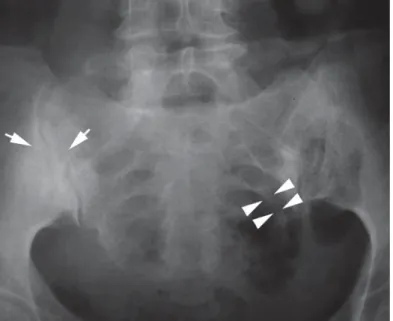
sis were normal and HLA B27 an-tigen was negative. There was marked sclerosis of the right sacro-iliac joint on the plain radiographs of the pelvis (Figure 1). Sacroiliac MRI was performed to reveal pos-sible sacroiliitis. MRI showed a lin-ear low signal intensity area, most-ly evident on T1-weighted images on the left sacral wing (Figure 2a) with associated bone marrow ede-ma on the left sacral wing and ili-ac bone on FSEIR images (Figure 2b) which were found to be con-cordant with stress fractures. MRI also revealed low signal intensity areas without accompanying bone marrow edema on the bilateral il-iac wings (Figure 2a,b) and these were consistent of with healed old fractures. In the retrospective evaluation of the plain pelvic ra-diographs, a linear fissure on the left sacral wing was also noticed (Fig.1). In the further biochemical analyses which were performed to evaluate the high alkaline phos-phatase level, there was also an increase in the serum calcitonin (15.9pg/ml), osteocalcin (44.7 ng/ ml) and parathormone (232 pg/ ml) levels and a decrease in 25-Dihydroxy vita-min D (6mg/L). Serum calcium level was within the normal lim-its (9.2 mg/dl). The patient was presumed to have secondary h y p e r p a r a t h -yroidism due to serum vita-mine D insuffi-ciency and de-pot vitamine D and Calcimax® t r e a t m e n t was admin-istered. Con-trol laborato-ry analyses in a four month period revealed an improvement in the serum par-athormone and alkaline phos-phatase levels although they were not within the normal limits. The serum calcium level had also in-creased slightly over the normal limits. Ultrasonographic and scin-tigraphic examinations revealed a parathyroid adenoma which was removed surgically. No postoper-ative complications occured. The final diagnosis was parathyroid
adenoma and primary hyperpar-athyroidism. In a one year follow-up the patient was symptoms free, and her biochemical analyses were within normal limits.
Discussion
Primary hyperparathyroidism refers to the excessive production of parathormone which leads to in-creased resorption of bone, and usually causes hypercalcemia. In 80-85% of the patients, prima-ry hyperparathyroidism is due to benign parathyroid adenomas. Hyperplasia of the parathyroid glands, and rarely parathyroid car-cinoma are other causes of prima-ry hyperparathyroidism. Patients may be asymptomatic or may have nonspecific symptoms including muscle weakness, myalgia, thirst, polyuria, etc. Diagnosis is usual-ly made by biochemical screening showing the increased levels of parathormon which leads to hy-percalcemia, hypophosphatemia, and hypercalciuria. Sestamibi scintigraphy scan, single photon emission computed tomography imaging, and/or neck ultrasound would help the surgeon to dem-onstrate the presence and loca-Figure 1. Plain pelvic radiogram showing sclerosis on the right sacroiliac
joint (arrows). In the retrospective evaluation of the radiogram, an indis-tinct linear fissure on the left sacral wing was also noticed (arrow heads).
Figure 2. Coronal T1-weighted MR image shows a linear low signal intensity area (a, arrows), with associated bone marrow edema on the left sacral wing and iliac bone on coronal FSEIR image (b, arrows), which were found to be concordant with stress fractures. There is also a low signal intensity line without accompanying bone marrow edema on the right iliac wing (arrow heads on a and b) consistent of with healed old fracture.
Journal of Ankara University Faculty of Medicine 2008, 61(4)
209
Nuray Haliloğlu, Esra Özkavukçu, Gülden Şahin tion of the parathyroid adenoma or the abnormal gland before sur-gery (1,2).
It should be kept in mind that an in-cidentally diagnosed insufficiency fracture may be an outcome of un-diagnosed endocrinological dis-orders as well as other benign or malignant diseases. Postmenopau-sal osteopenia, prolonged corti-costeroid treatment, pelvic irradi-ation, Paget disease, osteopetro-sis, metabolic bone diseases like osteomalacia and hyperparathy-roidism may be causes of insuffi-ciency fractures (3). Multipl frac-tures may rarely be the onset of primary hyperparathyroidism and they usually represent late presen-tation of the disease (2). Chron-ic vitamin D defChron-iciency as seen in our patient, is reported to be a risk factor for aggrevating the parathy-roid adenoma growth, parathor-mone secretion and increased bone turnover (4,5).
The sacrum and iliac bones are the major weight-bearing structures of the body. Pelvic ring is the main site of insufficiency fractures and sacrum is mostly involved (6,7).
Sacral fractures frequently occur in elderly women and the most common risk factor is osteoporo-sis. Low back pain with or without associated groin, hip or buttock pain is usually the leading clinical symptom. Physical examination and plain radiographs are indefi-nite or misleading in the vast ma-jority of the cases. Most of the pa-tients do not recall a major trauma and many of them are diagnosed incidentally by computed tomog-raphy (CT), MRI, skeletal scintig-raphy or positron emission tom-ography (PET) studies performed for other reasons (8). The patient we present here also had unsus-pected fractures which were diag-nosed definitely by MRI, and there was no need for further radiologi-cal examination.
The sensitivity of the plain films has been reported as 34-37%. On the contrary, scintigraphy has a high-er sensitivity (92.6%-97.3%) in the relevant literature and it sufficient-ly shows the sacral fracture (7). The typical CT appearance of a stress
fracture is a focal callus formation and endosteal thickening around
a fracture line (8). CT scan should not be the initial examination technique and must be saved for the patients whose MRI imaging does not demonstrate the fracture line (7,9).
As MRI is widely being used for the patients with hip pain radiologists must be aware of the MR appear-ance of sacral stress fractures. The fracture line is typically hypoin-tense on both T1- and T2-weight-ed images and in most of the cases there is a area of high signal inten-sity surrounding the fissure on T2-weighted sequences. This hyper-intense area resembles inflamma-tion or bone marrow edema seen in acute fracture (6).
In conclusion; MRI is a highly sensi-tive and spesific technique in the diagnosis of stress fractures. When multipl insufficiency fractures are detected, primary hyperparathy-roidism should be remembered in the differrential diagnosis, and serum biochemical screening and neck examination are important investigations for detecting the underlying parathyroid adenoma.
REFERENCES
1. DeLellis RA, Mazzaglia P, Mangray S. Primary hyperparathyroidism a cur-rent perspective. Arch Pathol Lab Med 2008;132:1251–1262.
2. Hussain A, Mahmood H, Geddoa E, Hamish M, Reda AH. Parathyroid ade-noma and hypercalcemia in a patient presenting with multiple pathologi-cal fractures of the long bones. Int J Surg 2008;6:465-468.
3. Peh WJG, Khong PL, Yin Y, et al. Ima-ging of pelvic insuffiency fractures. Radiographics 1996;16:335-348. 4. Raef H, Ingemansson S, Sobhi S, et
al. The effect of vitamin D status on the severity of bone disease and on
the other features of primary hyper-parathyroidism (pHPT) in a vitamin D deficient region. J Endocrinol In-vest 2004;27:807-812.
5. Moosgaard B, Vestergaard P, He-ickendorff L, et al. Plasma 25-hydroxyvitamin D and not1,25-dihydroxyvitamin D is associated with parathyroid adenoma secretion in primary hyperparathyroidism: a cross-sectional study. Eur J Endocri-nol 2006;155:237-44.
6. Kanberoglu B, Kantarci F, Cebi D, et al. Magnetic resonance imaging in osteomalacic insufficiency fractu-res of the pelvis. Clin Radiol 2005; 60:105-11.
7. Soubrier M, Dubost JJ, Boisgard S, et al. Insufficiency fracture. A survey of 60 cases and review of the literature. Joint Bone Spine 2003;70:209-18. 8. Fayad LM, Kamel IR, Kawamoto S,
et al Distinguishing stress fractures from pathologic fractures: a multi-modality approach. Skeletal Radiol 2005; 34:245–259.
9. Groves AM, Cheow HK, Balan KK, et al. 16-Detector multislice CT in the detection of stress fractures: a com-parison with skeletal scintigraphy. Clin Imag 2006;30:149.