Ankara Üniv Vet Fak Derg, 57, 267-270, 2010
Short Communication / Kısa Bilimsel Çalışma
Dermal squamous cell carcinoma (avian keratoacanthoma) in
parakeets
M. Ziynet YILDIRIM1, Sibel E. BILGIHAN OZTURK2, Zeynep PEKCAN3, Osman KUTSAL4,
Sevil ATALAY VURAL4
1District Branch of Ministry of Agriculture and Rural Affairs, İkizce, Ordu; 2Pet Veterinary Hospital, Bahçelievler, Ankara; 3Department of Surgery, Faculty of Veterinary Medicine, Kırıkkale University, Kırıkkale; 4Department of Pathology, Faculty of
Veterinary Medicine, Ankara University, Ankara.
Summary: Tumor suspected operation materials obtained from eight parakeets are evaluated clinically, pathologically and
immunohistochemically. The age of the parakeets varied from seven months to nine years old. Four were male, one was a female and the sexes of the rest three were not identified. Six of the operation materials were taken from the above the tail while the other two from beneath the wings. Evaluated tumors were observed to be hard in consistency and whitish color on cut surface. At histopathological examination; it is seen that hyperchromatic nucleated, big, ovoid shaped, pale eosinophilic cytoplasmated cells had formed groups in dermis. Keratinized horn pearl formations are identified in the centers of most of these cell groups. Within the horn pearl formations hyperkeratotic, parakeratotic changes and calcifications are found. In some cases inflammation areas consisting of heterophil leukocytes, lymphocytes and plasma cells in the dermis are observed. Also in histopathological examination of one case, tubercle formations made of coagulation necrosis in the center surrounded by heterophil leukocytes, macrophages, epithelioid cells, lymphocytes and foreign body giant cells are found as well as tumoral formations. Groups of cudgel shaped, red colored bacteria are found in Ziehl-Neelsen staining of the sections. Tissues are stained for proliferating cell nuclear antigen (PCNA) and p53 proteins immunohistochemically in order to interpret mitotic activity and malignancy of the tumor. TUNEL method is applied to the tissues to evaluate apoptosis.
Key words: Dermal squamous cell carcinoma, histopathology, immunohistochemistry, keratoacanthoma, parakeet.
Muhabbet kuşlarında dermal yassı hücreli kanser (keratoacanthoma)
Özet: Tümör şüpheli sekiz muhabbet kuşundan alınmış operasyon materyalleri klinik, patolojik ve immunhistokimyasal olarak
değerlendirildi. İncelenen muhabbet kuşları 7ay- 9 yaş arasındaydı ve dördü erkek, biri dişi iken üç tanesinin cinsiyeti bildirilmemişti. Operasyon materyallerinin altısı kuyruk üzerinden ikisi ise kanat altından alınmıştı. İncelenen tümörlerin sert kıvamlı ve kesit yüzlerinin ise beyazımtırak renkte olduğu gözlendi. Tümöral kitlelerin histopatolojik incelenmesinde; dermiste hiperkromatik çekirdekli, büyük, ovoid şekilli, solgun eozinofilik sitoplazmalı hücrelerin gruplar oluşturduğu dikkati çekti. Bu hücre gruplarının bir çoğunun orta kısımlarında keratinize glob korne yapıları belirlendi. Glob korne yapıları içerisinde hiperkeratotik ve parakeratotik değişiklikler ile kalsifikasyonlara rastlandı. Olguların bazılarında dermiste nötrofil lökosit, lenfosit ve plazma hücrelerinden oluşan yangısal alanlar görüldü. Ayrıca bir olguda, saptanan tümörden başka aynı hayvandan alınan kitlelerin histopatolojik incelenmesinde ise; ortada koagulasyon nekrozu alanının etrafında heterofil lökositler, histiyositler, epiteloid hücreler, lenfositler ve yabancı cisim dev hücrelerinin oluşturduğu tüberkül yapısına rastlandı. Yapılan Ziehl-Neelsen boyamasında kırmızı renkli, çomak şekilli bakteri kümeleri görüldü.
Mitotik aktiviteleri ve malignitelerini yorumlamak üzere dokular PCNA ve p53 proteinleri ile immunhistokimyasal olarak boyandı. Apoptozisin değerlendirilmesi için TUNEL metodu uygulandı.
Anahtar sözcükler: Dermal yassı hücreli kanser, histopatoloji, immunohistokimya, keratoacanthoma, muhabbet kuşu.
True squamous cell carcinoma is encountered rarely in birds and is mainly reported from aged birds of various species at different locations such as legs, pharyngeal cavity and head and is a malignant tumor of kerotinocytes, forming irregular masses or cords that proliferate downwards and invade cutis and subcutis (8). On the other hand; dermal squamous cell carcinoma
(DSCC; avian keratoacanthoma) is a neoplastic skin lesion with unknown etiology (5, 6, 7, 11). DSCC is generally encountered in broiler chickens during slaughtering (10) and is differed from true squamous cell carcinoma by not showing malignancy and regression with the time progress (5). The etiology of naturally occurring DSCC is not known but experimentally
M. Ziynet Yildirim - Sibel E. Bilgihan Ozturk - Zeynep Pekcan - Osman Kutsal - Sevil Atalay Vural 268
repeated application of methylcholanthrene and some viral agents such as Avipox and Avian Leucosis Viruses are thought to play a role (4, 5). Although the pathogenesis is not clear; the lesions are considered to have started from hyperplasic feather follicles and infiltration of the peripheral follicles into the dermis or formation of small follicular cysts whose walls are made of kerotinocytes. An obvious fibroplasia is observed with many inflammatory cells surrounding the lesion (5).
Macroscopically these lesions look like crater-shaped ulcers with raised margins (10). Small nodular lesions may also occur and mimic enlarged feather follicles (4, 5).
In histopathological examination nodular lesions are seen as proliferations of or cysts originated from feather follicle epithelium or hyperplastic feather follicles consisting of hyperkeratotic feathers. Ulcereated areas are encountered as keratin, bacteria, exfoliated epithelium and inflammatory cells. Epithelial edges surround the keratin in the middle. Kerotinocytes in the outer layer either keratinize to the middle or join the dermal fibroplasia. Inside the fibroblastic area surrounding the lesion keratinocytes, heterophil leukocytes, macrophages and perivascular lymphocytes are observed (5).
The p53 tumor suppressor gene is a transcription factor that regulates the expression of genes involved in cell-cycle control, apoptosis in cells with defective deoxyribonucleic acid (DNA), cellular differentiation and genetic instability. In malignancies, point mutations occur on this gene and this mutated p53 gene looses its suppressor function, coding mutated p53 protein which accumulates in affected cells of malign tumors while this property is not observed in benign ones. For this p53
protein is demonstrated by immunohistochemistry (IHC) in various carcinomas and sarcomas (1, 3)
Proliferating cell nuclear antigen (PCNA), whose levels in the nucleus are increased in the S-G2 and M phases of mitosis, is a nuclear nonhistone protein functioning in the initiation of cell proliferation by augmenting DNA polymerize (9).
Apoptosis or programmed cell death is a form of cell death which can be either physiologic or pathologic and is characterized by cell shrinkage, rapid condensation of nuclear chromatin and cytoplasm, convolution of the cell and subsequent separation of the cell into apoptotic bodies (2).
Recently, parakeets are chosen more commonly as pet animals as a part of our daily city lifes. As these animals stay with us for longer periods; tumors or tumoral-like lesions are encountered more frequently in veterinary clinics. Due to acquired literature, this study is believed to be the first DSCC case reported from parakeets as DSCC cases were previously reported mainly from chicken.
That’s why DSSC cases encountered in eight parakeets that were sent to the Pathology Department of Veterinary Medicine of Ankara University between June 2003 and December 2005 are evaluated clinically, pathologically and immunohistochemically for the veterinarians working in the field.
Case materials consisted of eight parakeets that were brought to the Surgery Clinics of Veterinary Medicine Faculty of Ankara University by their owners with complains of disorder in the feathers and nodular formations with an history of 2 weeks to 2 months. The age of the parakeets were as follows; 7 months (case
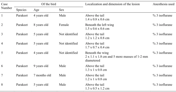
Table 1. Clinico-pathological information of the parakeets. Tablo 1. Muhabbet kuşlarının klinikopatolojik bilgileri.
Of the bird Case
Number Species Age Sex Localization and dimension of the lesion Anesthesia used 1 Parakeet 4 years old Male Above the tail
1.4 x 0.8 x 0.6 cm % 3 isoflurane
2 Parakeet 8 years old Female Beneath the left wing 1.5 x 0.6 x 0.6 cm
% 3 isoflurane 3 Parakeet 5 years old Not identified Above the tail
1.2 x 1.2 x 0.8 cm
% 3 isoflurane 4 Parakeet 5 years old Not identified Above the tail
1.7 x 0.7 x 0.4 cm
% 3 isoflurane 5 Parakeet 4 years old Not identified Beneath the wing
2 x 1.1 x 1.8 cm and 5 more masses of 1-2 mm diametered
% 3 isoflurane
6 Parakeet 9 years old Male Above the tail 1.3 x 1 x 0.8 cm
% 3 isoflurane 7 Parakeet 7 months old Male Above the tail
1.2 x 1 x 0.8 cm % 3 isoflurane
8 Parakeet 5 years old Male Above the tail
Ankara Üniv Vet Fak Derg, 57, 2010 269
number 7), 4 years (case number 1 and 5), 5 years (case number 3, 4 and 8), 8 years (case number 2) and 9 years (case number 7) old. Four were male (case number 1, 6, 7 and 8), one was a female (case number 2) and the sexes of the rest (case number 3, 4 and 5) were not recorded (Table 1).
The birds are anesthetized with 3% isoflurane, nodular lesions are removed and sutured with 6-0 absorbable material. The duration of the operations varied from half an hour to one hour. The birds are reanimated and wide-ranged antibiotics are used in order to prevent post operative complications. Elizabeth collar is used during the healing process.
Operation materials are stored in 10 % neutral formaldehyde solutions and sent to the Pathology Department of Veterinary Medicine of Ankara University to be examined. The tumoral masses are processed routinely, cut 5µ and stained with hematoxylin and eosin (HxE). Ziehl-Neelsen staining is performed to one of the sections in which also a tubercle formation is encountered. Standard avidin-biotin complex peroxidase (ABC-P) method (Dako Corporation) with p53 protein and Proliferating Cell Nuclear Antigen (PCNA) (Dako Corporation) and TUNEL method (Roche, Enzo) are also applied to the sections as instructed in their catalogues. Negative controls are run by omitting the primary antibodies.
Clinical findings: In 6 cases the masses were
observed above the tail and in 2 cases they were beneath the wings. It is observed that the nodular formations located beneath the wings impeded flying and the ones above the tail were ulcerated (Figure 1a).
Macroscopical findings: Operation materials (Table
1) were yellowish white colored and lobulated on cut surface (Figure 1b). In case number five; 1-2 mm diametered 5 more masses were also encountered.
Microscopical findings: Hyperchromatic nucleated,
big, ovoid shaped, pale eosinophilic cytoplasmated cells had formed groups in dermis. Keratinized horn pearl formations are identified in the centers of most of these
Fig. 1a. Appreance of tumor above the tail in a parakeet before surgery (arrow) (case no: 8). Fig. 1b. Cut surface of the tumoral mass taken from above the tail (case no: 5).
Fig. 2a. Microscopical appereance of tumoral cell groups (thick arrow) and newly started keratinization (thin arrow) (case no: 6). HxE. x100.
Fig. 2b. Wide keratinized areas (k) (case no:3). HxE. x 40.
Fig. 2c. Inflammatory cells. Lymphocytes (thick arrow) and heterophil leucocytes (thin arrow) (case no:7). HxE. x 100. Fig. 2d. Calcification (arrow) (case no: 5). H xE. x 40.
Fig. 3. (a). Negative staining with p53 protein (case no: 1), (b). PCNA (case no:6); (c). Intense staining by TUNEL (case no: 6) and (d) (case no: 4) x 100.
Şekil 1a. Bir muhabbet kuşunda cerrahi girişimden once kuyruğun üstündeki kitlenin görünümü (ok) (vaka no: 8) Şekil 1b. Kuyruk üzerinden alınan tümoral kitlenin kesit yüzü (vaka no: 5)
Şekil 2a. Tümöral hücre gruplarının mikroskobik görünümü (kalın ok) ve yeni başlayan keratinizasyon (ince ok) (vaka no: 6) Şekil 2b. Geniş keratinize alanlar (k) (vaka no: 3) HE, x40
Şekil 2c. Yangı hücreleri: Lenfositler (kalın ok) ve heterofil lökosit (ince ok) (vaka no: 7) HEx100 Şekil 2d. Kalsifikasyon (ok) (vaka no: 5) HEx40
Şekil 3. (a) p53 proteini ile negatif boyanma (vaka no: 1), (b) PCNA (vaka no: 6), (c) TUNEL ile yoğun boyanma (vaka no: 6) ve (d) (vaka no: 4) x100
M. Ziynet Yildirim - Sibel E. Bilgihan Ozturk - Zeynep Pekcan - Osman Kutsal - Sevil Atalay Vural 270
cell groups but the degree of keratinization varied from case to case. In some cases it has newly started (Fig 2a) while in some wide keratinized areas (Fig 2b) are observed. Within the horn pearl formations hyperkeratotic and parakeratotic changes are found. In 4, 5 and 7 numbered cases, inflammatory cells mainly composed of heterophil leukocytes, lymphocytes (Fig 2c) and plasma cells are observed. Calcifications (Fig 2d) as well as hyperkerototic and parakeratotic changes within keratinized horn pearl formations are seen in these cases. In histopathological examination of other small masses in case number 5, tubercle formations made of coagulation necrosis in the center surrounded by heterophil leukocytes, macrophages, epithelioid cells, lymphocytes and foreign body giant cells are found as well as tumoral formations. Groups of cudgel shaped, red colored bacteria are found in Ziehl-Neelsen staining of the sections.
Immunohistochemical findings: There were no
staining with p53 protein (Fig 3a) and PCNA (Fig 3b). On the contrary neoplastic cells were intensely stained by TUNEL (Fig 3c, Fig 3d).
Having similar histological appearance with the squamous cell carcinoma of mammalian species, showing different characteristics; DSCC cases are mainly encountered in broilers during slaughtering (5, 6, 7, 8, 10). It is interesting to observe DSCC in eight parakeets when considered that they are mainly reported from broilers.
Due to acquired literature knowledge DSCC cases are mostly reported from young animals. (6, 7, 10). It is hard to make a comment as there were not enough materials but in our cases the birds were four or older than four years old mainly except for only one case with seven months old.
Pectoral, dorsopelvic or femoral localizations of DSCC were reported from broilers (7) but in six paarkeets the lesions were encountered above the tail while in the other two they were beneath the wings.
Dermal squamous cell carcinoma cases are known to regress with the time progress (5). We were not able to make a comment as all the diagnoses were performed on operation materials.
In the present study the tissue sections are stained immunohistochemically but there were no staining with p53 protein and PCNA. Not obtaining positively staining with these markers is found to confirm the literature knowledge stating that dermal squomaus cell carcinomas showing benign characteristics are different from real squamous cell carcinomas showing malignity in birds. At the same time negative staining with PCNA is interpreted as the rarity or the absence of cellular proliferation. Intensely staining of apoptotic cells is found. Apoptosis, which is observed both in physiological or pathological conditions, occurs due to control mechanisms of death cell gene or genes and this, at the same time also confirms the negativity of p53 protein staining.
The diseases of parakeets are faced more frequently in veterinary medicine as these birds are nowadays commonly preferred pet animals. Eight cases observed in parakeets and found histopathologically similar with the DSCC cases reported mainly from broilers between June 2003 and December 2005 are presented to the knowledge of the people working in this area.
The authors would like to thank Dr. Aly Fadly, Dr. Isabel Maria Gimeno and Dr. Arun Pandiri for their kind interest, support and help.
References
1. Chu LL, Rutteman GR, Kong JMC, Ghahremani M, Schmeing M, Misdorp W, Garderen E, Pelletier J (1998): Genomic organization of the canine p53 gene and
its mutational status in canine mammary neoplasia. Breast
Cancer Res Treat, 50,11-25.
2. Cooper BJ (2002): Disease at the cellular level. 16-75. In DO Slauson, BJ Cooper (eds), Mechanisms of Disease. Mosby, Missouri.
3. Davis PL, Iglehart JD (1998): p53 Detection in Breast
Cancer. 319-330. In M Hanausek, Z Walaszek (eds), Tumor
Marker Protocols. Humana Press: Totowa, New Jersey. 4. Fallavena LCB, Canal CW, Salle CTP, Morares HLS,
Rocha SLS, Pereira RA, Silva AB (2002): Presence of
avipoxvirus DNA in avian dermal squamous cell carcinoma. Avian Pathol, 31, 241-246.
5. Hafner S, Goodwin MA (1997): Dermal squamous cell
carcinoma. 1044-1046. In BW Calnek, HJ Barnes, CW
Beard, LR McDougal, YM Saif (eds) Diseases of Poultry 10th edn. Ames, Iowa State Press.
6. Hafner S, Harmon BG, Rowland GN, Steward RG, Glisson JR (1991): Spontaneous Regression of “Dermal
Squamous Cell Carcinoma” in Young Chickens. Avian
Dis, 35, 321-327.
7. Hafner S, Harmon BG, Steward RG, Rowland GN (1993): Avian Keratoacanthoma (Dermal Squamous Cell
Carcinoma) in Broiler Chicken Carcasses. Vet Pathol, 30,
265-270.
8. Reece RL (1997): Tumors of Unkown Etiology. 489-510. In BW Calnek, HJ Barnes, CW Beard, LR McDougal, YM Saif Diseases of Poultry 10th edn. Ames, Iowa State Press. 9. Taylor CL (1994): Principal antibodies applicable to
paraffin sections. 83-84. In CR Taylor, RJ Cote. Immunomicroscopy: A Diagnostic Tool for the Surgical Pathologist 2 nd edn. WB Saunders Company: Pennsylvania. 10. Turnquest RU (1979): Dermal Squamous Cell Carcinoma
in Young Chickens. American Journal of Vet Res, 40,
1628-1633.
11. Weinstock D, Correa MT, Rives DV, Wages DP (1995):
Histopathology and Epidemiology of Condemnations due to Squamous Cell Carcinoma in Broiler Chickens in North Carolina. Avian Dis, 39, 676-686.
Geliş tarihi: 01.12.2008 / Kabul tarihi: 15.06.2009
Address for correspondence
Dr. M. Ziynet Yıldırım İkizce İlçe Tarım Müdürlüğü İkizce/ORDU