Address for Correspondence/Yazışma Adresi: Müge Çoban Karataş MD, Başkent University Faculty of Medicine, Department of Ophthalmology, Adana, Turkey Phone: +90 530 410 42 81 E-mail: [email protected] Received/Geliş Tarihi: 25.03.2014 Accepted/Kabul Tarihi: 04.07.2014
Objectives: Autologous dermis fat graft (DFG) is being used in both primary and secondary socket surgeries. In the present study, we aimed to evaluate patients’ satisfaction and possible intra- and postoperative complications in patients who had DFG transplantation. Ma te ri als and Met hods: In this retrospective study, the results of 17 patients who were operated between October 2008 and October 2012 were evaluated. Of these cases, 7 had primary and 10 had secondary DFG. Patient satisfaction was evaluated by asking the patients to fill out a questionnaire graded from 1 (not satisfied) to 4 (very satisfied). Additionally, the incidence of complications and requirement for another operation was noted.
Re sults: The average patient age was 30.5±17.9 years. Patients with primary grafts were 100% satisfied with the outcome and could wear their prosthesis without any discomfort. In this group, one patient had delay in epithelialisation of the graft and ptosis, which was treated with frontal sling surgery and artificial tears. In patients with secondary grafts, 6 patients (60%) were satisfied with the outcome. Four patients were not satisfied from the result. One had inferior lid laxity; however, after lateral tarsal strip surgery, she could wear her prosthesis. Another patient developed inferior forniceal adhesion. He was treated with mucous membrane grafting and artificial tears and could wear his prosthesis. One patient had infection and contraction of the socket due to inappropriate postoperative medication use. Following repeated DFG transplantation, he was able to wear his prosthesis. Another patient had fat atrophy prior to secondary DFG transplantation and developed atrophy of the graft following surgery. Her family refused additional surgery. This patient could not wear any prosthesis.
Conclusion: According to our results, we believe that DFG transplantation is successful in primary implantation. In secondary cases, correct patient selection is important to achieve good outcome. (Turk J Ophthalmol 2015; 45: 65-70)
Key Words: Dermis-fat graft, patient satisfaction, socket surgery
Amaç: Otolog dermis-yağ grefti (DYG) primer ve sekonder soket cerrahilerinde kullanılmaktadır. Biz bu çalışmamızda DYG implantasyonu geçirmiş olguları, hasta memnuniyeti ve cerrahi sırası ve sonrasındaki komplikasyonlar açısından değerlendirmeyi amaçladık.
Ge reç ve Yön tem: Retrospektif planlanan çalışmamızda Ekim 2008 ile Ekim 2012 arasında opere olan 17 olgu incelendi. Bunlardan 7 olguya primer, 10 olguya sekonder DYG implante edilmişti. Hastaların memnuniyeti bir sorgulama formunda derecesi 1 (memnuniyetsiz) ile 4 (çok memnun) arasında bir değeri seçmesi istenerek değerlendirildi. Ayrıca komplikasyonlar, sıklıkları ve farklı bir cerrahi ihtiyaçları not edildi.
Bulgular: Olguların yaş ortalaması 30,5±17,9 yıl idi. Primer DYG implante edilmiş olguların memnuniyeti %100 düzeyindeydi ve protezlerini bir rahatsızlık olmadan kullanıyorlardı. Bu grupta sadece bir hastada epitelizasyonda gecikme ve pitozis gelişmişti ve tedavisinde için frontal askılı pitoz cerrahisi uygulanmış ve beraberinde suni gözyaşları kullanılmıştı. Sekonder DYG uygulanmış olgularda 6 hasta (%60) sonuçtan memnundu. Memnun olmayan 4 olgudan birinde alt kapak laksisitesi gelişmişti, lateral tarsal şerit cerrahisi ile olgu protezini rahat kullanabilmişti. Bir diğer hastada alt fornikste yapışıklık gelişmişti. Hastaya müköz membran grafti ve suni göz yaşı tedavisi uygulanması sonrasında protezini kullanabildi. Bir olguda cerrahi sonrasında ilaçların uygun olmayan şekilde kullanımı sebebi ile enfeksiyon ve sokette kontraksiyon gelişti. Tekrar DYG sonrası hasta protezini takabildi. Bir diğer hastada sekonder DYG implantasyonu öncesinde orbitada yağ atrofisi vardı ve cerrahiden sonra da greft atrofisi gelişti. Hastanın ailesi başka bir müdahaleyi kabul etmedi. Bu hasta protez kullanamadı.
Sonuç: Çalışmamızın sonuçlarına göre DYG transplantasyonunun primer olgularda başarıyla uygulanabildiği düşüncesindeyiz. Sekonder olgularda, başarılı sonuçlar elde etmek için hasta seçiminin iyi yapılması önem taşımaktadır. (Turk J Ophthalmol 2015; 45: 65-70)
Anah tar Ke li me ler: Dermis yağ grefti, hasta memnuniyeti, soket cerrahisi Summary
Özet
Başkent University Faculty of Medicine, Department of Ophthalmology, Adana, Turkey
Müge Çoban Karataş, Rana Altan Yaycıoğlu, Handan Canan
Orbital Dermis-Fat Graft Transplantation: Results in
Primary and Secondary Implantation
Orbital Dermis Yağ Greft İmplantasyonu: Primer ve
Sekonder İmplant Sonuçları
Introduction
Enucleation is the removal of the entire globe. It is indicated in cases of intraocular malignancy, penetrating ocular trauma with irreversible loss of vision, and blind disfigured eye.1 An ideal implant is described as chemically
and biologically inert, simple in construction and easy to insert. It should be without sharp edges, safe from migration in the orbit and extrusion, and capable of providing good motility.2
An anophthalmic socket has characteristics significantly different from the normal orbit. Immediately after removal of the globe, multiple, irreversible orbital phenomena take place, the handling of which affects the long-term socket outcome and appearance. Alloplastic orbital implants are associated with potential complications, including exposure and extrusion.3,4 Orbital implant exposure remains a
significant cause of morbidity in patients undergoing enucleation and evisceration. The exposure allows bacterial colonization of implant, causing a chronic inflammatory infiltrate.5,6 Porous implants were inserted in 80% (3012
of 3777) of the cases with ocular implant identified from 49 publications. The difference in exposure rate between coralline hydroxyapatite (4.9%) and porous polyethylene (8.1%) implants is primarily related to a higher reported complication rate of uncovered porous polyethylene implants, particularly in retinoblastoma patients.7
A dermis-fat graft (DFG) offers the advantages of relative availability and safe and stable orbital volume replacement following enucleation.8
In this retrospective study, we report the complications and functional and cosmetic results of primary and secondary DFG transplantation.
Materials and Methods
In this retrospective study, the records of 17 patients who were operated between October 2008 and October 2012 in our clinic were evaluated. There were 11 male and 6 female patients. All the operations were done by the same surgeon (RAY) under general anesthesia. Of these cases, 7 had primary and 10 had secondary DFG transplantation. For primary cases, the patient was placed in a lateral position on his contralateral hip for harvesting the graft from the gluteal region. After marking the donor site which lies 5 cm superior of the middle point between the anterior superior iliac crest and tuberositas ischiadica of the femur, a circle with a diameter of 2.5 cm was created. The epidermis was incised and removed with care without damaging the
dermis, and DFG with approximately 2.5 cm depth was harvested. The donor site was closed subcutaneously with absorbable 2-0 vicryl and skin with 2-0 silk sutures in a mattress fashion.
After repositioning the patient, in primary cases, the eyeball was enucleated and double-armed 6-0 vicryl sutures were placed in each rectus muscle. The DFG was transferred into the socket with dermis facing out. The four rectus muscles were sutured to the edges of the dermis at 3, 6, 9, and 12 o’clock positions. Tenon capsule was fixed to the dermis with two separate sutures in each quadrant and consequently conjunctiva was sutured in a circular fashion to the dermis. Some parts of the dermis remained bare at the end of operation with expectation of the exposed dermis to epithelialize by the conjunctiva eventually. At the end of the procedure, a conformer was placed into the socket in front of the transplant which remained in place until the prosthesis was prepared approximately 6-8 weeks postoperatively.
For secondary cases socket preparation was necessary. Eroding, extruding, or mobilized orbital implants were explanted. Space for the graft was created with blunt dissection in the contracted socket. Consequently, DFG was transferred to the socket as explained above.
The primary outcome measures were patients’ satisfaction which was evaluated by a questionnaire graded from 1 (not satisfied), 2 (mildly satisfied), 3 (moderately satisfied) and 4 (very satisfied). Grades 1 and 2 were accepted as dissatisfaction and grades 3 and 4 were accepted as satisfaction for the data analysis. Secondary outcome measures the incidence of complications and requirement for another operation was investigated.
Results
The average patient age was 30.5±17.9 years. The median follow-up time was 39±25.4 months. For adults, trauma, and for children, retinoblastoma were the most common primary diagnosis (Table 1). Socket contraction was the most common preoperative diagnosis in secondary DFG patients. One patient had diabetes and hypertension (Patient #1). Only one of the secondary cases (Patient #9) had a history of radiotherapy to the orbit after enucleation due to retinoblastoma.
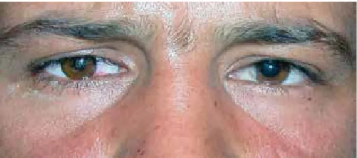
Seven patients had primary DFG, and 100% were satisfied with the outcome and could wear their prosthesis without any discomfort (Figure 1). In this group, one patient had postoperative complications (one minor and one major) (Patient #13). He had delay in epithelialization of the graft and ptosis. Ptosis was treated with frontal sling operation.
Epithelialization was complete in two weeks of using artificial tears after a period of 6 weeks of delayed healing. Finally, he could wear his prosthesis without any problems with good aesthetic outcome and was graded as grade 3 (moderately satisfied) (Table 1). The other 6 patients were complication-free and started using their prosthesis after an average of 8 weeks (range 6-12 weeks) following surgery and were graded as grade 4 (very satisfied) (Table 1).
Ten patients had secondary DFG transplantation. Of these, 6 patients (60%) were satisfied with the outcome and were graded as grades 3 and 4 (Table 1). Four patients were dissatisfied (Grade 1-2, Table1) secondary to postoperative complications. One patient had lower lid laxicity and lateral tarsal strip was performed (Patient #2). Another patient developed contraction in the lower fornix and was treated with mucous membrane grafting along with copious lubrication (Patient #7). The third patient had infection and contraction of the socket due to inappropriate use of medication following surgery (Figure 2) (Patient #1). Three months later, he underwent another DFG with successful outcome. Eventually, these three patients could
wear prosthesis comfortably after additional treatments. The last patient had fat atrophy prior to secondary DFG, and developed atrophy of the DFG 3 months following surgery (Patient #9, Table 1). Her family did not accept additional surgery. Finally, she was unable to wear any prosthesis.
Discussion
Dermis-fat graft has been used successfully for primary enucleations, superior sulcus augmentation, enophthalmos, and contracted sockets.8,9,10 They are known to be
advantageous to alloplastic materials in respect of foreign body reaction, toxic reactions, infection, and late exposure following conjunctival erosions. Additionally, in cases of conjunctival shrinkage and forniceal shortening, DFGs provide additional epithelial lining.11,12
In our study, we retrospectively investigated the long-term outcomes of primary and secondary DFGs. The final functional and aesthetic outcome and the rate of complications were evaluated. We found out that all of the primary DFG patients were satisfied with the aesthetic and functional outcome. On the contrary, only 60% of the secondary DFG patients were satisfied with the result.
Nentwich et al.13 retrospectively analyzed 173 primary
and 66 secondary DFG patients in their study. Of the primary DFG patients, 83%, and of the secondary DFG patients, 57% were highly satisfied from the result. Major complications were uncommon. In this large sample, DFG proved to be an effective and safe method for the reconstruction of anophthalmic sockets.
Atrophy in DFG may occur. Smith et al. reported in their series that significant atrophy of primary grafts does not occur very frequently; it is more common in cases of secondary implantation, particularly in cases of chemically injured and severely contracted sockets.14 In general, 5-10%
volume loss is assumed normal.12
Guberina et al. revealed 52 consecutive patients who underwent autogenous DFG in anophthalmic sockets during the past five years.11 The results of the study show that
81% of the cases had no change in the volume of the graft during a mean follow-up period of 2.5 years, ranging from a minimum of six months to a maximum of five years. The remaining 19% underwent graft resorption to a varying degree. They considered DFGing a procedure of choice in cases of extruded or migrated implants, an alternative to a synthetic allograft in primary enucleations, and a useful procedure in selected cases of enophthalmic and contracted socket.11 We also had one patient in our small group who
had fat atrophy of intraorbital structures before the surgery
Figure 2. Sixty–two-year-old male patient who had infection and contraction of the socket due to inappropriate use of medication following surgery
Figure 1. Thirty-five-year-old male patients after primary dermis-fat graft (DFG) who achieved good aesthetic outcome and could wear his prosthesis without any discomfort
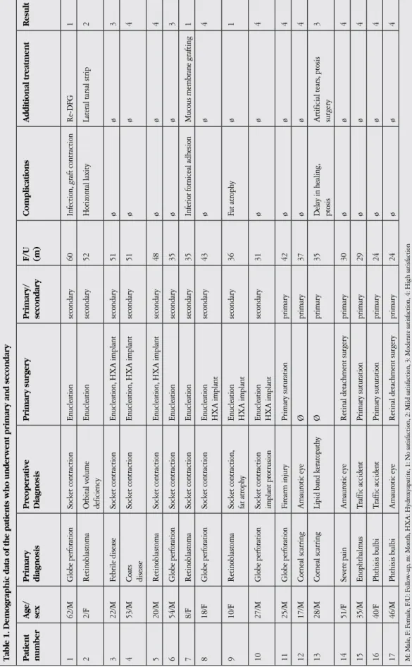
Table 1. Demographic data of the patients who underwent primary and secondary Patient number Age/ sex Primary diagnosis Preoperative Diagnosis Primary surgery Primary/ secondary F/U (m) Complications Additional treatment Result 1 62/M Globe perforation Socket contraction En ucleation secondary 60
Infection, graft contraction
Re-DFG
1
2
2/F
Retinoblastoma
Orbital volume deficiency
En
ucleation
secondary
52
Horizontal laxity
Lateral tarsal strip
2 3 22/M Febrile disease Socket contraction En
ucleation, HXA implant
secondary 51 ø ø 3 4 53/M Coats disease Socket contraction En
ucleation, HXA implant
secondary 51 ø ø 4 5 20/M Retinoblastoma Socket contraction En
ucleation, HXA implant
secondary 48 ø ø 4 6 54/M Globe perforation Socket contraction En ucleation secondary 35 ø ø 3 7 8/F Retinoblastoma Socket contraction En ucleation secondary 35
Inferior forniceal adhesion
Mucous membrane grafting
1 8 18/F Globe perforation Socket contraction En ucleation HXA implant secondary 43 ø ø 4 9 10/F Retinoblastoma
Socket contraction, fat atrophy
En ucleation HXA implant secondary 36 Fat atrophy ø 1 10 27/M Globe perforation
Socket contraction implant protrusion
En ucleation HXA implant secondary 31 ø ø 4 11 25/M Globe perforation Firearm injury Primary suturation primary 42 ø ø 4 12 17/M Corneal scarring Amaurotic eye Ø primary 37 ø ø 4 13 28/M Corneal scarring
Lipid band keratopathy
Ø primary 35 Delay in healing , ptosis Artificial tears , ptosis surgery 3 14 51/F Severe pain Amaurotic eye
Retinal detachment surgery
primary 30 ø ø 4 15 35/M Enophthalm us Traffic accident Primary suturation primary 29 ø ø 4 16 40/F Phthisis bulbi Traffic accident Primary suturation primary 24 ø ø 4 17 46/M Phthisis bulbi Amaurotic eye
Retinal detachment surgery
primary 24 ø ø 4 M: Male , F: Female , F/U: Follow-up
, m: Month, HXA: Hydroxyapatite
and also developed fat atrophy in three months after the DFG. This patient had received radiation therapy following enucleation due to retinoblastoma and was suffering from severe postenucleation syndrome. The history of radiotherapy and the lack of fat in the orbit is possibly the reason of fat atrophy following secondary DFG. The implanted dermofat tissue needs vascularisation following transplantation. And, we believe that the lack of healthy vasculature led to this atrophy. Thus, we believe that DFG is not recommended in cases with severe fat atrophy in the orbit. This may seem in contrast with the fact that DFG is the preferred treatment in cases with superior sulcus defects due to fat atrophy. However, these cases are suffering from volume loss in fat tissue not total fat atrophy. And in cases with volume loss, further DFG might be required.15
One of the major complications is loss of the transplanted tissue due to necrosis after infection or of an unknown cause. Fortunately, this is not a common complication.16,17 Poor
socket vasculature with socket contracture, prior history of severe orbital trauma, chronic orbital inflammation, postoperative orbital volume loss with fat atrophy, or surgical removal of posterior orbital volume at the time of enucleation are predisposing factors for graft ulceration and necrosis.15 We had one patient from the secondary DFG
group who had infection and contraction of the socket. He was also suffering from diabetes and hypertension that could affect wound healing. He had a severely traumatized globe after perforation with socket contraction. Enucleation was performed as primary surgery. We believe that patients’ lack of cooperation and inappropriate use of medication following surgery may also have contributed to graft necrosis. Thus, we would like to emphasize the importance of patient awareness of appropriate medication following surgery.
Following enucleation surgery, the loss of volume and rotation of intraorbital contents can result in superior sulcus deepening, enophthalmos, ptosis, ectropion, and lower lid laxity, which are known as postenucleation syndrome.17 We
had a patient in the secondary DFG group with lower lid laxity which was corrected by lateral tarsal strip. She was able to wear her prosthesis after this corrective surgery.
Ptosis is also a reported complication after anophthalmic socket reconstruction.18,19 Several factors might lead
to postoperative ptosis including damage to the levator muscle or aponeurosis, oculomotor nerve damage during preparation, deficit in the volume, and change in the pivots around which levator muscle works. We had one patient in the primary DFG group who had mild ptosis before surgery. Although he could wear prosthesis after DFG, he needed ptosis correction to achieve a better cosmetic result.
Various alloplastic orbital implants, preferably with a spherical configuration, are employed for the routine care of enucleated socket. Autologous DFG represents a safe alternative to alloplastic orbital implants. Loss of transplant or other serious complications were only rarely observed. Due to its high degree of safety concurrent with excellent functional and cosmetic results, DFG is particularly advantageous for young patients. In cases of complicated orbits and contracted sockets, DFG often represents the only promising option.20,21
In conclusion, a DFG is an effective means of replacing orbital volume and affording motility of ocular prosthesis. It is associated with low morbidity and a satisfactory cosmetic result.14 Complications are usually minor. We suggest that
DFG could be used routinely as primary orbital implant. The outcome in motility, prosthesis fitting, cosmetic appearance and patient satisfaction are encouraging. Most complications occurred in patients with severely traumatized sockets who had undergone earlier an extensive ocular surgery or who had a systemic disease contributing to defective wound healing. In secondary cases, appropriate patient selection is important to achieve good outcome.
Concflict of Interest: The authors reported no conflict
of interest related to this article.
References
1. Soll DB. Management of the anophthalmic socket and techniques of enucleation, evisceration and exentration:surgical procedures and management of complications. In Duane TD (ed): Duane’s Clinical Ophthalmology, Vol 5 Philadelphia, Lippincott Williams and Wilkins, 1992, pp. 407-423. 2. Soll DB. The anophthalmic socket. Ophthalmology. 1982;85:205-208. 3. McCord CD. The extruding implant. Trans Am Acad Ophthalmol
Otolaryngol. 1976;81:587-590.
4. Dryden R, Leibson J. Postenucleation orbital implant extrusion. Arch Ophthalmol. 1978;96:2064-2065.
5. Quaranta-Leoni FM, Moretti C, Sposato S, Nardoni S, Lambiase A, Bonini S. Management of porous orbital implants requiring explantation: a clinical and histopathological study. Ophthal Plast Reconstr Surg. 2014;30:132-136. 6. McElnea EM, Ryan A, Fulcher T. Porous orbital implant exposure: the
influence of surgical technique. Orbit. 2014;33:104-108.
7. Custer PL, Trinkaus KM. Porous implant exposure: Incidence, management, and morbidity. Ophthal Plast Reconstr Surg. 2007;23:1-7.
8. Hintschich CR, Beyer-Machule CK. Dermis fatty tissue transplant as primary and secondary orbital implant. Complications and results. Ophthalmologe. 1996;93:617-622.
9. Smith B, Bosniak SL, Lisman RD. An autogenous kinetic dermis-fat orbital implant:An updated technique. Ophthalmology. 1982;89:1067-1071. 10. Shore JW, Burks R, Leone CR Jr, McCord CD Jr. Dermis-fat graft for orbital
reconstruction after subtotal exentration. Am J Ophthalmol. 1986;102:228-236.
11. Guberina C, Hornblass A, Meltzer MA, Soarez V, Smith B. Autogenous dermis-fat orbital implantation. Arch Ophthalmol. 1983;101:1586-1590. 12. Bosniak SL. Reconstruction of the anophthalmic socket:state of the art. Adv
Ophthal Plast Reconstr Surg. 1987;7:313-348.
13. Nentwich MM, Schebitz-Walter K, Hirneiss C, Hintschich C. Dermis Fat Grafts as Primary and Secondary Orbital Implants. Orbit. 2014;33:33-38.
14. Smith B, Bosniak S, Nesi F, Lisman R. Dermis-fat orbital implantation:118 cases. Ophthalmic Surg. 1983;14:941-943.
15. Shore JW, McCord CD Jr, Bergin DJ, Dittmar SJ, Maiorca JP, Burks WR. Management of complications following dermis-fat grafting for anophthalmic socket reconstruction. Ophthalmology. 1985;92:1342-1350.
16. Aguilar GL, Shannon GM, Flanagan JC. Experience with dermis-fat grafting:an analysis of early postoperative complications and methods of prevention. Ophthalmic Surg. 1982;13:204-209.
17. Amornvit P, Rokaya D, Shrestha B, Srithavaj T. Prosthetic rehabilitation of an ocular defect with post-enucleation socket syndrome: A case report. Saudi Dent J. 2014;26:29-32.
18. Massry GG, Holds JB. Coralline hydroxyapatite spheres as secondary orbital implants in anophthalmos. Ophthalmology. 1995;102:161-166.
19. Nunery WR, Heinz GW, Bonnin JM, Martin RT, Cepala MA. Exposure rate of hydroxyapatite spheres in the anophthalmic socket:histopathologic correlation and comparison with silicone sphere implant. Ophthal Plast Surg. 1993;9:96-104.
20. Hintschich C. Dermis-fat graft. Possibilities and limitations. Ophthalmologe. 2003;100:518-524.
21. Hintschich CR, Altan-Yaycioglu R. Management of post-enucleation socket syndrome. In: Spaeth G, Danesh-Meyer H, and Kampik A(eds). Ophthalmic Surgery, Principles and Practice, fourth edition, Elsevier, London, UK. Bölüm 56, pp 450-461.