© 2018. This is an open access article distributed under the Creative Commons Attribution-NonCommercial-NoDerivatives 4.0 License. (http://creativecommons.org/licenses/by-nc-nd/4.0/)
The effect of ligand-to-Eu
3+charge-transfer transitions
(LMCT) on the photoluminescence intensity of M
2SiO
4: Eu
3+(M = Ca, Zn) type phosphors
ESRAÖZTÜRK∗, ERKULKARACAOGLUDepartment of Metallurgical and Materials Engineering, Faculty of Engineering, Karamano˘glu Mehmetbey University, Karaman, 70200, Turkey
In this study, silicate systems, M2SiO4(M = Ca, Zn) were produced by solid state reaction and doped with 1 mol% Eu3+
rare-earth ion. Their heat treatments, which were conducted at 1200 °C and above for minimum 3 hours under an open atmo-sphere, were applied according to the DTA/TG results. Powder X-ray diffraction XRD analyses were performed to determine the phase properties of the phosphor systems after the sintering process. It was proved that the structures of two of the phosphor systems were well formed in except that the Zn2SiO4had some ZnO secondary phases. The expected photoluminescence (PL)
results were presented and the transitions of the Eu3+ions were observed for both phosphors. Keywords: silicate phosphor; solid state reaction method; luminescence; Eu3+
1.
Introduction
For a long time, considerable effort has been given to the development of inorganic phosphors due to their wide applications for coatings in lamps (fluorescent), light emitting diodes (LEDs), cath-ode ray tubes, flat panel displays, electrolumines-cent and optoelectronic devices, radiation detectors in medical imaging systems, etc. [1,2].
Silicate-based inorganic phosphors offer supe-rior properties due to their stable crystal structure, thermal stability, wide energy band gap, low cost, non-toxicity, multi-color phosphorescence, high re-sistance to acid, alkali and oxygen, transparency in the ultraviolet (UV) to visible range, wide energy band gap (5.5 eV), etc. [1,3].
Among the rare-earth (RE) ions, the divalent (Eu2+) and threevalent (Eu3+) europium play an important role in blue to red emission, depending on the structure. The optical spectra of the Eu2+ and Eu3+ centers in solids or host structures can be easily differentiated. The photoluminescence (PL) properties of the Eu3+ ions originate from
∗E-mail: [email protected]
the intra-configurational 4f6→ 4f6parity forbidden
transitions [2, 4]. The corresponding emission spectrum comprises narrow and intense lines in the red region, the energies of which are prac-tically independent of the host lattice. Moreover, the PL properties of Eu2+ ions result from the 4f65d1 → 4f7 parity allowed transition and the
emission spectrum shows a broad band whose en-ergy greatly depends on the crystal environment. Therefore, the Eu2+ ion provides good PL prop-erties in that the color depends on the host lattice and can vary from UV to red region. It can also be mentioned that materials containing both Eu2+ and Eu3+ions are potentially very interesting phos-phors with a high color rending index (CRI) [4].
The structural properties of orthosilicate (Ca2SiO4), which is a well-known silicate type
structure, have been investigated in detail. It was found that Ca2SiO4has five polymorphs which can
be divided into hexagonal α, orthorhombic α0H,
α0L, γ and monoclinic β forms. The two phases which are observed at ambient conditions are phase γ, which is thermodynamically more stable and can therefore be obtained at higher temperatures, and phase β. The β → γ transition temperature is about 550 °C at ambient pressure [4].
cathode ray tubes (CRTs), plasma display panels (PDPs), laser crystals, upconversion luminescent materials, etc, due to its unique luminescent proper-ties, a wide energy band gap (5.5 eV), and excellent chemical stability. This structure shows polymor-phic phases α and β; the α-phase exhibits a sta-ble structure, while the β-phase has a metastasta-ble crystal structure [1,3,5–7].
When all this information is taken into account, these two silicate type hosts can be said to exhibit good thermal and chemical stability, and the Eu3+ ion is an ideal red phosphor activator due to its 4f6 electronic configuration. Therefore, in this study, Eu3+ doped silicate-type red color emitting phos-phors, namely Ca2SiO4:Eu3+ and Zn2SiO4:Eu3+,
which can be effectively excited by UV-source, were successfully synthesized by means of solid state reaction method.
2.
Experimental
2.1. Materials and methods
In this research, the solid state reaction method was chosen to synthesize the phosphor systems, and high purity CaCO3 (99.9 %), ZnO (99.9 %),
SiO2 (99.9 %) and Eu2O3 (99.99 %) raw
mate-rials were used to form M2SiO4:1 % mol Eu3+
(M = Ca, Zn). The constituents were thoroughly mixed and ground in an agate mortar to obtain a ho-mogeneous mixture. The dry-mixed compositions were then pre-heated at 800 °C for 2 hours tak-ing into consideration the DTA/TG results. Then the phosphor systems, namely Ca2SiO4:Eu3+ and
Zn2SiO4:Eu3+, were sintered in pure alumina
cru-cibles at different sintering conditions. The sin-tering processes were performed at 1200 °C for 2 h and at 1220 °C for 8 h under an open at-mosphere to get the Zn2SiO4:Eu3+ system
phos-phor. To obtain the Ca2SiO4:Eu3+ system
phos-phor, a sintering process at 1250 °C for 3 h was car-ried out. Then the sintered phosphors were cooled
mogravimetric (TG) analysis (Seiko Instruments Inc./Exstar TG/DTA 6200) at a heating rate of 10 °C/min from room temperature to 1300 °C were conducted to analyze the decomposition and oxi-dation process of the phosphor systems. After the heat treatments, which were applied taking into ac-count the DTA/TG results, a BRUKER AXS D8 ADVANCE model X-ray diffractometer (XRD), run at 40 kV and 30 mA (CuKα = 1.5406 Å radiation) in a step-scan mode (0.02°/2θ), was employed to determine the phases after the sin-tering processes. Finally, the photoluminescence (PL) properties, and the excitation and emission spectra of the phosphors were analyzed by a flu-orometer (Photon Technology International, PTI, QuantaMaster™ 30).
3.
Results and discussion
3.1. Thermal analysis
Among the phosphors used in this study, only the Ca2SiO4:Eu3+ system contained a
degrad-able reactant, namely CaCO3. Despite this, the
DTA/TG analyses were carried out between 50 °C and 1300 °C in both phosphor systems (Fig.1and Fig.2).
The major weight loss between 600 °C and 900 °C is related to the decomposition of CaCO3
or the elimination of CO2 in the system (Fig. 1).
The decomposition reaction under heating is given as follows:
CaCO3 4
−→ CaO +CO2 (1)
The endothermic peak at 800 °C is associated with the decomposition of CaCO3 which
trans-forms into CaO [8]. The TG curve shows a total mass loss equal to 33.5 %, which is almost similar to the calculated mass loss (∼33.7 %).
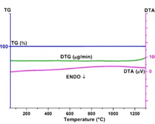
As shown in Fig.2, it is clearly seen that the TG curve does not show any significant weight loss in the applied temperatures range [9]. This is not sur-prising as the sample had no carbonate or hydrate
Fig. 1. DTA/TG curves of Ca2SiO4:Eu3+.
Fig. 2. DTA/TG curves of Zn2SiO4:Eu3+.
phases. Therefore, there was also no physically ad-sorbed water. Furthermore, the raw material such as ZnO, led to the complete formation of the phosphor system. The DTA curve does not exhibit any de-fined endothermic/exothermic peaks, as expected.
3.2. X-ray diffraction (XRD) analysis
Heat treatments of the samples were carried out in accordance with to the thermal analysis results. Therefore, to eliminate the volatile components, a pre-sintering at 800 °C for 2 h was applied to the samples. The major sintering processes determined the phases which developed.
The XRD pattern of Ca2SiO4:Eu3+ is shown
in Fig. 3. All the lines have been indexed by
Fig. 3. XRD pattern of Ca2SiO4:Eu3+ sintered at
1250 °C for 3 h.
considering the patterns of standard β-Ca2SiO4
(PDF 00-033-0302). Since the ionic radius of Eu3+ (0.095 nm) is close to that of Ca2+ (0.099 nm) and the ionic radius of Si4+ (0.054 nm) is very small compared with Eu3+, the Eu3+rare-earth as a dopant ion has not significantly influenced the host or the XRD patterns. Therefore, it is obvi-ous that the Eu3+ ions substitute for Ca2+ sites in β -Ca2SiO4. In this system, the β-Ca2SiO4 has
a monoclinic structure with the lattice parameters a = 9.31 Å, b = 6.76 Å, c = 5.51 Å, α = γ = 90° and β = 94.46°, based on the space group of P21/n (14) and two different Ca sites, Ca (1)
and Ca (2) [10,11].
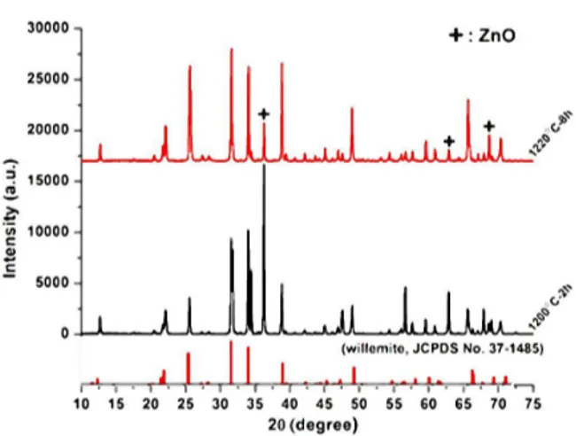
The XRD patterns of Zn2SiO4:Eu3+ sintered at
different temperatures are given in Fig.4.
The diffraction pattern of Zn2SiO4:Eu3+ that
was sintered at 1220 °C for 8 h is well matched with the PDF 00-037-1485 card except for some secondary ZnO lines. The phosphor system has a trigonal-hexagonal structure with the lattice param-eters a = 13.94 Å, b = 13.94 Å, c = 9.31 Å, α = 90°, β = 90° and γ = 120°. The point to be noted here is that the last applied sinter-ing temperature (1220 °C) is still not sufficient to form the single Zn2SiO4 phase. Therefore,
in-creasing the sintering temperature and/or time may cause complete elimination of the ZnO secondary phase as the Zn and Si elements on the surface
Fig. 4. XRD patterns of Zn2SiO4:Eu3+ sintered at
1200 °C for 2 h and 1220 °C for 8 h.
appear to be mobile enough to move and diffuse inside the porous body and cause the formation of the Zn2SiO4phase [1,12].
3.3. Photoluminescence properties
The samples show PL properties with the excitation and emission bands. Fig. 5 shows the PL spectra of the Ca2SiO4:Eu3+ phosphor
excited at 282 nm.
Fig. 5. PL spectra of Ca2SiO4:Eu3+phosphor.
The broad and the most intense excitation band near 282 nm is the charge-transfer state (CTS) band, because of an electron transferred from the oxygen 2p orbital to the empty 4f orbital
All the emission bands are attributed to tran-sitions of the Eu3+ ion: at 580 nm to 610 nm from5D0→7F1 which is the most intense and is
caused by magnetic dipole transitions between the
5D
0 → 7F1 states, at 610 nm to 635 nm from 5D
0 → 7F2 which result from electronic dipole
transitions between the 5D0 →7F2 states, and at
707 nm from 5D0→ 7F4. The 5D0-7F2 (613 nm)
transition is forbidden (f-f intra-configurational transition) for the Eu3+ ions located on the sites that deviate from the inversion center of symme-try. Therefore, it shows a significant and intense red emission band at 615 nm [1,2].
The PL spectra of the Zn2SiO4:Eu3+ phosphor
under excitation at 465 nm and maximum emission at 618 nm occur in the red region (Fig.6).
Fig. 6. PL spectra of Zn2SiO4:Eu3+phosphor.
It has been proved that all the excitation and emission bands are attributed to transi-tions of Eu3+, so this study revealed similar
re-sults to the Ca2SiO4:Eu3+ system phosphor. It
should be noted that the maximum excitation of the Zn2SiO4:Eu3+ system phsophor is different
excitation of Zn2SiO4:Eu3+at 282 nm, attributed to
ligand-to-Eu3+(O2−–Eu3+) charge-transfer transi-tion (LMCT), was induced because of the elec-tron transfer from 2p orbitals of O2− ions to 4f shells of Eu3+ ions, the Ca2SiO4:Eu3+ revealed
the excitation maximum at 465 nm which was associated with 7F0 → 5D2 transitions of Eu3+
in the host lattice [1,13].
4.
Conclusions
The hosts, Ca2SiO4 and Zn2SiO4, which were
activated with Eu3+, were prepared successfully by the solid state reaction method according to the DTA/TG results. While the Ca2SiO4:Eu3+
sys-tem phosphor was well crystallized and indexed, the Zn2SiO4:Eu3+ phosphors had some secondary
phases which were indexed as ZnO, although the sintering temperature and time of sintering were increased. The PL analysis revealed that the two phosphors with different hosts exhibit similar emis-sions in the red region that are attributed to transi-tions of the Eu3+ion. The point to be considered is that the PL intensities of the two phosphor systems are quite different from each other, i.e. the Ca2SiO4
system is more intense than the Zn2SiO4one.
Fur-thermore, the ligand-to-Eu3+ charge-transfer tran-sition (LMCT) with an excitation band at 282 nm gives the most dominant peak for Zn2SiO4:Eu3+
in comparison to the other excitation bands in the same system. Therefore, there is very high en-ergy transfer to Eu3+ transitions causing steadily growing emission intensity.
Acknowledgements
The authors would like to thank the Karamano˘glu Mehmetbey University, the Scientific Research Projects Com-mission (BAP), the Project Number 17-M-14, Republic
of Turkey, for their financial support. The authors are grateful to Prof. Dr. Adil Denizli from Hacettepe University, Depart-ment of Chemistry and Biochromatography and Biodiagnos-tics Research Group for their kind help.
References
[1] BASAVARAJ R.B., NAGABHUSHANA H., PRASAD
D.B., SHARMA S.C., PRASHANTHA S.C., NAGAB
-HUSHANAB.M., Optik, 19 (2015), 1745.
[2] ÖZTÜRK E., KARACAOGLU E., J. Therm. Anal.Calorim., 120 (2015), 1139.
[3] SUNITHAD.V., NAGABHUSHANAH., SHARMAS.C., SINGHF., NAGABHUSHANAB.M., DHANANJAYAN., SHIVAKUMARAC., CHAKRADHARR.P.S., J. Lumin., 143 (2013), 409.
[4] BARAN A., BARZOWSKA J., GRINBERG M., MAHLIK S., SZCZODROWSKI K., ZORENKO Y., Opt. Mater., 35 (2013), 2107.
[5] XIEQ., ZHANP., WANGW., LIZ., ZHANGZ., J. Al-loy. Compd., 642 (2015), 131.
[6] WUY., WANGNY., HED., FUM., CHENZ., LIY., J. Lumin., 130 (2010), 1768.
[7] SUF., MAB., DINGK., LIG., WANGG., CHENW., JOLYA.G., MCCREADYD.E., J. Lumin., 116 (2006), 117.
[8] HSUC.K., Thermochim. Acta, 392 – 393 (2002), 157. [9] FARAGH.K., HANAFIZ.M., DAWYM., ABD ELAZIZ
E.M., CJPAS, 4 (2010), 1303.
[10] HAOZ., ZHANGJ., ZHANGX., LUOY., ZHANGL., ZHAOH., J. Lumin., 152 (2014), 40.
[11] LUOY.Y., JOD.S., SENTHILK., TEZUKAS., KAKI
-HANAM., TODAK., MASAKIT., YOOND.H., J. Solid State Chem., 189 (2012), 68.
[12] YE R., MA R., ZHANG C., GAO Y., HUA Y., DENGD., LIUP., XUS., J. Alloy. Compd., 566 (2013), 73.
[13] LI Y.-C., CHANG Y.-H., LIN Y.-F., CHANG Y.-S., LINY.-J., J. Alloy. Compd., 439 (2007), 367.
Received 2017-10-31 Accepted 2018-05-02