RESEARCH ARTICLE
Retrospective evaluation of canine neoplasms in Konya region between 2006 and 2010
Semih Altan
1, Fahrettin Alkan
2, Yılmaz Koç
2, Özgür Özdemir
3, Orhan Yavuz
31Dicle Üniversitesi Veteriner Fakültesi, Cerrahi Anabilim Dalı, Diyarbakır, 2Selçuk Üniversitesi Veteriner Fakültesi, Cerrahi Anabilim Dalı, 3Selçuk Üniversitesi Veteriner Fakültesi, Patoloji Anabilim Dalı, Konya, Türkiye
Received: 26.08.2013, Accepted: 30.10.2013 *semih.altan@dicle.edu.tr
Özet
Altan S, Alkan F, Koç Y, Özdemir Ö, Yavuz O. Konya ve yöre-sinde köpeklerde 2006-2010 yılları arasında rastlanılan tü-mörlerin retrospektif değerlendirilmesi. Eurasian J Vet Sci, 2013, 4, 185-191
Amaç: Bu çalışmada 2006-2010 yılları arasında üniversi-te hayvan hastanesine getirilen 58 köpeküniversi-te klinik, radyolo-jik, operatif ve histopatolojik muayeneleri neticesinde teşhis edilen neoplazma vakalarının geriye dönük değerlendirilme-si yapıldı.
Gereç ve Yöntem: Tümöral vakalar köpeklerin ırk, yaş ve cinsiyetine, tümörlerin özellikleri ve yerleşim yerleriyle bir-likte tedavi şekillerine göre değerlendirildi.
Bulgular: Tümörlerin %48.3’üne cerrahi uygulama, %20 .7’sine kemoterapi, %5.2’sine hem cerrahi hem kemotera-pi ve %13.8’ine ise levamizol ve/veya otovaksinasyon uygu-landı. Köpeklerin %12’sine ise ötenazi uyguuygu-landı. Kemotera-pi, transmissible venereal tümör vakalarında yapılırken, pa-pilloma vakalarında levamizol ve otovaksinasyon yapıldı. Tü-mörlerin %53.4’ü benign karakterde iken %46’6’sı malign karakter gösterdiği belirlendi.
Öneri: Bu değerlendirme ile Konya ve yöresindeki köpekler-de gözlenen tümöral vakalar ve bunların tedavi sonuçları or-taya konmuştur. Ayrıca, venereal tümörler kemoterapi uygu-lamasıyla (vinkristin), papillomlar ise levamizol ve otovaksi-nasyon işlemiyle tedavi edilebilir.
Anahtar kelimeler: Köpek, retrospektif değerlendirme, ne-oplazma, kemoterapi, cerrahi.
Abstract
Altan S, Alkan F, Koc Y, Ozdemir O, Yavuz O. Retrospective evaluation of canine neoplasms in Konya region between 2006 and 2010. Eurasian J Vet Sci, 2013, 4, 185-191
Aim: It was conducted that a retrospective evaluation of cas-es diagnosed as neoplasms according to operative, clinical, histopathological and radiological examinations in 58 dogs that were examined at University Animal Hospital between 2006 and 2010.
Materials and Methods: The tumoral cases had been evalu-ated according to race, gender, age of dogs, characteristics and location of the neoplasms, and applications of treatment.
Results: Surgical treatment was applied in 48.3% of the cases, chemotherapy was applied in 20.7%, both surgical treatment and chemotherapy were applied in 5.2% of cases, and levamisole and/or autovaccination were aplied 13.8% of cases. Euthanasia was applied in 12% of cases. Chemothera-py was used primarily on transmissible venereal tumors, and papillomatosis cases were treated with levamisole and auto-vaccination. Among the diagnosed neoplasms, 53.4% were benign and 46.6% were malignant.
Conclusions: This retrospective evaluation revealed differ-ences in the treatment methods and results of dog neoplasia in Konya region. Moreover, venereal tumors could be treated with chemotherapy (vincristine), while papillomas could be treated with levamisole and/or autovaccination.
Keywords: Canine, retrospective evaluation, neoplasia, chemoterapy, surgery
Introduction
Neoplasms are described as abnormal cellular growths with constant, irregular, and faster growth attributes that dis-tinguish them from normal tissue. Although any tissue can develop into a neoplasm via the effects of various agents (viruses, mutagenic compounds, radiation etc), the general attribute of all neoplasms is a genomic defect in the cells. Neoplasms are classified according to host breed, age and gender as well as tissue, organ and system of origin. Benign and malignant are defined according to growth and attri-butes of a neoplasm. The incidence of neoplasms in dogs is highest in the mammary gland, followed by the cutaneous, urogenital, and head–neck regions, respectively (Morris and Dobson 2001, Cullen et al 2002, Vural et al 2007).
The treatment and prognosis of a patient with a neoplasm are strictly linked to the nature of the neoplasm. Thus, it is imperative to understand the histological structure, dimen-sions and anatomical location of neoplasms. Surgery, ra-diotherapy and chemotherapy are the three methods used to treat human neoplasms. The most effective treatment method for solid neoplasms in animals is surgery (Morris and Dobson 2001).
Numerous studies have evaluated neoplasms in dogs in Tur-key according to host breed, gender, age, location and charac-teristics (Pamukçu and Ertürk 1962, Ertürk et al 1971, Erer and Kıran 1993, Sönmez and Özmen, 1996, Gülçubuk and Gürel 2003).
We evaluated cases with a neoplasm diagnosis according to clinical, radiological, histopathological and operational diag-noses in dogs treated at the University Animal Hospital be-tween 2006 and 2010.
Materials and Methods
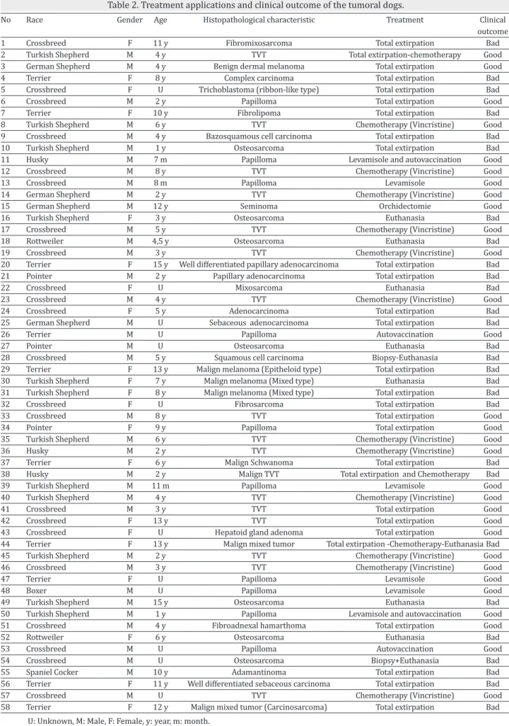
We retrospectively evaluated cases with a diagnosis of a neo-plasm in various tissues among 58 dogs of various ages and breeds that presented at the University Animal Hospital be-tween 2006 and 2010 according to the clinical, radiological, histopathological and operational diagnoses. The cases were treated with surgery, surgery accompanying chemotherapy, adjuvant therapy (levamisole) and autovaccination or eutha-nasia with consent of the owners, according to the histopath-ological diagnosis (Table 2).
Chemotherapy was the most applied to transmissible ve-nereal tumor (TVT). For the chemotherapy, vincristine
sul-was used at 3 mg/kg SC (Actipar, Alke ilaç, İstanbul, Turkey), together with/without autogenic vaccination. The treatment applications were presented in Table 2.
Results
The locations and origins of the neoplasms from the dogs are presented in Table 1. Surgery was applied to 28 of the 58 (48.3%) canines with a neoplasm diagnosis, whereas che-motherapy (Vincristine) was performed in 20.7% of cases. Surgery with chemotherapy in 5.2% of cases and levamisole and/or autovaccination in 13.8% of cases were performed. Approximately 12% of cases were euthanized at the request of the owner of the animal.
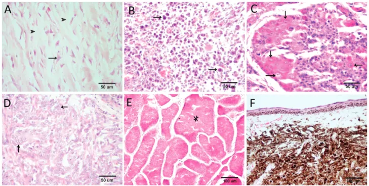
Among the diagnosed neoplasms, 53.4% were benign (Fig-ure 1D) and 46.6% were malignant (Figs. 1A-C). Of the be-nign neoplasms, 52% were TVT, 32% were papillomas, and 16% were other benign neoplasms (seminoma, benign der-mal melanoma, fibrolipoma, hepatoid gland adenoma and fibroadnexal hamarthoma) (Figures 1 and 2). Of the malig-nant neoplasms, 37% were sarcomas, 30% were carcinomas, 11% were malignant melanomas, 8% were malignant mixed neoplasms, (Figure 2) and 15% were other malignant neo-plasms (malignant TVT, trichoblastoma, adamantinoma and malignant schwannoma).
Of the 58 dogs with a neoplasm, the ages of 12 could not be determined. The mean age of the remaining 46 canines was 6.1±4.1 years, whereas 11% were <1 year old, 52% were 1–5 years, 17% were 6–10 years and 20% were >10 years. The locations of the neoplasms were genital-perineal-ingui-nal (36%), head and neck region (33%), extremities (15.5%) and abdominal-thoracic region (15.5%). Of the neoplasms, 67.2% were epithelial, 20.7% were mesenchymal, 6.9% were melanocyte neoplasms, 3.5% were mixed neoplasms, and 1.7% were odontological neoplasms (Table 1).
Of the 58 dogs with a neoplasm diagnosis, 38% were a cross-breed, 17% were Terriers, 17% were Turkish Shepherds, 10% were German Shepherds, 5% were Pointers and Hus-kies, 4% were Rottweiler, and 2% were Boxers and Cocker Spaniels. Among the dogs with a neoplasm diagnosis, 21 were female and 37 were male (Table 2).
Discussion
The ages of 12 of the 58 subject dogs could not be deter-mined. This was possibly due to the inclusion of unrecorded
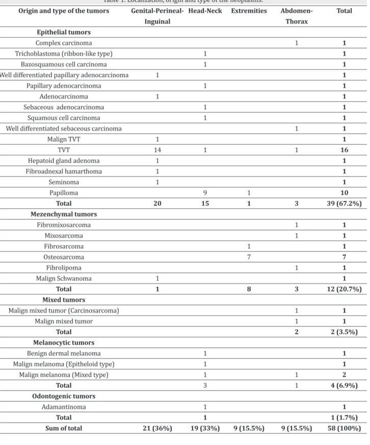
Table 1. Localization, origin and type of the neoplasms. Origin and type of the tumors
Epithelial tumors Complex carcinoma Trichoblastoma (ribbon-like type)
Bazosquamous cell carcinoma Well differentiated papillary adenocarcinoma
Papillary adenocarcinoma Adenocarcinoma Sebaceous adenocarcinoma
Squamous cell carcinoma Well differentiated sebaceous carcinoma
Malign TVT TVT
Hepatoid gland adenoma Fibroadnexal hamarthoma Seminoma Papilloma Total Mezenchymal tumors Fibromixosarcoma Mixosarcoma Fibrosarcoma Osteosarcoma Fibrolipoma Malign Schwanoma Total Mixed tumors
Malign mixed tumor (Carcinosarcoma) Malign mixed tumor
Total Melanocytic tumors Benign dermal melanoma Malign melanoma (Epitheloid type)
Malign melanoma (Mixed type) Total Odontogenic tumors Adamantinoma Total Sum of total Genital-Perineal-Inguinal 1 1 1 14 1 1 1 20 1 1 21 (36%) Head-Neck 1 1 1 1 1 1 9 15 1 1 1 3 1 1 19 (33%) Extremities 1 1 1 7 8 9 (15.5%) Abdomen-Thorax 1 1 1 3 1 1 1 3 1 1 2 1 1 9 (15.5%) Total 1 1 1 1 1 1 1 1 1 1 16 1 1 1 10 39 (67.2%) 1 1 1 7 1 1 12 (20.7%) 1 1 2 (3.5%) 1 1 2 4 (6.9%) 1 1 (1.7%) 58 (100%)
No Race Gender Age Histopathological characteristic Treatment Clinical outcome 1 Crossbreed F 11 y Fibromixosarcoma Total extirpation Bad 2 Turkish Shepherd M 4 y TVT Total extirpation-chemotherapy Good 3 German Shepherd M 4 y Benign dermal melanoma Total extirpation Good 4 Terrier F 8 y Complex carcinoma Total extirpation Bad 5 Crossbreed F U Trichoblastoma (ribbon-like type) Total extirpation Bad 6 Crossbreed M 2 y Papilloma Total extirpation Good 7 Terrier F 10 y Fibrolipoma Total extirpation Bad 8 Turkish Shepherd M 6 y TVT Chemotherapy (Vincristine) Good 9 Crossbreed M 4 y Bazosquamous cell carcinoma Total extirpation Bad 10 Turkish Shepherd M 1 y Osteosarcoma Total extirpation Bad 11 Husky M 7 m Papilloma Levamisole and autovaccination Good 12 Crossbreed M 8 y TVT Chemotherapy (Vincristine) Good
13 Crossbreed M 8 m Papilloma Levamisole Good
14 German Shepherd M 2 y TVT Chemotherapy (Vincristine) Good 15 German Shepherd M 12 y Seminoma Orchidectomie Good 16 Turkish Shepherd F 3 y Osteosarcoma Euthanasia Bad 17 Crossbreed M 5 y TVT Chemotherapy (Vincristine) Good 18 Rottweiler M 4,5 y Osteosarcoma Euthanasia Bad 19 Crossbreed M 3 y TVT Chemotherapy (Vincristine) Good 20 Terrier F 15 y Well differentiated papillary adenocarcinoma Total extirpation Bad 21 Pointer M 2 y Papillary adenocarcinoma Total extirpation Bad
22 Crossbreed F U Mixosarcoma Euthanasia Bad
23 Crossbreed M 4 y TVT Chemotherapy (Vincristine) Good 24 Crossbreed F 5 y Adenocarcinoma Total extirpation Bad 25 German Shepherd M U Sebaceous adenocarcinoma Total extirpation Bad
26 Terrier M U Papilloma Autovaccination Good
27 Pointer M U Osteosarcoma Euthanasia Bad
28 Crossbreed M 5 y Squamous cell carcinoma Biopsy-Euthanasia Bad 29 Terrier F 13 y Malign melanoma (Epitheloid type) Total extirpation Bad 30 Turkish Shepherd F 7 y Malign melanoma (Mixed type) Euthanasia Bad 31 Turkish Shepherd F 8 y Malign melanoma (Mixed type) Total extirpation Bad 32 Crossbreed F U Fibrosarcoma Total extirpation Bad
33 Crossbreed M 8 y TVT Total extirpation Good
34 Pointer F 9 y Papilloma Total extirpation Good 35 Turkish Shepherd M 6 y TVT Chemotherapy (Vincristine) Good 36 Husky M 2 y TVT Chemotherapy (Vincristine) Good 37 Terrier F 6 y Malign Schwanoma Total extirpation Bad 38 Husky M 2 y Malign TVT Total extirpation and Chemotherapy Bad 39 Turkish Shepherd M 11 m Papilloma Levamisole Good 40 Turkish Shepherd M 4 y TVT Chemotherapy (Vincristine) Good
41 Crossbreed M 3 y TVT Total extirpation Good
42 Crossbreed F 13 y TVT Total extirpation Good 43 Crossbreed F U Hepatoid gland adenoma Total extirpation Good 44 Terrier F 13 y Malign mixed tumor Total extirpation -Chemotherapy-Euthanasia Bad 45 Turkish Shepherd M 2 y TVT Chemotherapy (Vincristine) Good 46 Crossbreed M 3 y TVT Chemotherapy (Vincristine) Good
47 Terrier F U Papilloma Levamisole Good
48 Boxer M U Papilloma Levamisole Good
49 Turkish Shepherd M 15 y Osteosarcoma Euthanasia Bad 50 Turkish Shepherd M 1 y Papilloma Levamisole and autovaccination Good 51 Crossbreed M 4 y Fibroadnexal hamarthoma Total extirpation Good 52 Rottweiler F 6 y Osteosarcoma Euthanasia Bad
increasing (Cotchin 1954, Erer and Kıran 1993, Gülçubuk and Gürel 2003). However, we identified few neoplasms in dogs of this age (Table 2). Approximately 52% of benign neo-plasms in this study were TVT, which occurs only in dogs at 2–5 years of age (Purohit 2009). In 13 of the 17 TVT cases, the dogs were <6 years old, which supports our view. Fur-thermore, the incidence of neoplasm was higher in dogs of ages 1–5 years and >10 years. In another study (Erer and Kıran 1993), 48.1% of 34 dogs with a neoplasm diagnosis were ages 6–10, compared to 20% in our study. Similarly, in another study (Gülçubuk and Gürel 2003), the incidence of neoplasm was 4% in dogs of age <1 year and 52% among dogs of age 6–10 years. However, in the present study, the neoplasm incidence was 11% among dogs of age <1 year and 17% among dogs of age 2–6 years. We found malignant neo-plasms generally in dogs >10 years of age, whereas benign neoplasms were seen at any age (Table 2). According to this retrospective evaluation, age is thus not the sole indicator of the incidence of neoplasm among dogs. According to the study conducted by Erer and Kıran (1993) in Konya region, TVT cases found as 14.7%, whereas in our study, TVT cases found 29.3%. The other data presented was similar to Erer and Kıran (1993).
The incidence of malignant neoplasms in previous studies is higher than that of benign neoplasms (Mialot and Lagadic 1990, Eren and Kıran 1993, Sönmez and Özmen 1996, Gül-çubuk and Gürel 2003), but here only 46.6% of neoplasms were malignant, which was possibly due to the high-quality care and environmental conditions in which the dogs were kept (Table 1). In this study, the incidence of neoplasm was higher among Turkish Shepherds and Terriers out of cross-breeds (Table 2). This is possibly due to the national breed-ing status and popularity of Turkish Shepherds in Turkey. Terriers and Turkish Shepherds were the most susceptible to neoplasms, and the number of malignant neoplasm cases among Terriers was higher (n=7) when compared to Turk-ish Shepherds, whereas benign neoplasms were higher in number among Turkish Shepherds (n=6). The average age of Turkish Shepherd (5 ± 4.1 years) is shorter than that of Ter-rier (11±2.9 years). Over the age of 10 is the period during which malignant neoplasms occur most commonly (Morris and Dobson 2001, Cullen et al 2002). Indeed, the susceptibil-ity of Terriers were more susceptible to mammary and cu-taneous neoplasms (Muller et al 2000, Gülçubuk and Gürel 2003). Owing to five of the seven malignant neoplasms cases which were mammary, it is supporting above mentioned reports (Table 2). The most common neoplasm in Turkish Shepherds was TVT. This was most likely due to the outdoor living environment of the Turkish Shepherd, which can lead to uncontrolled sexual behavior and the spread of disease. Osteosarcoma is most common in larger breeds (Erer et al 1998, Cullen et al 2002). In this study, osteosarcomas were
identified in three Turkish Shepherds, two Rottweiler (Fig. 1A and 2A), a Pointer, and a crossbreed, which supports pre-vious reports (Table 2).
Some reports suggest that females are more susceptible to neoplasms than males (Mialot and Lagadic 1990, Sönmez and Özmen 1996, Gülçubuk and Gürel 2003), whereas others (Cotchin 1954, Cotchin 1959, Erer and Kıran 1993) refute any such association. In our study, 36% of diagnosed neoplasm cases were in females and 64% were in males. However, sub-jects were evaluated at the surgery clinic; thus female genital system neoplasms (TVT, mamma, etc.), which are addressed by the Department of Obstetrics and Gynecology, were not evaluated. This could lead to inaccurate conclusions regard-ing the incidence of neoplasms in females. Most cases of fe-male genital neoplasms involve the mammary gland (Cotchin 1954, Cotchin 1959, Köküuslu and Akkayan 1972, Erer and Kıran 1993, Gülçubuk and Gürel 2003). In this study, the most common neoplasms among females were mammary neoplasms (48%), which support previous reports (Fig. 1B). TVT that is mainly located genital region was often observed among males (Morris and Dobson 2001, Cullen et al 2002). A previous study (Sönmez and Özmen 1996) of 89 canines found TVT cases in 31 (35%) dogs; 15 females and 16 males. In this study, 17 TVT cases were found in the 58 cases (29.3%), which comprised 1 female and 16 male. While the incidence rates were higher, and in agreement with previ-ous reports (Sönmez and Özmen 1996), the actual incidence of TVT among females could not be determined as the dogs were lost to follow-up at the Department of Obstetrics and Gynecology.
Oral papillomatosis is a common disease of puppies (Koç et al 2001). Oropharyngeal neoplasms were found to comprise 21% of papillomas in a study (Vural et al 2007). However, in this study, approximately 53% of head–neck region neo-plasms were papillomas. Furthermore, the high frequency of papillomas in dogs <1 year of age supports the opinions of Vural et al (2007).
Surgery, radiotherapy, and surgery accompanying chemo-therapy are the three main treatment methods for plasms. The most effective treatment method for solid neo-plasms among animals is thought to be surgery (Morris and Dobson 2001). In this study, approximately half of the cases underwent surgery, 20.7% chemotherapy, and 5.2% both surgery and chemotherapy.
Conclusions
In conclusion, the number of cases of neoplasms is increas-ing due to the increase in the Konya region pet population. Neoplasms such as TVT and papilloma are generally treated successfully, whereas malignant neoplasms are not treated
Figure 2. Histopathological samples of various tumor tissues. (A) Osteosarcoma. Atypical osteoblast (arrow) and osteoid tissues (arrow head), (B) Malign melanoma (Mixed type). Malignant cells containing melanin (arrow), (C) Sebaceous adenocarcinoma. Atypical epithelial cells (ar-rows), (D) Papillary adenocarcinoma. Atypical epithelial cells (ar(ar-rows), (E) Hepatoid gland adenoma. Benign epithelial cells and glandular for-mations (asterisk), (F). Benign dermal melanoma. Dermal cells containing melanin. H&E.
appropriately. The majority of neoplasms occurred in Turk-ish Shepherds and Terriers, and the majority of malignant cases were in Terriers.
Acknowledgment
This article was presented as poster presentation at 17th Fe-cava Eurocongress in Istanbul/Turkey in 7-10 September 2011 funding by "University of Selçuk Scientific Research Project Office" with 11701574 project number.
References
Cotchin E, 1954. Neoplasia in the dog. Vet Rec, 66, 879-884. Cotchin E, 1959. Some tumors of dogs and cats of
comparati-ve comparati-veterinary and human interest. Vet Rec, 71, 1040-1054. Cullen JM, Page R, Misdorp W, 2002. An Overview of Cancer
Pathogenesis, Diagnosis, and Management In: Tumors in Domestic Animals, Ed; Moulton JE, fourth edition, Black-well, Iowa, USA, pp: 3-44.
Erer H, Kıran MM, 1993. Tumors of dogs between the years 1985-1992 in Konya. Eurasian J Vet Sci, 9, 87-89.
Erer H, Elma E, Hatipoğlu F, Alkan F, 1998. Kangal ırkı bir kö-pekte osteosarkom olgusu. Eurasian J Vet Sci, 14, 107-112. Ertürk E, Tanzer F Bulucu M, 1971. Dog and cat tumors exa-mined during the period of 1964-1970 at the department of pathological anatomy of the faculty of veterinary medi-cine. A Ü Vet Fak Derg, 18, 383-386.
Gülçubuk A, Gürel A, 2003. Dog tumors observed in Istanbul between the years of 1995 to 2000. İ Ü Vet Fak Derg, 29; 83-91.
Koç Y, Alkan F, Kul M, Hatipoğlu F, 2001. The use of levami-sole in oral papillamatosis of the dogs. J Turk Vet Med As-soc, 13, 46-49.
Köküuslu C, Akkayan C, 1972. The Primary mammary tumors of the cats and dogs examined between the years 1968 and 1972 in Ankara. A Ü Vet Fak Derg, 19, 502-516.
Mialot M Lagadic M, 1990. Epidemiologie descriptive des tu-meurs du Chien et du Chat. J Rec Med Vet, 166, 937-947. Morris J, Dobson J, 2001. Small Animal Oncology. Blackwell,
Oxford, UK.
Muller GH, Kirk RW, Scott DW, 2000. Neoplastic and Non-Neoplastic tumors. In: Muller’s and Kirk’s Small Animal Dermatology, Sixth edition, Saunders, Philedelphia, USA, pp; 1236-1414.
Pamukçu AM, Ertürk E, 1962. Tumors types of dogs in Anka-ra. A Ü Vet Fak Derg, 9, 1-9.
Purohit GN, 2009. Canine Transmissible Venereal Tumor: A Review. The Internet Journal of Veterinary Medicine, 6, 1. Accessed at: 10 June 2013.
Sönmez G, Özmen Ö, 1996. Bursa’da 1988-1996 yılları ara-sında incelenen köpek tümörleri. U Ü Vet Fak Derg, 15, 69-76.
Vural SA, Keleş H, Özsoy ŞY, Özkul İA, 2007. Oropharyngeal tumors between 1977 to 2005 at dog and cats: a retrospec-tive study. A Ü Vet Fak Derg, 54, 197-203.