Original Paper
© 2015 The Author(s) Published by S. Karger AG, Basel Gazi Universitesi, Eczacılık Fakültesi, Farmakoloji Anabilim Dalı,
06330, Etiler, Ankara, (Turkey)
Tel.+90-312 2023127, Fax +90-312 2235018, E-Mail [email protected] Fatma Akar (PhD)
Long-Term Dietary Fructose Causes
Gender-Different Metabolic and Vascular
Dysfunction in Rats: Modulatory Effects of
Resveratrol
Mehmet Bilgehan Pektaşa,c Gökhan Sadib Fatma Akarc
aDepartment of Medical Pharmacology, Faculty of Medicine, Afyon Kocatepe University, Afyonkarahisar, bDepartment of Biology, K.Ö. Science Faculty, Karamanoglu Mehmetbey University, Karaman,
cDepartment of Pharmacology, Faculty of Pharmacy, Gazi University, Ankara, Turkey
Key Words
Fructose • Resveratrol • Gender • Metabolic and vascular function • Insulin • eNOS • iNOS • SIRT1
Abstract
Background/Aims: There is limited knowledge on the gender differences in the effects of
dietary fructose. In the current study, we investigated whether long-term fructose intake impacts metabolic parameters and vascular reactivity differently between male and female rats. Moreover, we tested whether resveratrol has a gender-specific effectiveness on the alterations.
Methods: Male and female rats were divided into four groups as control; resveratrol; fructose
and resveratrol plus fructose. Fructose was given to the rats as 10% solution in drinking water for 24 weeks. All rats were fed with the standard diet with or without resveratrol. Results: High-fructose diet increased plasma insulin, triglyceride and VLDL levels as well as omental weights in both genders. Long-term dietary fructose causes marked increase in body weight of males, but not females. Dietary fructose impaired endothelial relaxation to acetylcholine and intensified contraction to phenylephrine in the aortas of male and female rats, but differently it also reduced insulin-induced vasodilation in aortas of female rats. These changes were associated with decreased expression levels of endothelial nitric oxide synthase (eNOS) mRNA and protein, but increased in inducible NOS (iNOS), in aortas of male and female rats. Dietary fructose suppressed expression levels of sirtuin 1 (SIRT1) and insulin receptor substrate-2 (IRS-2) mRNA in aortas from female rats. Resveratrol supplementation efficiently restored fructose-induced metabolic and vascular dysfunction in both genders probably by regulating eNOS and iNOS production. Moreover, the augmentations in SIRT1 and IRS-2 mRNA in females and IRS-1 mRNA in males may possibly contribute to the beneficial effects of resveratrol as well.
Conclusion: Long-term fructose intake may differently affect metabolic and vascular function
Introduction
The increased consumption of fructose may contribute to the worldwide high prevalence of metabolic syndrome, which is characterized by hyperlipidemia, hyperinsulinemia, hypertension and obesity. The majority of metabolic syndrome studies have been performed on male animals. In relation to the effect of gender on diet-induced metabolic disturbances, it has been demonstrated that the signs of metabolic disorder display a higher incidence in males than females which is possibly explained with the presence of estrogen [1–3]. Differently, obesity was reported to exist in both genders due to high-fat and caloric diet [4–6]. However, little is known about the gender dependent effects of dietary fructose on metabolic and vascular functions. Female rats on fructose diet, but not their male counterparts, were shown to be protected from hypertension and hyperinsulinemia despite the presence of elevated triglycerides [7]. A human study reported that dietary fructose causes elevations in fasting and postprandial plasma triacylglycerol concentrations in healthy men compared to the women [8].
Several studies have shown that high-fructose diet leads to metabolic disorder characterized by insulin resistance, hypertriglyceridemia, endothelial dysfunction and hepatic steatosis in rats without any weight gain [9]. Previous data from our laboratory have also shown that high-fructose diet, given as high-fructose corn syrup (HFCS), causes metabolic disturbances associated with endothelial dysfunction, vascular insulin resistance, down-regulation of insulin receptor substrate-1 (IRS-1), endothelial nitric oxide synthase (eNOS) and sirtuin 1 (SIRT1) as well as up-regulation of inflammatory markers such as inducible NOS (iNOS) and NADPH oxidase in aortic tissues of male rats [10, 11]. In abovementioned studies, we also found that HFCS induces metabolic syndrome in the absence of obesity. However, we had not investigated the influences of high-fructose diet in female rats. The gender-different influence of dietary fructose and its modification by resveratrol have not been reported in the literature. The investigation of gender-dependent differences on dietary fructose-induced metabolic disturbances will provide new insights to understand the related mechanisms and to design a rational therapeutic intervention strategy. Therefore, in the current study, we studied whether long-term fructose intake (10% beverage for 24 weeks) differently influences the metabolic parameters and vascular reactivity in conjunction with IRS-1/2, eNOS, SIRT1 and iNOS expressions in male and female rats. Moreover, we also evaluated whether resveratrol ameliorates the changes induced by fructose intake through gender-specific mechanisms. In a recent study performed with healthy male and female rats, we documented that resveratrol modulated vascular responsiveness to estrogen, diversely between genders [12]. In relation to the current study, our previous studies illustrated that resveratrol improves dietary HFCS-induced vascular insulin resistance, endothelial dysfunction, hypertriglyceridemia and hyperinsulinemia in male rats [10, 11, 13].
Materials and Methods
Chemicals
Chemicals were purchased from Sigma Chemical Co. (St. Louis, MO) unless otherwise stated. Insulin was from Eli Lilly. Fructose was obtained from Danisco Sweeteners OY (Finland) and trans-resveratrol was from Herb-Tech (ROC). The purity of resveratrol was tested by HPLC followed with LC-MS and 98% of the constituent was determined as trans-resveratrol.
Animals and diets
All protocols for animal use were approved by the Ethical Animal Research Committee of Gazi University (G.U. ET-10.045). Four week old male and female Wistar rats were housed under temperature- and humidity-controlled rooms (20 – 22°C) with a 12-h light-dark cycle. The animals were fed with a standard rodent chow diet that composed of 62% starch, 23% protein, 4% fat, 7% cellulose, standard vitamins and salt mixture. At the end of acclimation for 1 week, male and female rats were randomly divided into four
groups as control; resveratrol; fructose and resveratrol plus fructose (resveratrol + fructose). Fructose was given to the rats as 10% solution in drinking water ad libitum for 24 weeks. Resveratrol was mixed with powdered material of standard rodent chow at a dose of 500 mg/kg and then the combination was pressed to make a chow pellet in the production company. The chow was stocked in temperature-controlled (4oC) and dark rooms. All rats were fed with the standard diet with or without resveratrol ad libitum for 24
weeks. Body weights, food and liquid intakes were recorded weekly during the follow-up period. The daily resveratrol ingestion was calculated from the amount of chow intake. The daily fructose consumption was determined by measuring the liquid intake. At the end of follow-up period, the rats were anesthetized with a mixture of ketamine–xylazine (100 and 10 mg/kg, respectively, i.p.) and thereafter, blood samples were rapidly collected via cardiac puncture. The thoracic aortas and whole omentum were dissected, washed and some part of the tissues were blotted dry and frozen in liquid nitrogen and stored at −85 °C.
Measurement of metabolic parameters in the plasma
Cardiac blood samples of non-fasted rats were immediately centrifuged at +4 °C and 10,000g for 30 min. Samples were stored at −85 °C until analysis. Plasma triglyceride, very low density lipoprotein (VLDL), high density lipoprotein (HDL) and total cholesterol levels were determined by standard enzymatic techniques. Insulin levels were assessed by using ELISA kit (Mercodia) according to the manufacturer’s instructions. Glucose levels were measured using a glucometer (Roche Diagnostics) in blood collected from the tail veins of rats.
Measurement of vascular reactivity in the thoracic aorta
The thoracic aortas were isolated and immediately transferred to a cold Krebs solution that contained (mM): NaCl 118, KCl 4.73, KH2PO4 1.2, MgSO4.7H2O 1.2, CaCl2 2.5, NaHCO3 25, glucose 11 and EDTA 0.026. The aortic rings were suspended between a tissue holder and isometric force–displacement transducer (EMKA, Paris) in a 5 ml organ bath filled with Krebs solution at 37°C and oxygenated by 95% O2 and 5% CO2. Special care was taken to preserve the endothelium during the preparation of the aortic rings. Functional integrity of the endothelium was checked with the application of acetylcholine (10-6 M) on phenylephrine
(10-6 M)-precontracted aortic rings and preparations demonstrating <70% relaxations in control rats from
both genders were discarded [10, 11]. The cumulative concentration–response curves of phenylephrine (10-9–10-4 M) were constructed in the aortic rings. The relaxant effects of acetylcholine (10-9 to 10-4 M) and
insulin (10-9 to 3x10⁻⁶ M) were studied in the rings constricted submaximally with phenylephrine (10-6 M). Determination of gene expressions of IRS-1, IRS-2, eNOS, iNOS and SIRT-1 with real time polymerase chain reaction
Total RNAs were isolated from arterial tissues using the RNeasy total RNA isolation kit (Qiagen, Venlo, Netherlands) as described according to the manufacturer protocol. After isolation, amount and quality of the total RNA were determined using spectrophotometry at 260/280 nm. 1 µg of total RNA were reverse transcribed to cDNA using commercial first strand cDNA synthesis kit (Thermo Scientific, USA) as described by the supplier. Gene expressions of IRS-1, IRS-2, eNOS, iNOS and SIRT-1 were determined with real time PCR as we described in detail previously [13]. Relative expressions of genes with respect to internal control; glyceraldehyde 3-phosphate dehydrogenase (GAPDH) were calculated with the efficiency corrected advance relative quantification tool provided by the LightCycler® 480 SW 1.5.1 software.
Immunoblot analysis of IRS-1, eNOS and iNOS proteins
Frozen aortic samples were homogenized in two volumes of homogenization medium containing 50 mM Tris, 150 mM NaCl, 5 mM EDTA, 1% (w/w) Triton x-100, 0.26% (w/v) sodium deoxycholate, 50 mM sodium flouride, 0.1 mM sodium ortovanadate and 0.2 mM PMSF by using Tissue RuptorTM (Qiagen,
Venlo, Netherlands) homogenizer and centrifuged at 1500g for 10 min at 4 °C. Total protein concentrations of the resulted supernatants were determined by Lowry method [14]. Equal amounts of protein (75 µg) from different groups were separated by SDS-PAGE and electroblotted onto PVDF membranes. The blotted membranes were blocked with 3% BSA. eNOS (anti-eNOS rabbit IgG, Abcam, 1:1000), iNOS (anti-iNOS rabbit IgG, Abcam, 1:1000) and IRS-1 (anti-IRS1 rabbit IgG, Abcam, 1:1000) antibodies were used for the priming for 2 h with constant shaking. As an internal control, GAPDH proteins were also primed with their primary antibody (anti-GAPDH rabbit IgG, Santa Cruz, 1:2000). Horseradish peroxidase (HRP) conjugated secondary
antibodies (Goat Anti-rabbit IgG-HRP conjugate; Santa Cruz, USA, 1:10,000) was incubated for 1 h and the blots were incubated in ClarityTM Western ECL (Bio-Rad Laboratories, Hercules CA, USA) substrate solution
for 5 min. Images of the blots were obtained using the ChemiDocTM MP Chemiluminescence detection system
(Bio-Rad Laboratories, Hercules CA, USA) equipped with a CCD camera. The relative expression of proteins with respect to GAPDH was calculated using the ImageLab4.1 software.
Statistical analysis
Data was given as mean ± standard error of the mean (SEM); n is the number of rats. The relaxations to acetylcholine and insulin were expressed as percent decreases of the precontraction induced by phenylephrine. Contractile responses to phenylephrine were calculated as a percentage of the KCl (40 mM)-induced contraction. The concentration–response curves to agonists were compared at each concentration for differences. The maximal response (Emax) and potency (EC50) of the agents were determined by using Prism 5.02 GraphPad software. EC50 values were given as −log M. Gene and protein expression data was normalized to the mean of the control groups, which was arbitrarily set to 100% and the relative changes were given as percent changes over control. Statistical comparisons were performed using unpaired Student’s t test or one-way ANOVA followed by the Bonferroni post hoc test. P values smaller than 0.05 were considered as statistically significant.
Results
Resveratrol intake
Resveratrol consumption of the rats was calculated to be approximately 36 mg/kg body weight/day in males and 40 mg/kg body weight/day in females in the healthy conditions. In fructose-treated groups, resveratrol intake was calculated as 29 mg/kg body weight/day in male and 27 mg/kg body weight/day in female rats.
The effects of dietary fructose and resveratrol on body weight, omental fat weight, caloric intake and metabolic parameters
As shown in Table 1, the total caloric intakes of fructose-treated male and female rats were higher than those of their matching controls, while unchanged after resveratrol supplementation. Fructose-treated male rats gained significantly higher weight than control group, whereas the body weight of fructose-treated female rats showed a tendency towards an increase compared to controls, but was not found significant. Resveratrol-treated healthy male and female rats consumed more total calories, however gained less weight than their matching controls. Notably, dietary fructose increased omental fat mass in both male and female rats which responded to resveratrol supplementation with a significant reduction (Fig. 1).
Fig. 1. The ratio of omental fat weight to body weight from the control, resveratrol (Res), fructose (Fruc) and resveratrol+fructose (Res+Fruc)-treated male and female rats. Val-ues are expressed as mean ± SEM, n = 6–10. * P < 0.05, significantly different from control.
# P < 0.05, significantly different from
fructose-treated rats. † P < 0.05, significantly different
Furthermore, dietary fructose elevated plasma triglyceride, VLDL and insulin levels in both genders. Resveratrol supplementation significantly attenuated the elevated levels of triglyceride, VLDL and insulin in rats fed with fructose (Table 1). Dietary fructose did not increase blood glucose levels in both genders but resveratrol supplementation caused a tendency towards reduction. Resveratrol significantly raised HDL levels in female rats. Total cholesterol levels were higher in female than in male rats after the treatments.
The effects of dietary fructose and resveratrol on relaxation response to acetylcholine and insulin in aortic rings
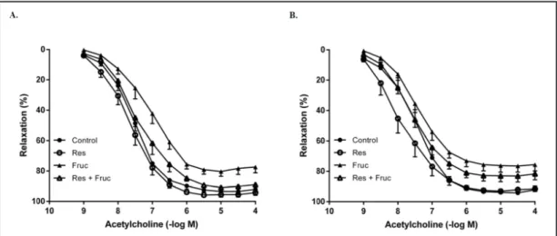
Dietary fructose decreased acetylcholine-induced relaxations, especially in terms of maximal responses (Emax), in aortic rings from both genders when compared to the controls.
The relaxations, in terms of EC50 and Emax, were improved after resveratrol supplementation
in fructose-treated male and female rats (Figs. 2A and B, Table 2); while an endothelium-independent relaxation response to sodium nitroprusside (10-10 to 10-5 M) was found
indifferent between the groups (data not shown).
Table 1. The effects of resveratrol (Res), fructose (Fruc) and resveratrol plus fructose (Res+Fruc) consumptions on body weight, food intake, liquid intake and metabolic parameters of male and fe-male rats. Values are expressed as mean ±SEM, n = 6–12. * P < 0.05, significantly different from control.
# P < 0.05, significantly different from fructose-treated rats. † P < 0.05, significantly different from male
Table 2. The potency (EC50; –log M) and maximum relaxation (Emax)
val-ues to acetylcholine in endothelium-intact aortic rings from the control, resveratrol (Res), fructose (Fruc) and resveratrol+fructose (Res+Fruc)-treated male and female rats. Values are expressed as mean ±SEM, n =
6–12. * P < 0.05, significantly different from control. # P < 0.05,
signifi-cantly different from fructose-treated rats. † P < 0.05, significantly
Insulin-induced relaxations showed a gender-dependent discrepancy in rat aortic rings, which were more pronounced in those of females. Resveratrol extensively promoted the maximal relaxation to insulin, especially in males. Dietary fructose importantly weakened insulin-induced relaxations in the aortic rings of females. Conversely, resveratrol supplementation enhanced the relaxations to insulin in both male and female rats fed with fructose (Figs. 3A and B, Table 3).
Table 3. Maximum relaxation (Emax) values to insulin in
endothe-lium-intact aortic rings from the control, resveratrol (Res), fruc-tose (Fruc) and resveratrol+frucfruc-tose (Res+Fruc)-treated male and female rats. Values are expressed as mean ±SEM, n = 6–12.
* P < 0.05, significantly different from control. # P < 0.05,
sig-nificantly different from fructose-treated rats. † P < 0.05,
signifi-cantly different from male
Fig. 2.The concentration-response curves for acetylcholine in the endothelium-intact aortic rings from the control, resveratrol (Res), fructose (Fruc) and resveratrol+fructose (Res+Fruc)-treated male (A) and female (B) rats. Values are expressed as means ± SEM. n=6-12.
Fig. 3. The concentration-response curves for insulin in the endothelium-intact aortic rings from the con-trol, resveratrol (Res), fructose (Fruc) and resveratrol+fructose (Res+Fruc)-treated male (A) and female (B) rats. Values are expressed as means ± SEM. n=6-12.
The effects of dietary fructose and resveratrol on contractile response to phenylephrine in aortic rings
Phenylephrine-induced contractions were more pronounced in male than in female rats, in terms of Emax. Dietary fructose strengthened the contractions to phenylephrine in aortic rings of male and female rats, while resveratrol intake produced a tendency to reduction in the contractions. On the other hand, resveratrol supplementation in fructose diet-fed rats suppressed the augmented responsiveness to phenylephrine, in terms of Emax and EC50, especially in males (Figs. 4A and B, Table 4).
The effects of dietary fructose and resveratrol on IRS-1, IRS-2, SIRT1, eNOS and iNOS gene and protein expressions
The gene and protein expression levels of IRS-1, eNOS and iNOS expression in the aortic samples from male and female rats were established by real-time PCR and Western Blot analysis, respectively. Accordingly, resveratrol supplementation increased IRS-1 mRNA expression in aortas of male rats from control and fructose-treated groups, whereas in females, it produced an increase in control group, but only a trend towards elevation in fructose-treated group. In contrary, resveratrol supplementation markedly increased IRS-2 mRNA expression in the aortas of female rats obtained from either control or fructose-treated groups, without any significant influence in those values of male rats. Moreover, a reduction on IRS-2 mRNA expression was noted in aortas of female rats fed with fructose when compared to control. However, no change was found in IRS-1 protein levels between the groups from both genders (Figs. 5A1/2 and 6A1/2 and 6B1/2).
Table 4. The potency (EC50; –log M) and maximum contraction (Emax) values to phenyl-ephrine in endothelium-intact aortic rings from the control, resveratrol (Res), fruc-tose (Fruc) and resveratrol+frucfruc-tose (Res+Fruc)-treated male and female rats.
Val-ues are expressed as mean ±SEM, n = 6–11. * P < 0.05, significantly different from control. # P < 0.05, significantly different from fructose-treated rats. † P < 0.05, significantly
different from male
Fig. 4. The concentration-response curves for phenylephrine in the endothelium-intact aortic rings from the control, resveratrol (Res), fructose (Fruc) and resveratrol+fructose (Res+Fruc)-treated male (A) and female (B) rats. Values are expressed as means ± SEM. n=6-11.
Fig. 5. IRS-1 (A), IRS-2 (B), eNOS (C), iNOS(D) and SIRT1 (E) mRNA levels in aortas from the control, resveratrol (Res), fructose (Fruc) and resveratrol+fructose (Res+Fruc)-treated male (1) and female (2) rats. Data was nor-malized with corresponding GAPDH. Each bar represents at least four rats. *P < 0.05, significantly different from control; #P < 0.05, significantly different from fructose.
Despite the increment in the expression of IRS-1 mRNA, unchanged protein content would suggest a translational control mechanism regulating the IRS-1 amount independent of its mRNA contents. Expressions of eNOS mRNA and protein levels were decreased in aortas of male and female rats fed with fructose, when compared to controls. Resveratrol supplementation elevated eNOS mRNA and protein levels in healthy rats and normalized the reduced levels in both male and female rats fed with fructose (Figs. 5C1/2 and 6A1/2 and 6C1/2). Dietary fructose increased the expressions of iNOS mRNA and protein levels
Fig. 6. Representative Western blot images (A) for IRS-1, eNOS and iNOS protein expressions in aortas from the control, resveratrol (Res), fructose (Fruc) and resveratrol+fructose (Res+Fruc)-treated male (1) and female (2) rats. IRS-1 (B), eNOS (C) and iNOS(D) protein levels were quantified using densitometry and nor-malized with corresponding GAPDH. Each band presented in a row derived from the same experiment and blots were processed in parallel experiments. Each bar represents at least four rats. *P < 0.05, significantly different from control; #P < 0.05, significantly different from fructose.
in aortas from male and female rats fed with fructose, when compared to controls. On the other hand, resveratrol supplementation significantly suppressed the increased expressions of iNOS in aortas from male and female rats fed with fructose (Figs. 5D1/2 and 6A1/2 and 6D1/2). Fructose feeding reduced the expression of SIRT1 mRNA levels in aortas only from female rats fed with fructose which is restored by resveratrol (Figs. 5E1/2).
Discussion
In our recent studies, we demonstrated that fructose intake is a potential risk factor in the development of cardiovascular and metabolic disorders, which can be alleviated by resveratrol supplementation, in male rats. However, the gender-different influence of dietary fructose and its modification by resveratrol have not been examined yet. The present study originally evaluated gender-dependent differences on dietary fructose-induced metabolic disturbances and their exerted modulation by resveratrol. Herein, we showed that long-term fructose consumption has gender-different influence on body weights of rats, but not on abdominal fat accumulations. Although, the other metabolic parameters and vascular functions appear to be equally affected from dietary fructose in both genders, there were some gender differences including vasorelaxation to insulin, IRS-2 and SIRT1 expressions. Resveratrol supplementation improves the disturbances related to chronic fructose consumption in both male and female rats, probably by regulating eNOS and iNOS productions. The gender-dependent effects of fructose could be an important topic to argue when considered along with high fructose intake in the current human diet.
In animal studies, it is of interest to note that fructose given in chow to induce a metabolic disturbance is much higher than that is present in a regular solid human diet; besides 50% of total sugar consumption in human is coming from soft drinks [9, 15]. Therefore, in this study, fructose was given to rats in the drinking water as a dilution of 10% for 6 months period to mimic the sugar concentration of regular soft drinks that have 7–13% carbohydrate content [16] and thus provide a right reflection of the dietary pattern in humans.
Previous examinations showed that dietary fructose consumption in rats causes a metabolic disorder associated with hyperinsulinemia, hypertriglyceridemia, vascular and hepatic dysfunction, although the body weights have not been usually increased [9]. Female rats on fructose diet were shown to be protected from hypertension and hyperinsulinemia, despite the elevated levels of triglycerides, differently from male counterparts, whereas the body weight changes have not been implicated [7]. In the current study, we found that female rats gained less weight than male rats with a chronic fructose diet. The reason for unchanged body weights in the females is not clear, since a higher caloric intake was detectable, compared to the control female rats. However, omental fat tissue weights were increased in both genders similarly on high-fructose diet, proposing that the local fat accumulation could be a more accurate parameter to decide the existence of metabolic disorder. Of note, in female rats, relatively slight weight gain induced by fructose diet does not seem to prevent development of metabolic dysfunction as evidenced by the presence of hyperinsulinemia and hypertriglyceridemia. These findings revealed that metabolic changes are more likely linked to abdominal fat accumulation, but independent from the general obesity. In addition, the control females appeared to have more omental fat mass than males, signifying the gender characteristics. In our previous studies, the weight gaining has not been established in male rats consumed 10% HFCS in drinking water for 10-12 weeks [10, 11]. The discrepancy between these former and the present studies can be explained by the differences in experimental protocol design, in particular with feeding durations to induce metabolic disorder as well as the mixture of HFCS that contains 56% fructose and 37% glucose. Previously, in a high-fat diet-induced obesity model, the male mice were shown to gain more body weight and had severe insulin resistance and adipocyte hypertrophy than those of the females [3, 17]. In these studies, the protection with females to metabolic
abnormalities could be reasonably affirmed to female hormone, estrogen. Overall, the type of diet may differently affect metabolic parameters between males and females.
eNOS plays a pivotal role in the modulation of vascular tone and its down-regulation implicates several vasculopathies including metabolic syndrome and diabetes [18]. The effects of insulin are mediated by the binding to insulin receptors / IRS-1, IRS-2, which triggered downstream signaling pathways. Further studies confirmed the existence of a functional connection between insulin signaling and eNOS or SIRT1 [19, 20]. In this study, we showed that dietary fructose reduced relaxations to acetylcholine, but intensified contractions to phenylephrine, in aortas of male and female rats, probably due to low production of nitric oxide, as evidenced by decreased eNOS mRNA and protein expressions. These results are partially in line with the findings of short-term fructose application (10% in drinking water for five weeks) in male rats which decreased the expression levels of IRS-1 and eNOS proteins in the aorta [21, 22]. Herein, fructose diet did not change IRS-1 expression in aortas from both genders, but, decreased IRS-2 mRNA expression only in those of female rats. Parallely, fructose diet diminished insulin-induced vasodilation in the aortas of female rats, which was already more apparent than those of males, signifying the gender characteristics. Suppressed SIRT1 expression by fructose could also play a role in the diminished relaxation of the females, as described in our previous study [10]. Previously, the lack of IRS-2 has been determined to cause dyslipidemia, hypertension and insulin resistance in mice [23]. Our recent studies showed that dietary HFCS (10% in drinking water) consumption did not decrease insulin-induced relaxation and IRS-1/2 mRNA expressions in male rats. However, a blunted endothelial relaxation to acetylcholine and intensified contractions to both endothelin-1 and to angiotensin were determined in male rats on HFCS diet [10, 11]. These results suggested that the relaxation response to insulin shows a specific permanence against sugar rich diets regarding to other endogenous factors in the males. On the other hand, an increased vascular inflammation as evidenced by the induction of iNOS expression, likewise in our previous study [11], may also contribute to the deteriorated vascular and endothelial function, i.e. response to phenylephrine and acetylcholine, both in male and female rats. Previously, the induction of iNOS was proposed to contribute to the endothelial dysfunction in streptozotocin-induced diabetic rats [24]. It has been suggested that inflammatory mediators may possibly constitute a link between metabolic irregularity and vascular insulin resistance [25]. Moreover, high-fat diet feeding was shown to cause early onset of the inflammation process particularly in the vasculature than the other tissues such as skeletal muscle and liver, indicating a higher sensitivity of vascular systems to harmful effects of nutritional factors [26]. Collectively, suppressed eNOS and provoked iNOS expressions could be an underlying mechanism that influences endothelial and vascular dysfunction initiated by high-fructose diet both in male and female genders.
Resveratrol-supplemented healthy male and female rats consumed more total calories, but gained less weights than the control rats, which may be attributed to its caloric restriction mimetic effect as determined in others and ours previous studies [11, 27, 28]. Supportingly, we found that resveratrol supplementation, in both male and female rats that fed with fructose diet, decreased the body weight gain, similar to mice that fed with a high-fat diet. Furthermore, resveratrol leads to a significant decrease in omental weight in association with the improvement of hyperinsulinemia and hypertriglyceridemia, which shows its favorable effects on metabolic functions. Moreover, enhancement in HDL levels by resveratrol in female rats may be ascribed to its gender specific effect.
Resveratrol was previously shown to contribute to the improvement of insulin resistance, probably via SIRT1-dependent mechanisms, in male mice on a high-fat diet and in other conditions in vitro [28-31]. Our recent study demonstrated that resveratrol increases vascular insulin sensitivity possibly by promoting eNOS, whereas suppressing iNOS expression, in male rats fed with HFCS [11]. In concert with these findings, augmentation in the expression of eNOS and SIRT1 proteins with resveratrol was associated with the amelioration of endothelial and vascular functions in HFCS-drinking male rats [10].
Herein, resveratrol improved the vascular responses to acetylcholine and insulin, as well as phenylephrine, in aortas from fructose-fed male and female rats possibly due to increased eNOS, while decreased iNOS expression. In relation to the interaction between insulin signaling pathway and resveratrol, it was shown that resveratrol did not affect expression levels of IRS-1 and IRS-2 [32], consistent with our previous results in the vasculature of male rats [11]. However, resveratrol reduced IRS-1/2 serine/threonine phosphorylations, which are the feedback inhibitors of insulin signaling pathway, and thus promoted the insulin sensitivity [33]. Resveratrol was also shown to restore hepatic IRS1-mediated signaling and increase insulin sensitivity in IRS2-deficient mice [34]. Moreover, we recently reported that resveratrol enhances the expression levels of IRS-1 and IRS-2 in livers of male rats fed with HFCS, thereby exerting alleviation on hepatic dysfunction [13]. In the current study, long-term resveratrol treatment (for six month) demonstrates a differential impact on the expression levels of IRS1 and IRS2 mRNA, as well SIRT1, between males and females which could have an efficient and distinctive role in its favorable effects. These findings indicate that resveratrol communicates with insulin signaling and its downstream pathways, as represented here via eNOS and SIRT1. Besides, on the basis above findings, it is reasonable to assume that the action of resveratrol on insulin signaling may display some sort of difference depending on metabolic status and gender as well as cell and tissue types. In conclusion, the principal mechanism to improve the effects of resveratrol may involve the regulation of eNOS and iNOS expressions, as the dietary fructose had reciprocally affected these molecules.
In summary, our results demonstrate that, long-term fructose intake may affect metabolic and vascular function between male and female rats differently. Fructose-induced metabolic disorders are more likely related to abdominal fat accumulation, but independent from the general obesity, in females. Resveratrol supplementation has potential to alleviate the disturbances in both genders. The differences observed between male and female rats in response to dietary fructose and their modification by resveratrol can be attributed to the existing different levels of sex hormones in both genders using a speculative approach. To understand the clinical implication of the current findings, more investigations need to be performed on male and female human subjects.
Abbreviations
Diab (diabetic); EC50 (half maximal effective concentration (potency)); Emax (maximal response); eNOS, (endothelial nitric oxide synthase); Fruc (fructose); GAPDH (glyceraldehyde 3-phosphate dehydrogenase); HDL (high density lipoprotein); HFCS (high-fructose corn syrup); HRP (horseradish peroxidase); IR (insulin receptor); IRS-1 and IRS-2 (insulin receptor substrate–1/2); NAPDH (nicotinamide adenine dinucleotide phosphate); PAGE (polyacrylamide gel electrophoresis); PVDF (polyvinylidene floride); qRT-PCR (quantitative real-time PCR); Res (resveratrol; SDS (sodium dodecyl sulfate); SIRT1 (sirtuin1); VLDL( very low density lipoprotein).
Acknowledgments
This study was supported by a grant from Gazi University Research Fund (BAP 02/2010-27).
Disclosure Statement
References
1 Horton TJ, Gayles EC, Prach PA, Koppenhafer TA, Pagliassotti MJ: Female rats do not develop sucrose-induced insulin resistance. Am J Physiol 1997;272:R1571–1576.
2 Gómez-Pérez Y, Amengual-Cladera E, Català-Niell A, Thomàs-Moyà E, Gianotti M, Proenza AM, Lladó I: Gender dimorphism in high-fat-diet-induced insulin resistance in skeletal muscle of aged rats. Cell Physiol Biochem 2008;22:539–548.
3 Stubbins RE, Holcomb VB, Hong J, Núñez NP: Estrogen modulates abdominal adiposity and protects female mice from obesity and impaired glucose tolerance. Eur J Nutr 2012;51:861–870.
4 Coatmellec-Taglioni G: Sexual Dimorphism in Cafeteria Diet-Induced Hypertension Is Associated with Gender-Related Difference in Renal Leptin Receptor Down-Regulation. J Pharmacol Exp Ther 2003;305:362–367.
5 Clark C, Smith W, Lochner A, du Toit EF: The effects of gender and obesity on myocardial tolerance to ischemia. Physiol Res 2011;60:291–301.
6 Barron AM, Rosario ER, Elteriefi R, Pike CJ: Sex-specific effects of high fat diet on indices of metabolic syndrome in 3xTg-AD mice: implications for Alzheimer’s disease. PLoS One 2013;8:e78554.
7 Galipeau D, Verma S, McNeill JH: Female rats are protected against fructose-induced changes in metabolism and blood pressure. Am J Physiol Heart Circ Physiol 2002;283:2478–2484.
8 Bantle JP, Raatz SK, Thomas W, Georgopoulos A: Effects of dietary fructose on plasma lipids in healthy subjects. Am J Clin Nutr 2000;72:1128–1134.
9 Tran LT, Yuen VG, McNeill JH: The fructose-fed rat: a review on the mechanisms of fructose-induced insulin resistance and hypertension. Mol Cell Biochem 2009;332:145–159.
10 Akar F, Uludağ O, Aydın A, Aytekin YA, Elbeg S, Tuzcu M, Sahin K: High-fructose corn syrup causes vascular dysfunction associated with metabolic disturbance in rats: protective effect of resveratrol. Food Chem Toxicol 2012;50:2135–2141.
11 Babacanoglu C, Yildirim N, Sadi G, Pektas MB, Akar F: Resveratrol prevents high-fructose corn syrup-induced vascular insulin resistance and dysfunction in rats. Food Chem Toxicol 2013;60:160–167. 12 Soylemez S, Gurdal H, Sepici A, Akar F: The effect of long-term resveratrol treatment on relaxation to
estrogen in aortae from male and female rats: Role of nitric oxide and superoxide. Vascul Pharmacol 2008;49:97–105.
13 Sadi G, Ergin V, Yilmaz G, Pektas MB, Yildirim OG, Menevse A, Akar F: High-fructose corn syrup-induced hepatic dysfunction in rats: improving effect of resveratrol. Eur J Nutr 2015;54:895-904.
14 Lowry OH, Rosebrough NJ, Farr AL, Randall RJ: Protein measurement with the Folin phenol reagent. J Biol Chem 1951;193:265–275.
15 Vos MB, Kimmons JE, Gillespie C, Welsh J, Blanck HM: Dietary fructose consumption among US children and adults: the Third National Health and Nutrition Examination Survey. Medscape J Med 2008;10:160. 16 Ventura EE, Davis JN, Goran MI: Sugar content of popular sweetened beverages based on objective
laboratory analysis: focus on fructose content. Obesity (Silver Spring) 2011;19:868–874.
17 Nishikawa S, Yasoshima A, Doi K, Nakayama H, Uetsuka K: Involvement of sex, strain and age factors in high fat diet-induced obesity in C57BL/6J and BALB/cA mice. Exp Anim 2007;56:263–272.
18 Muniyappa R, Sowers JR: Role of insulin resistance in endothelial dysfunction. Rev Endocr Metab Disord 2013;14:5–12.
19 Sadi G, Pektaş MB, Koca HB, Tosun M, Koca T: Resveratrol improves hepatic insulin signaling and reduces the inflammatory response in streptozotocin-induced diabetes. Gene 2015;570:213–220.
20 Sun C, Zhang F, Ge X, Yan T, Chen X, Shi X, Zhai Q: SIRT1 improves insulin sensitivity under insulin-resistant conditions by repressing PTP1B. Cell Metab 2007;6:307–319.
21 Zhao CX, Xu X, Cui Y, Wang P, Wei X, Yang S, Edin ML, Zeldin DC, Wang DW: Increased endothelial nitric-oxide synthase expression reduces hypertension and hyperinsulinemia in fructose-treated rats. J Pharmacol Exp Ther 2009;328:610–620.
22 Xu X, Zhao CX, Wang L, Tu L, Fang X, Zheng C, Edin ML, Zeldin DC, Wang DW: Increased CYP2J3 expression reduces insulin resistance in fructose-treated rats and db/db mice. Diabetes 2010;59:997–1005. 23 Kubota T, Kubota N, Moroi M, Terauchi Y, Kobayashi T, Kamata K, Suzuki R, Tobe K, Namiki A, Aizawa S,
Nagai R, Kadowaki T, Yamaguchi T: Lack of insulin receptor substrate-2 causes progressive neointima formation in response to vessel injury. Circulation 2003;107:3073–3080.
24 Nagareddy PR, Xia Z, McNeill JH, MacLeod KM: Increased expression of iNOS is associated with endothelial dysfunction and impaired pressor responsiveness in streptozotocin-induced diabetes. Am J Physiol Heart Circ Physiol 2005;289:2144–2152.
25 Koh KK, Han SH, Quon MJ: Inflammatory markers and the metabolic syndrome: insights from therapeutic interventions. J Am Coll Cardiol 2005;46:1978–1985.
26 Kim F, Pham M, Maloney E, Rizzo NO, Morton GJ, Wisse BE, Kirk EA, Chait A, Schwartz MW: Vascular inflammation, insulin resistance, and reduced nitric oxide production precede the onset of peripheral insulin resistance. Arterioscler Thromb Vasc Biol 2008;28:1982–1988.
27 Pearson KJ, Baur JA, Lewis KN, Peshkin L, Price NL, Labinskyy N, Swindell WR, Kamara D, Minor RK, Perez E, Jamieson HA, Zhang Y, Dunn SR, Sharma K, Pleshko N, Woollett LA, Csiszar A, Ikeno Y, Le Couteur D, Elliott PJ, Becker KG, Navas P, Ingram DK, Wolf NS, Ungvari Z, Sinclair DA, de Cabo R: Resveratrol delays age-related deterioration and mimics transcriptional aspects of dietary restriction without extending life span. Cell Metab 2008;8:157–168.
28 Zhang J, Chen J, Yang J, Xu CW, Pu P, Ding JW J, iang H: Resveratrol attenuates oxidative stress induced by balloon injury in the rat carotid artery through actions on the ERK1/2 and NF-kappa B pathway. Cell Physiol
Biochem 2013;31:230-241.
29 Lagouge M, Argmann C, Gerhart-Hines Z, Meziane H, Lerin C, Daussin F, Messadeq N, Milne J, Lambert P, Elliott P, Geny B, Laakso M, Puigserver P, Auwerx J: Resveratrol improves mitochondrial function and protects against metabolic disease by activating SIRT1 and PGC-1alpha. Cell 2006;127:1109–1122. 30 Yao Q-P, Qi Y-X, Zhang P, Cheng B-B, Yan Z-Q, Jiang Z-L: SIRT1 and Connexin40 Mediate the normal shear
stress-induced inhibition of the proliferation of endothelial cells co-cultured with vascular smooth muscle cells. Cell Physiol Biochem 2013;31:389–399.
31 Liu X, Shao K, Sun T: SIRT1 regulates the human alveolar epithelial A549 cell apoptosis induced by Pseudomonas aeruginosa lipopolysaccharide. Cell Physiol Biochem 2013;31:92–101.
32 Fröjdö S, Cozzone D, Vidal H, Pirola L: Resveratrol is a class IA phosphoinositide 3-kinase inhibitor. Biochem J 2007;406:511–518.
33 Fröjdö S, Durand C, Molin L, Carey AL, El-Osta A, Kingwell BA, Febbraio MA, Solari F, Vidal H, Pirola L: Phosphoinositide 3-kinase as a novel functional target for the regulation of the insulin signaling pathway by SIRT1. Mol Cell Endocrinol 2011;335:166–176.
34 González-Rodríguez A, Mas Gutierrez JA, Sanz-González S, Ros M, Burks DJ, Valverde AM: Inhibition of PTP1B restores IRS1-mediated hepatic insulin signaling in IRS2-deficient mice. Diabetes 2010;59:588–599.