Araştırma/Research
Evaluation of the distal tibial fractures treated with intramedullary nailing in terms of
malalignmentSerdar YÜKSEL
Bağcılar Training and Research Hospital, Department of Orthopedics and Traumatology
Abstract:
Background: The aim of the present study was to evaluate outcomes of distal tibia fractures AO/OTA 43 A1, A2, A3 whom we have treated through intramedullary nailing especially for alignment disorders.
Materials and Methods: Fifty-five patients with distal tibia fractures AO/OTA type 43A1, A2, A3 (38 males, 17 females, mean age 41.3±15.3 years, mean follow-up period 35.1±9 weeks) who were treated through intramedullary nailing were evaluated retrospectively. Fracture union status, union time, complications and alignment disorders of the patients were evaluated. Olerud-Molander score was used for clinical evaluation.
Results: Union was achieved in all patients. Mean Olerud-Molander score was 88.5 ± 8. Mean varus angle was 0.6 ± 1.4°, mean valgus angle was 2.1 ± 1.9°, mean recurvatum angle was 0.7 ± 1.2°, mean procurvatum angle was 0.3 ± 0.7°, mean rotation angle was 0.5 ± 1.4°. Two patients (3.6%) had varus malunion; 6 patients (10.9%) had valgus malunion, 1 patient (1.8%) had recurvatum malunion and 3 patients (5.5%) had rotation malunion.
Conclusion: Successful results are obtained by intramedullary nailing treatment of distal tibia fractures. However, alignment problems, valgus alignment disorder in particular are not a rare complication (10.9%). A careful evaluation during surgical treatment and different techniques may help to prevent this complication.
Keywords: tibial fracture, fracture fixation, tibia.
DOI: 10.30569/adiyamansaglik.449026 Yazışmadan Sorumlu Yazar
Serdar YÜKSEL
Bağcılar Training and Research Hospital, Department of Orthopedics and Traumatology
Tel : +90 5363107468
E-mail: [email protected]
Geliş Tarihi: 30.07.2018 Kabul Tarihi: 08.08.2018
969
İIntramedüller çivi ile tedavi edilmiş distal tibia kırıklarının dizilim bozukluğu
açısından incelenmesi
Özet
Amaç: Çalışmamızda intramedüller çivileme ile tedavi edilmiş AO/OTA tip 43 A1, A2, A3 distal tibia kırıklarının dizilim bozukluğu açısından sonuçlarını değerlendirmeyi amaçladık.
Yöntem: İntramedüller çivileme ile tedavi edilmiş AO/OTA tip 43A1, A2, A3 distal tibia kırığı olan 55 hasta (38 erkek, 17 kadın, ortalama yaş 41.3±15.3, ortalama takip süresi 35.1±9 hafta); retrospektif olarak değerlendirildi. Hastaların kırık kaynama durumu, kaynama zamanı, komplikasyonlar ve kaynama durumları değerlendirildi. Klinik değerlendirme için Olerud-Molander skoru kullanıldı.
Bulgular: Tüm hastalarda kaynama sağlandı. Ortalama Olerud-Molander skoru 88.5 ± 8 idi. Ortalama varus açılanması 0.6 ± 1.4°, ortalama valgus açılanması 2.1 ± 1.9°, ortalama rekurvatum açılanması 0.7 ± 1.2°, ortalama prokurvatum açılanması 0.3 ± 0.7°, ortalama rotasyon açısı ise 0.5 ± 1.4° olarak bulundu. İki hastada (%3.6) varus, 6 hastada (%10.9) valgus, 1 hastada (%1.8) rekurvatum and 3 hastada (%5.5) rotasyonda yanlış kaynama tespit edildi.
Sonuç: İntramedüller çivileme ile tedavi edilmiş distal tibia kırıklarında başarılı sonuçlar elde edilmiştir. Bununla birlikte, dizilim problemleri, valgus dizilim bozukluğu nadir görülen komplikasyonlar değildir(%10.9). Bu komplikasyonların engellenmesinde; cerrahi tedavi esnasında dikkatli değerlendirme ve farklı teknikler gerekebilmektedir.
Anahtar Kelimeler: tibia kırığı, kırık fiksasyonu, tibia.
Introduction
There is not any consensus about surgical treatment of distal tibia fractures (1-4).
The most
common treatment method preferred is intramedullary nailing (IMN) through minimally
invasive plaque osteosynthesis (MIPPO). Fixation with minimal invasive plaque provides a
more intact structure and an easier treatment method for implementation technique. However,
infection, wound problems were frequently reported (5,6).
Nevertheless, intramedullary
nailing has many advantages for surgical treatment of distal tibia fractures (less soft tissue
dissection, lower infection rates, closed method implementation option etc.). However,
metaphysial dilatation, shortening on distal side of the fracture as well as alignment problems
appear as important problems in this procedure (1,7-10). Recently developed nailing systems
970
which enable more nailing and more distal side procedures both increase biomechanical
stability and alignment problems are tried to be prevented (11).
However, malalignment still appears as an important complication of intramedullary nailing
procedure. Our hypothesis in the present study is that successful outcomes are obtained
through intramedullary nailing procedure for surgical treatment of distal tibia fractures;
however, alignment problems should not be ignored and such complications may be reduced
by being careful. Therefore, our aim in the present study to address the outcomes obtained in
the fractures AO/OTA 43A1, A2, A3 that we have treated through intramedullary nailing,
especially for malalignment.
Material and Method
All tibia fractures treated in our hospital between January 2008 and October 2014 were
evaluated retrospectively. Cases with distal tibia fractures without extension to the ankle who
were treated by intramedullary nailing (IMN) and followed-up at least for one year were
enrolled into the study. The cases treated with therapy procedures other than intramedullary
nailing (plate osteosynthesis, external fixator etc.), with extension to the ankle and grade 3
open fractures according to Gustilo Anderson were excluded.
Sixty four patients with AO/OTA 43A1, A2 and A3 fractures who were treated through IMN
and followed-up for at least one year were evaluated. Nine patients could not be contacted
during follow-ups. Consequently, 55 patients (38 males, 17 females, mean age 41.3±15.3)
with AO/OTA A1, A2, A3 fractures were included into our study.
Surgical technique
The procedures investigated in the present study were performed by 3 different surgeons.
Early irrigation, debridement, antibiotherapy and skeletal traction were applied for open
971
fractures. After the wound was observed as clean and available for surgery, permanent
surgical treatment was implemented. The fracture of the fibula was assessed first during
surgical approach. For fibula fractures considered as syndesmosis injury and instable, the
fibula was fixated with a plate. IMN procedure was performed through a medial parapatellar
approach while the knee was at flexion by 90° in all cases. The guidewire was observed on
medial side of both anteroposterior and lateral planes to prevent malunion in the cases and
intramedullay nailing was performed accordingly. Fractures with a sufficient distance on the
distal side, 3 locking screws were inserted whereas 2 locking screws were inserted for the
cases without sufficient distance. Polar screw was not inserted in none of the cases. The
patients were not allowed to load for 6 weeks after the procedure. Then, they were allowed to
load partially and complete loading on the leg was allowed after callus was observed in at
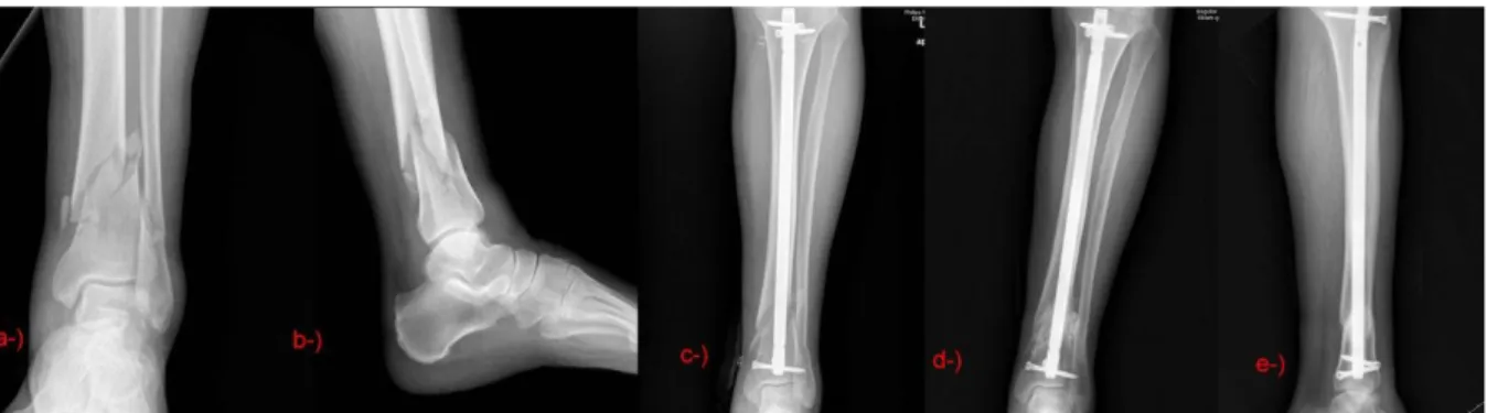
least one cortex radiologically after 10 weeks (Figure 1).
Figure 1: Seventy-year-old male AO/OTA type 43A3 a, b) Preoperative X-rays c) Early postoperative X-ray d, e) Postoperative X-ray at 19 th month
Ages, genders, fracture sides, injury mechanisms, fracture type, additional fracture, smoking
status, diabetes history of the patients as well as number of the screws applied onto the distal
side were evaluated. During postoperative period, follow-up period, union status, union time,
infection status, malunion and anterior knee pain were all evaluated. Malunion evaluation
included radiological assessment of varus, valgus, recurvatum, procurvatum as well as
radiological assessment of rotation. Rotation assessment was performed by comparing the
972
tibial tubercle, tibial crest and midline of the ankle with the healthy side (4).
A deformity of 5
degrees and above on any plane radiologically and clinically was considered as malunion.
4Clinical evaluation of the patients were done by Olerud Molander scoring.
Statistical Analysis
The statistical analysis was performed by SPSS 22.0 software programme (IBM Corporation,
Armonk, NY,USA). The average, standard deviation, median minimum, median maximum,
frequency and ratio values were used for descriptive statistics of the data. Distribution of the
variables were measured by using Kolmogorov-Smirnov test. Mann-Whitney U test was used
for quantitative analysis of the data. A p value of <0,05 was considered as statistically
significant.
Results:
Mean age of 55 patients (38 males, 17 females) was 41.3±15.3. Trauma history of the patients
was falling in 33 (60%) patients, sports injury in 4 (7.2%) patients and traffic accident in 18
(32.7%) patients. The fracture type included AO/OTA type 43A1 in 24 (43.6%) patients,
43A2 in 18 (32.7%) patients and 43A3 in 13 (23.6%) patients. Eight (14.5%) patients had
Gustilo Andersion type 1 fracture whereas 8 (14.5%) patients had Gustilo Anderson type 2
fracture. Closed fracture was detected in 39 (70.9%) patients. Fifty-two (94.5%) patients had
fibula fracture. Plate fixation was applied in 5 (9.1%) patients only. Three screws were placed
on distal side of the nail in 35 (63.6%) patients whereas 2 screws were placed on distal side of
the nail in 20 (36.4%) patients. Detailed characteristics of the patients were presented in
Table 1.
Mean follow-up period was 35.1±9 weeks. Complete (100%) union was provided in all
patients. Mean union period was 14.2±1.9 weeks. Mean Olerud Molander score was 88.5±8.
Superficial infection developed in 5 (9.1%) patients and 2 (3.6%) patients developed deep
973
infection (Table 2). Infection regression was achieved through wound care and oral
antibiotherapy in the patients with superficial infection. After fracture union was achieved by
antibiotic pressure in the patients with deep infection, the intramedullary nails were removed.
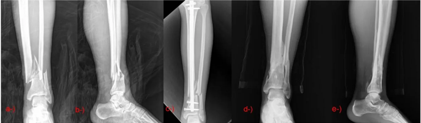
(Figure 2). Anterior knee pain was detected in 15 (27.3%) patients postoperatively.
Figure 2: Thirty-one-year-old male AO/OTA type 43A3 a, b) Preoperative X-rays c) Early postoperative X-ray d,e)Postoperative X-ray at 34 th month, removal of the nail after fracture healing due to deep infection
Alignment problems were as follows; mean varus angle 0.6±1.4 degrees; mean valgus
angle was 2.1±1.9 degrees; mean recurvatum angle was 0.7±1.2 degrees; mean procurvatum
angle was 0.3±0.7 degrees and mean rotation angle was 0.5±1.4 degrees. Two (3.6%) patients
developed varus malunion; 6 (10.9%) patients developed valgus malunion; one (1.8%) patient
developed recurvatum malunion; 3 (5.5%) patients developed rotation malunion (Table 2).
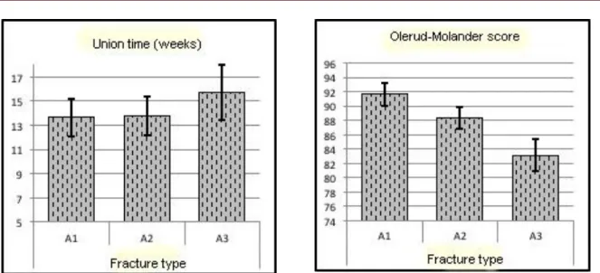
There was not any significant association between Olerud Molander score and fracture union time and wound status, fibula fixation, and number of distal screws (p>0.05) (Table 3,4 ). Union time was significantly longer in AO/OTA 43A3 fracture type (p<0.05) (Figure 3). Furthermore, Olerud Molander score was found significantly lower in AO/OTA 43A3 fracture type (p<0.05) (Figure 4).
974
Figure 3: Association between fracture type and union time Figure 4: Association between fracture type and Olerud Molander score
Table 1: Characteristics of patients
Min.-Max. Median Average.±SD/n-%
Age (year) 19,0 - 82,0 40,0 41,3 ± 15,3 Gender Female 17 30,9% Male 38 69,1% Side Left 35 63,6% Right 20 36,4% Trauma mechanism Falling 33 60,0% Sport injury 4 7,3% Traffic accident 18 32,7% Fracture Type(AO/OTA classification) A1 24 43,6% A2 18 32,7% A3 13 23,6%
Open fracture type (Gustilo Anderson classification) 0 39 70,9% 1 8 14,5% 2 8 14,5% Polytrauma 8 14,5% Smoking 14 25,5%
Diabetes mellitus history 6 10,9%
Fibula fracture 52 94,5%
Fibula osteosynhtesis 5 9,1%
Distal screw number 2
20
36,4%
975 Table 2: Results
Min-Max Median Average.±s.d./n-%
Fallow-up (months) 13,0 - 55,0 36,0 35,1 ± 9,0 Union Time (weeks) 11,0 - 18,0 14,0 14,2 ± 1,9 Olerud-Molander Score 70 - 100 90 88,5 ± 8,0 Varus malalignment (degrees) 0 - 6 0 0,6 ± 1,4 Valgus malalignment (degrees) 0 - 7 2 2,1 ± 1,9 Recurvatum malalignment (degrees) 0 - 5 0 0,7 ± 1,2 Procurvatum malalignment (degrees) 0 - 3 0 0,3 ± 0,7 Rotation malalignment (degrees) 0 - 5 0 0,5 ± 1,4 Varus Malunion (person)
2 3,6% Valgus Malunion (person)
6 10,9% Recurvatum Malunion (person)
1 1,8% Procurvatum Malunion (person)
0 0,0% Rotation Malunion (person)
3 5,5% Superficial Infection 5 9,1% Deep Infection 2 3,6%
976 Table 3: Parameters associated with union time
Union Time (weeks)
p
Min-Max Median Average.±s.d.
Fracture type
A1 11 - 18 14 13,6 ± 1,6
0,017 K
A2 12 - 17 13 13,8 ± 1,6 A3 12 - 18 16 15,7 ± 2,3 Open fracture type (Gustilo
Anderson classification) 0 12 - 18 14 14,3 ± 1,9 0,053 K 1 11 - 16 13 13,0 ± 1,6 2 13 - 18 14 14,9 ± 2,0 Fibula osteosynthesis No 11 - 18 14 14,1 ± 1,9 0,858 m Yes 12 - 18 13 14,6 ± 2,7
Distal screw numbers
II 12 - 18 14 14,1 ± 1,8
0,845 m III 11 - 18 14 14,2 ± 2,0
K Kruskal-wallis / m Mann-whitney u test
Table 4: Parameters associated with Olerud Molander score
Olerud-MolanderScore
p Min-Max Median Average .± s.d.
Fracture type
A1 80 - 100 90 91,7 ± 7,2
0,012 K
A2 80 - 100 85 88,3 ± 7,5 A3 70 - 95 85 83,1 ± 7,8 Open fracture type (Gustilo
Anderson classification) 0 70 - 100 90 90,0 ± 7,3 0,747 K 1 75 - 100 83 86,9 ± 11,3 2 75 - 90 83 83,1 ± 5,3 Fibula osteosynthesis No 70 - 100 88 88,6 ± 8,0 0,881 m Yes 75 - 95 95 88,0 ± 9,7
Distal screw numbers
II 75 - 100 90 89,5 ± 8,1
0,476 m III 70 - 100 85 88,0 ± 8,1
K
Kruskal-wallis / m Mann-whitney u test
DISCUSSION:
One of the most common problems experienced in IMN for treatment of distal tibia fracture is malalignment.In the present study, malalignment was detected in 16.3% (9 patients) of the patients (valgus malalignment, 10.9%). Previous studies reported more malalignment in IMN approach for treatment of distal tibia fracture when compared with fixation by plate. However, Polat et al. reported similar malalignment rates in their study where they compared MIPPO and IMN (12).Nevertheless,
977
malalignment on coronal plane was detected more in IMN group whereas malalignment on the sagittal plane was more in MIPPO groups (12).In the present study, valgus malalignment was detected more especially on coronal plane (10.9%). Authors of previous studies and meta-analyses suggested that cause of more malalignment detection in IMN procedure was comparison of open reduction and fixation by a plate. They defended that since anatomic reduction is achieved during open reduction, malalignment was observed less (12).However, similar malalignment rates were detected in MIPPO procedure where closed reduction methods are used and IMN procedure (12).
McArthur et al. reported in their study that a pseuodo-malalignment may appear unless a fluoroscopic imaging was performed at a complete vertical angle onto the tibia and ankle during intramedullary nailing (13).They detected a malalignment of 15 degrees in the measurements performed through imaging performed at 15 degrees of craniocaudal angle as well as vertical angle imaging (13).General positioning of the knee joint at flexion during intramedullary nailing prevents a complete vertical imaging in some cases. This inaccurate imaging may cause postoperative malalignment (13).One of the causes of malalignment which was observed more during postoperative period in the present study may be due to the procedure performed at knee flexion in all cases. The intramedullary nailing with suprapatellar insertion which is preferred during recent years is performed at semi-extension position (14).We believe that this new approach will be preferred more in the future since the ankle and distal side of the tibia can be imaged easier at semi-extension position. In the study conducted by Aviluce at al. where tibial IMN procedure through suprapatellar and infrapatellar insertion were compared, they reported lower malalignment rates in IMN procedure through suprapatellar insertion (15).
There are various studies related to IMN procedure for distal tibia fractures with intraarticular extension (3,11,16,17).This procedure may also be performed with newly designed nails which enable more screwing and more distal procedure (11). One of the most important issues in intramedullary nailing procedure of distal tibia fractures is that the guidewire inserted before nailing procedure is just on the middle at both coronal and sagittal planes (3). Unless this is achieved, the nail is placed centrally and causes intraoperative malalignment. Unless the guidewire is placed centrally, polar screw
978
procedures were started to be applied lately (3,9,10).Other procedures include calcaneal traction and percutaneous clamp procedures (3,11). These methods were usually preferred in the present study; therefore, polar screw procedure was not needed in any case.
There are different opinions about place of fibula fixation in treatment of distal tibia fractures. In the study carried out by Egol et al., significantly lower malalignment rates were detected in the patients whom fibula fixation was done (7).Moreover, a cadaver study indicated that fibula fixation in tibia and fibula fractures increase rotational stability (18).However, higher non-union rates were detected in the group where fibula fixation was done in distal tibia fractures (19,20).Number of the patients whom fibula fixation was done is quite lower in the present study (9.1%). We believe that less fibula fixation rates may be one of the causes for higher valgus malalignment cases. Further studies where higher number of patients are compared whether fibula fixation is done for similar fracture types are needed to be able to suggest the clear effect of fibula fixation on malalignment and union.
Limitations of the present study were as follows; the study was retrospective, number of the cases was less, the procedures were performed by different surgeons, rates of the patients whom fibula fixation was done were lower, therefore effects on outcomes could not be evaluated completely.
Consequently, successful functional and radiological outcomes may be achieved through IMN procedure for treatment of distal tibia fractures. However, malalignment, valgus malalignment in particular is an important complication of IMN procedure. We suggest that such complication may be prevented by a careful exploration during surgical treatment. Furthermore, we also believe that polar screw procedure as well as intramedullar nailing procedure with suprapatellar insertion where fluoroscopic evaluation may be done easier at semi-extension of the knee during the treatment may be the factors to reduce the malalignment.
979 REFERENCES
1. Casstevens C, Le T, Archdeacon MT, Wyrick JD. Management of extra-articular fractures of the distal tibia: intramedullary nailing versus plate fixation. J Am Acad Orthop Surg. 2012;20(11):675-83.
2. Aksekili MA, Celik I, Arslan AK, Kalkan T, Ugurlu M. The results of minimally invasive percutaneous plate osteosynthesis (MIPPO) in distal and diaphyseal tibial fractures. Acta Orthop Traumatol Turc. 2012;46:161-7.
3. Katsenis DL, Begkas D, Spiliopoulos G, Stamoulis D, Pogiatzis K. The results of closed intramedullary nailing for intra-articular distal tibial fractures. J Orthop Trauma. 2014;28:108-13.
4. Marcus MS, Yoon RS, Langford J, Kubiak EN, Morris AJ, Koval KJ, et al. Is there a role for intramedullary nails in the treatment of simple pilon fractures? Rationale and preliminary results. Injury. 2013;44:1107-11.
5. Li Y, Jiang X, Guo Q, Zhu L, Ye T, Chen A. Treatment of distal tibial shaft fractures by three different surgical methods: a randomized, prospective study. Int Orthop. 2014;38:1261-7.
6. Janssen KW, Biert J, van Kampen A. Treatment of distal tibial fractures: plate versus nail: a retrospective outcome analysis of matched pairs of patients. Int Orthop. 2007;31:709-14.
7. Egol KA, Weisz R, Hiebert R, Tejwani NC, Koval KJ, Sanders RW. Does fibular plating improve alignment after intramedullary nailing of distal metaphyseal tibia fractures? J Orthop Trauma. 2006;20:94-103.
8. Koval KJ, Clapper MF, Brumback RJ, Ellison PS, Jr., Poka A, Bathon GH, et al. Complications of reamed intramedullary nailing of the tibia. J Orthop Trauma. 1991;5:184-89
9. Mosheiff R, Safran O, Segal D, Liebergall M. The unreamed tibial nail in the treatment of distal metaphyseal fractures. Injury. 1999;30:83-90.
10. Moongilpatti Sengodan M, Vaidyanathan S, Karunanandaganapathy S, Subbiah Subramanian S, Rajamani SG. Distal tibial metaphyseal fractures: does blocking screw extend the indication of intramedullary nailing? ISRN Orthop. 2014;2014:542623.
11. Nork SE, Schwartz AK, Agel J, Holt SK, Schrick JL, Winquist RA. Intramedullary nailing of distal metaphyseal tibial fractures. J Bone Joint Surg Am. 2005;87:1213-21.
12. Polat A, Kose O, Canbora K, Yanik S, Guler F. Intramedullary nailing versus minimally invasive plate osteosynthesis for distal extra-articular tibial fractures: a prospective randomized clinical trial. J Orthop Sci. 2015;20:695-701.
13. McArthur JR, Makrides P, Wainwright D. Resemblance of valgus malalignment of the distal tibia in 15-degree craniocaudal radiographs. J Orthop Surg (Hong Kong). 2013;21:337-9.
14. Brink O. Suprapatellar nailing of tibial fractures: surgical hints. Curr Orthop Pract. 2016;27:107-12. 15. Avilucea FR, Triantafillou K, Whiting PS, Perez EA, Mir HR. Suprapatellar Intramedullary Nail Technique
Lowers Rate of Malalignment of Distal Tibia Fractures. J Orthop Trauma. 2016;30:557-60.
16. Beytemur O, Albay C, Adanir O, Yuksel S, Gulec MA. Is intramedullary nailing applicable for distal tibial fractures with ankle joint extension? Eklem Hastalik Cerrahisi. 2016;27:125-31.
17. Ristiniemi J, Luukinen P, Ohtonen P. Surgical treatment of extra-articular or simple intra-articular distal tibial fractures: external fixation versus intramedullary nailing. J Orthop Trauma. 2011;25:101-5.
980
18. Kumar A, Charlebois SJ, Cain EL, Smith RA, Daniels AU, Crates JM. Effect of fibular plate fixation on rotational stability of simulated distal tibial fractures treated with intramedullary nailing. J Bone Joint Surg Am. 2003;85:604-8.
19. Vallier HA, Le TT, Bedi A. Radiographic and clinical comparisons of distal tibia shaft fractures (4 to 11 cm proximal to the plafond): plating versus intramedullary nailing. J Orthop Trauma. 2008;22:307-11. 20. Varsalona R, Liu G. Distal tibial metaphyseal fractures: the role of fibular fixation. Strategies in trauma and