http://ejvs.selcuk.edu.tr www.eurasianjvetsci.org
RESEARCH ARTICLE
Comparison of the effects of intraocular pressure with phacoemulsification and
extra-capsular cataract extraction methods in dogs with cataract
Mustafa Arıcan
1, Hanifi Erol
2, Kurtuluş Parlak
1, Ümit Kamış
3, Nuri Yavru
11Department of Surgery, Faculty of Veterinary Medicine, University of Selcuk, 3Department of Ophtamology, Faculty of Medicine, Konya, 2University of Erciyes, Faculty of Veterinary Medicine, Department of Surgery, Kayseri, Turkey
Received: 17.05.2014, Accepted: 29.05.2014 *[email protected]
Özet
Arıcan M, Erol H, Parlak K, Kamış Ü, Yavru K. Kataraktlı köpeklerde fakoemülsifikasyon ve ekstrakapsüler katarakt ekstraksiyon yöntemlerinin intraoküler basınça etkilerinin karşılaştırılması.
Amaç: Bu çalışmada intraoküler lens (IOL) konularak veya konulmadan yapılan ekstrakapsüler ekstraksiyon (EKKE) ve fa-koemülsifikasyon (FAKO) yöntemlerinin 28 gün süre içinde göz içi basınçlarına etkilerini araştırılması amaçlanmıştır.
Gereç ve Yöntem: Materyal olarak her iki cinsiyetten farklı yaşlarda vücut ağırlıkları 10 ile 30 kg arası olan katarakt teşhisi konulan 20 köpek kullanıldı. Katarakt teşhisi, direkt ve indirekt oftalmoskop, ultrasonografi, biyomikroskop ile yapıldı. Katarakt olguları klinik muayene ve ultrason ile immatür (7 olgu), matür (7 olgu) ve hipermatür (6 olgu) olarak sınıflandırıldı. Köpekler 10’arlı iki gruba ayrıldı. Katarakt teşhisi konulan 10 köpeğe EKKE ve diğer 10 köpeğe ise FAKO operasyonları gerçekleştirildi. Her iki grup için 41 dioptrilik tek parça akrilik intraoküler lens kullanıldı.
Bulgular: Göz içi basınçı IOL kullanılmayan EKKE grubunda 14. günde en düşük seviyede ölçüldü. IOL uygulanan grupta ise düzensiz seyir gösterdi. Fakoemülsifikasyon IOL uygulanmayan grupta ise göz içi basınçları 21. günde en düşük seviyede ölçül-dü. Bununla birlikte 28. gün sonunda bütün değerlerin referans aralığında olduğu tespit edildi.
Öneri: Kataraktlı olgularda göz içi basınçları dikkate alındığında her iki cerrahi uygulamanında kullanılabileceği kanısına varılmıştır. Cerrahinin başarısı doğru hasta seçimi, doğru cerrahi teknik, doğru ekipman ile olmaktadır. Bunun yanı sıra katarakt immatür dönemde teşhis edilirse fakoemülsifikasyonun başarı oranı artmaktadır.
Anahtar kelimeler: Köpek, katarakt, fakoemülsifikasyon, göz içi basınç
Abstract
Arican M, Erol H, Parlak K, Kamis U, Yavru K. Comparison of the effects of intraocular pressure with phacoemulsification and extracapsular cataract extraction methods in dogs with cataract.
Aim: The aim of this study was to investigate the effects of in-traocularpressure (IOP) in extracapsular extraction (ECCE) and phacoemulsification (PHACO) methods with or without intraoc-ular lens (IOL) on dogs with cataract for a 28-day period.
Materials and Methods: Twenty adult dogs of both sexes with cataracts, weighing between 10 to 30 kg and at different ages were used as materials. Cataracts were diagnosed by direct and indirect ophthalmoscopy, ultrasonographic examination and bi-omicroscopy. Cataracts were classified as immature (7 animals), mature (7 animals) and hypermature (6 animals). Dogs were di-vided into two groups each consisting of 10 animals. Ten dogs with cataract were operated on for ECCE and the other group of 10 dogs underwent a phacoemulsification procedure. In the two groups, 10 animals were used for 41 dioptry single-piece acrylic intraocular lens.
Results: Intraocular pressure was felt at the lowest level 14 days after the operation in the ECCE without IOL implanted group. The IOL implanted group showed irregular levels. Intraocular pressure level was the lowest on the 21st day, without IOL im-planted group in phacoemulsification. However, all values re-mained within the reference values at the end of a 28-day period postoperatively.
Conclusions: Both surgeries could be used for cataract cases in terms of IOP effects. Selection of the patient, correct surgical technique and adequate equipment are important for the suc-cess of a surgery. But, it has been also concluded that sucsuc-cess of phacofragmentation surgery increased when the animals are in immature stages.
Keywords: Dog, cataract, phacoemulsification, intraocular pressure
Eurasian J Vet Sci, 2014, 30, 4, 188-194
DOI:10.15312/EurasianJVetSci.201447375
Eurasian Journal
Introduction
Cataracts are the most frequent causes of blindness in dogs. The remove of the lens for the repair of vision lost due to a cataract has become an ordinary procedure and technique in veterinary medicine (Honsho et al 2007). The success rate of cataract surgeries has risen significantly during the recent decades, especially due to the development of phacoemulsi-fication and intra ocular lens (IOL) implantation (Gelatt and Gelatt 2001, Kecova and Necas 2004). The use of intraocular lenses is currently the treatment of choice (Kecova and Necas 2004) IOL implantation improves visions and reduces the formation of posterior capsule opacity (PCO) after surgery (Gelatt and Gelatt 2001).
Cataract extraction procedures are known to cause of in-crease in intraocular pressure (IOP) within the first few hours after surgery in dogs (Honsho et al 2007). The reason of ocular hypertension in these cases that was to blockage of the iridocorneal angle by lenticular remnants, soluble proteins, pigments and vitreous humor, trauma during the surgical procedure. It was breakdown of the blood aqueous barrier and the presence of inflammatory cells, presence of viscoelastic material, hemorrhage and synechiae (Smith et al 1996, Sigle and Nasisse 2006).
A great advantage of ultrasound is that it can be used to evaluate the internal structure of the eye. The knowledge of the dimensions of optical components is required to better understand many studies and clinical problems related to vi-sion. Both A-mode and B-mode ultrasound have been used for ocular biometry (Paunksnis et al 2001). Ultrasonography is obligatory for a good selection of patients for cataract sur-gery because it offers valuable information especially of the posterior segment, which is not always visible by semiotic ophthalmic techniques.
The purpose of this research was to investigate the effects of IOP in extra capsular extraction (ECCE) and phacoemulsifi-cation methods with or without IOL on dogs with cataracts over a 28-day period. Ultrasonography was used to evaluate the condition of the lens and fundus of the eye with a cata-ract.
Materials and Methods
Animals
Twenty adult dogs of both sexes with cataracts, weighing between 10 to 30 kg and at different ages were used as ma-terials. The study was carried out according to the criteria of the Ethical committee of Selcuk University. The dogs were randomly selected on the basis of on assessment of their general clinical condition with significance on hematimetric indexes and biochemical outline. Cataracts were diagnosed
by direct and indirect ophthalmoscopy, Schirmer tear test, ultrasonographic examination and, for in some cases (im-mature cataracts), biomicroscopy. Cataracts were classified as immature (7 animals), mature (7 animals), and hyperma-ture (6 animals) (Figure 1) through clinical examination and ultrasonography. Dogs were divided into two groups each consisting of 10 animals. Ten dogs with cataract were oper-ated on for ECCE and the other group of 10 dogs underwent a phacoemulsification (PHACO) procedure. In the two groups, 10 animals (5 for ECCE and 5 for PHACO) were used for 41 dioptry single-piece acrylic intraocular lens. One eye (left eye) of all dogs with cataracts was used as material during the research period.
Ultrasonography examination
Ultrasonography was performed using ophthalmic exami-nation B mode probes convex mechanical transducer of 7.5 MHz (Esaote Piemedikal, Model 410477) (Martins et al 2010) (Figure 2). The transducer was positioned to scan the entire globe. The lens was evaluated with respect to echo-genicity. Alterations in the posterior segment were investi-gated and assessed. The lens of each eye was classified as having hyperechoic or hypoechoic areas. At the end of the examination, the eyes were rinsed with sterile 0.9% sodium chloride solution.
Preoperative procedures
The preoperative procedures were started 24 hours before the surgery with topical administration of corticosteroids/ antibiotics (0.1% Dexamethasone, Maxidex 0.1 mg 5 mL oft. sol/lomefloksasin, Okacin 3 mg 5 mL, Novartis) every 2 hours. Twenty-four hours before the treatment, daily ther-apy was initiated with (600 000 IU Pencilline G Procain flk 800.000) in IM. One hour before the surgery, sikloplegic/ midriatic (Siklopentolat HCl, Sikloplejin 1%, Fenilefrin HCl, Mydfrin® 2.5%) were instilled at 15 minute intervals the dogs were intravenously preanesthetized with xylazine hy-drochloride (Alfazyne 2%) and ketamin hyhy-drochloride (Al-famine 10%). After 10 minutes, anaesthesia was induced by isoflurane (AErrane, Baxter 2-4%).
Extracapsular extraction (ECCE)
Extracapsular extraction tecniques were followed by Gelatt and Gelatt (2001). The anterior chamber was entered with a blade (Figure 3). After cornea-scleral insicion, the anterior chamber was then filled with Trypan blue to stain the ante-rior crystalline capsule (Figure 4). Two minutes after appli-cation of the dye and its contact with the anterior capsule of the lens, the remaining dye was removed by washing the anterior chamber with balanced salt solution (BSS®, Alcon Laboratories). The anterior chamber was then supplied with a viscoelastic substance of hydroxypropylmethylcellulose
Figure 1. Cataracts were classified as immature, mature and hypermature through clinical exam-ination and ultrasonography.
Figure 3. The anterior chamber was entered with a blade.
Figure 2. Ultrasonography was performed using ophthalmic examination B mode probes convex mechanical transducer of 7.5 MHz.
Figure 4. The anterior chamber was then filled with trypan blue to stain the anterior crystalline capsule.
Figure 6. A wide incision was made in the limbus and the anterior lens capsule.
Figure 5. The anterior chamber was then filled with a viscoelastic substance of hydroxypropyl methylcellulose.
Figure 7. Nucleus and cortex were extracted
manually. Figure 9. Irrigation and aspiration were used towards the end of the surgery to remove the re-maining lens materials from the equatorial and posterior lens capsules.
Figure 8. A phacofragmentation tip was intro-duced through the corneal incision via the ante-rior lens capsule.
(Crown gel 2.4%, Nanotech ophtalmic) (Figure 5). A continu-ous curvilinear capsulorhexis was done with forceps. The corneal incision was maken large with corneal scissors. A wide incision was made in the limbus and the anterior lens capsule, nucleus and cortex were extracted manually (Fig-ures 6 and 7), followed by flushing to remove any remaining lens particles. Corneal incision was closed with interrupted nylon 8/0 sutures (Surgicryl®, Polyglycolic Acid). Dexameth-asone 0.1 mL was injected subconjunctivally. Topical admin-istration of corticosteroids/antibiotics (0.1% Dexametha-sone, Maxidex 0.1 mg 5 mL oft. sol /lomefloksasin, Okacin 3 mg 5 mL, Novartis) every 2 hours.
Phacoemulsification technique
After cornea-scleral insicion, the anterior chamber was then filled with Trypan blue to stain the anterior crystalline
cap-sule. Two minutes after application of the dye and its contact with the anterior capsule of the lens, the remaining dye was removed by washing the anterior chamber with balanced salt solution (BSS®, Alcon Laboratories). The anterior chamber volume was filled with viscoelastic material (hydroxypro-pylmethylcellulose (Crown gel 2.4%, Nanotech ophtalmic). Modified capsulorhexis penses were used to cut into the anterior lens capsule. Part of the anterior lens capsule was carefully torn and removed. A phacofragmentation tip (Al-con Universal USA S/N 1077 Al(Al-con Surgical, INC, IRVINE, CA) was established through the corneal incision via the anterior lens capsule (Figure 8). Irrigation and aspiration systems were used assisting the end of the surgery to eliminate the remaining lens materials from the equatorial and posterior lens capsules (Figure 9). Corneal incision was closed with in-terrupted nylon 8/0 sutures (Surgicryl®, Polyglycolic Acid). The anterior chamber was filled with balanced salt solution.
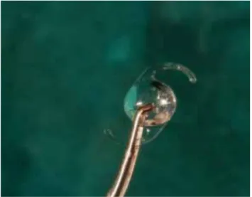
Figure 10. Acrylic hydrophilic monomer (Lens Teco, Lenstec) optic IOL of
41 D was used. Figure 11. The IOL optic was holding by forceps and inserted into the IOL cartridge
Figure 12. Eyes were examined postoperatively until at least 1 month after
the surgery. Figure 13. Preoperative and postoperative intraocular pressures were measured and recorded on the 7th, 14th, 21st and 28th days with a re-bound tonometer.
Postoperatively, similar procedures such as ECCE were per-formed for PHACO group.
Intraocular lenses (ILO)
Acrylic hydrophilic monomer (Lens Teco, Lenstec) optic IOL of 41 D was used. The IOL optic was holding by forceps and placed into the IOL cartridge (Figure 10). After supplying the capsular bag and anterior chamber with a hydroxypropyl-methylcellulose (Crown gel 2.4%, Nanotech ophtalmic), the IOL in the cartridge was put in the capsular bag with the IOL inserter without enlarging the corneal incision (Figure 11).
Postoperative care
Topical administration of antibiotics Okacin (lomefloksasin, Okacin 3 mg 5 mL, Novartis) was performed every 2 hours for two weeks. Corticosteroids (0.1% Dexamethasone, Maxi-dex 0.1 mg 5 mL oft. sol) were administered every 3 hours for two weeks and afterwards daily used. Carprofen (Rimadyl®, Pfizer Co., Ltd) 2 mg/kg was orally given once dexametha-sone had stopped. Medications and doses were changed based on intraocular complications. All dogs were instruct to wear Elizabethan collar at all times during the first 2 months to prevent self trauma. Eyes were examined postoperatively until at least 1 month after the surgery (Figure 12).
Intraocular pressure
Preoperative and postoperative intraocular pressures were measured and recorded on the 7th, 14th, 21st and 28th days with a rebound tonometer (TonoVet®, Figure 13). The cor-rect results for intraocular pressure were measured for the animal standing positions. The following changing was com-pared for intraoperative complications, variations in IOP and photophobia and blepharospasm, conjunctival hyperemia, corneal oedema, aqueous humor transparency, and iridocy-clitis for the surgical duration of each technique.
Statistical analysis
IBM SPSS 19 Pearson correlation test was used for the evalu-ation of postoperative vision after surgery was performed for
immature, mature and hypermature eyes. IOP values were compared between animals by replicated measures analysis of variance using IBM SPSS 19, one-way ANOVA and Duncan test. It should be noted that eyes were eliminated from the statistical analysis of the IOP changing since complications might interrupt with the results.
Results
Ultrasonography examination
Hypermature cataract with resorption of the axial regions of the anterior cortex and of the nucleus was observed in 6 animals. The axial diameter of the eyes with immature and mature cataracts was smaller in size compared to hyper-mature cataracts. But, there were no statistically significant differences. On clinical examination, posterior and anterior capsular cataract was detected in the dog with hypermature cataract in both eyes. Nucleus and posterior capsule of lens was hyperechoic echogenicity. However, the posterior cap-sule was impossible given the degree of opacification of the lenses in the other animals. Different ultrasonography altera-tions were observed in the posterior segment in some cases. Hypoechoic echogenicity of lens was detected in dogs with mature and hypermature cataract. The biometric measure-ments were shown in Table 1.
Operation results
Photophobia and blepharospasm were detected in both eyes immediately after the surgery. However, these situations were more severe in eyes submitted to ECCE. Conjunctival hyperaemia was demonstrated in all eyes, moderate at the beginning and becoming severe by the second hour after surgery. Moderate to severe corneal oedema was observed in the eyes submitted to ECCE. In the eyes submitted to phaco-emulsification, oedema was limited to region close to and above the suture lines. Ocular discomfort was observed in ECCE groups animals compared to those submitted to phaco-emulsification.
ECCE (-); Extracapsular extraction without IOL; ECCE (+); Extracapsular extraction with IOL; PHACO (-) Phacoemulsification without IOL; PHACO (+) Phacoemulsification with IOL; The letters a, b, c in each row shows statistical significance (P˂0.05).
ECCE (-) ECCE(+) PHACO (-) PHACO(+) Pre-operative 38.72±5.51a 15.36±0.92b 26.76±1.87a 29.12±3.95a 7. Day 27.84±4.03b 23.56±3.69a 12.20±1.30bc 18.52±1.90b 14. Day 16.52±1.56c 23.44±3.68a 11.52±1.41bc 17.56±1.65b 21. Day 23.96±2.23bc 13.72±1.06b 9.28±0.87c 18.80±1.47b 28. Day 21.08±1.72bc 16.76±1.37ab 14.32±1.33b 17.76±1.57b
Intraocular pressure
ECCE without IOL applied group was showed statistical differences (P<0.05) on the 7th and 14th days. Intraocular pressure was getting lower compared to the day 7th, 14th, 21st and 28th. But all the data were seen to be within normal values. The assessment regarding postoperative intraocular pressure was higher on the 7th and 14th days compared to the 21st day. But all the data were seen to be between nor-mal values. PHACO without IOL group also showed statistical differences on the 21st day, they were seen within normal values. PHACO with IOL applied group showed statistical dif-ferences in the range from 0 to the 7th, 14th, 21st and 28th days (P<0.05). The results were also seen within normal val-ues for this group in Table 2.
Discussion
The present study shows that there are a lot of advantages of phacoemulsification (Chahory et al 2003), such as short continuance of surgery, a low risk of vitreous loss, conserva-tion of the IOP during surgery and less extreme iridocyclitis, in addition to a smaller incision and smaller scar attended by greater corneal transparency and less astigmatism as sup-ported by previous studies (Lannek and Miller 2001, Sigle and Nasisse 2006, Yi et al 2006). In addition, during the sur-gery, corneal oedema, posterior capsule rupture and corneal thermal injuries were also observed.
In the study, IOP measurements were seen reference values for ECCE and PHACO with or without IOL groups. Hyperma-ture cataract cases showed a positive correlation with es-tablished anterior chamber depth, posterior lens surface to retina and a higher IOP. It is reported that IOP is known to stay on elevated during the first 24 hours after surgery, es-pecially in eyes submitted to phacoemulsification (Smith et al 1996, Lannek and Miller 2001, Sigle and Nasisse 2006, Yi et al 2006). Small incisions and highly viscous composition contribute to the incident of postoperative ocular hyperten-sion. Small incisions supported better cooptation of the sur-gical wound but ruined drainage of the viscoelastic agents, contributing to the happening of ocular hypertension as sup-ported by Chahory et al (2003). Decreased drainage of the aqueous humor due to mechanical obstruction of the draage corner with lenticular remnants, plasma proteins and in-flammatory cells, as well as narrowing of the trabecular net-work as a result of oedema, favour an increase in IOP. Yi et al (2006) showed that in the presence of intraocular inflamma-tion, the IOP demonstrates to decrease after some hours. In the present study, IOP started to be measured 7 days after the surgery and pressure was within reference levels for PHACO and ECCE groups. It is also in support of the result of previ-ous studies where IOP was getting normal values within 24 hours after surgery (Miller et al 1997, Gaiddon et al 2000).
In the present study, ultrasonography examination showed the differences in lens thickness in some stages of cataract. Axial thickness of lenses with immature and mature cata-racts was also demonstrated. Previous studies showed that while mature cataracts and hypermature cataracts increase in size through intumescences, immature cataracts could be decrease in thickness owing to loss of soluble lens mate-rial from the lens capsule (Hamidzada and Osuobeni 1999, Paunksnis et al 2001, Williams 2004). Biometry of the globe is normally assumed with a mode ultrasonography to con-cluded axial length prior to intraocular lens implantation. It has been reported that A-mode and B-mode ultrasonog-raphy to give axial measurements were not statistically sig-nificantly contrary and thus B-mode ultrasonography can be as accurate as A-mode biometry (Hamidzada and Osuobeni 1999, Paunksnis et al 2001, Martins et al 2010). In this study, B-mode ultrasonography was used. Lens thickness expands with age, a feature noted in previous reports in humans and in the dog. In the present study, thickness of lens with age was not compared because there were not enough cases for statistical analysis. But, this finding was repeated in several studies and it was shown that lens thickness in each group increased with age. On the other hand, the finding of de-creased anterior chamber depth in eyes with immature cata-ract suggests that narrow iridocorneal angle could be result in to increased intraocular pressure (Van der Woerdt et al 1993).
Conclusions
Both surgeries could be used for cataract cases in terms of IOP effects. Selection of the patient, correct surgical tech-nique, adequate equipment was important for the success of a surgery. But, it was also concluded that success of phaco-fragmentation surgery increased when the animals were in immature stages. There was a possibility of faster postopera-tive rehabilitation of patients with minimal intraoperapostopera-tive damage of ocular tissues with foldable materials. Thus, ultra-sonographic examination was always very helpful to support postoperative term.
Acknowledgments
We would like to thank for financial support by University of Selcuk (Scientific Research Projects (BAP) (Project No. 084001073).
References
Chahory S, Clerc B, Guez J, Sanaa M, 2003. Intraocular pressu-re development after cataract surgery: a prospective study in 50 dogs (1998-2000). Vet Ophthalmol, 6, 105-112. Gaiddon J, Lallement PE, Peiffer RLJr, 2000. Implantation of
a foldable intraocular lens in dogs. JAVMA, 216, 875-877. Gelatt KN, Gelatt, JP, 2001. Surgical procedures of the lens
and cataracts. In: Gelatt KN, Gelatt JP (eds.). Small Animal Ophthalmic Surgery: Practical Techniques for the Veteri-narian, Butterworth-Heinemann, Oxford, UK, pp: 286-334. Hamidzada WA, Osuobeni EP, 1999. Agreement between
A-mode and B-mode ultrasonography in the measurement of ocular distances. Vet Radiol Ultrasound, 40, 52-57. Honsho CS, Oria AP, Pigatto JAT, Laus JL, 2007. Modified
ext-racapsular extraction versus endocapsular phacoemul-sification: Intraoperative and immediate postoperative events. Braz J Vet Res Anim, 59, 105-113.
Kecova H, Necas A, 2004. Phacoemulsification and intraocu-lar lens implantation: Recent trends in cataract surgery. Acta Vet Brno, 73, 85-92.
Lannek EB, Miler, PE, 2001. Development of glaucoma after phacoemulsification for removal of cataracts in dogs: 22 cases (1987-1997). JAVMA, 218, 70-76.
Martins BC, Rodrigues Jr EF, Souza ALG, Almeida DE, Brito FLC, Canola JC, Laus DBJL, 2010. A and B mode ultraso-nography in preoperative evaluation of lens and posterior segment of dogs eyes with cataract. Pesquisa Vet Brasil, 30, 121-126.
Miller PE, Stanz KM, Dubielzig RR, Murphy CJ, 1997. Mecha-nisms of acute intraocular pressure increases after
pha-coemulsification lens extraction in dogs. Am J Vet Res, 5, 1159-1165.
Paunksnis A, Svaldeniene E, Paunksniene M, Babrauskiene V, 2001. Ultrasonographic evaluation of the eye parameters in dogs of different age. Ultragarsas, 2, 39.
Sigle KJ, Nasisse MP, 2006. Long-term complications after phacoemulsification for cataract removal in dogs: 172 ca-ses (1995-2002). JAVMA, 2006, 228, 74-79.
Smith PJ, Brooks DE, Lazarus JA, Kubilis PS, Gelatt KN, 1996. Ocular Hypertension following cataract surgery in dogs: 139 cases (1992-1993) JAVMA, 209, 105-111.
Van der Woerdt A, Wilkie DA, Myer CW, 1993. Ultrasonograp-hic abnormalities in the eyes of dogs with cataracts: 147 cases (1986-1992). JAVMA, 15: 838-841.
Williams DL, 2004. Lens morphometry determined by B-mode ultrasonography of the normal and cataractous canine lens. Vet Ophthalmol, 7, 91-95.
Yi NY, Park SA, Jeong MB, Kim WT, Kim SE, Chae JM, Seo KM, 2006. Phacoemulsification and acryl foldable intraocular lens implantation in dogs: 32 cases. J Vet Med Sci, 7, 281-285.