Original Investigation
Published Online: 22.05.2019
Necati KAPLAN
1, Numan KARAARSLAN
2, Ibrahim YILMAZ
3, Duygu YASAR SIRIN
4, Feride Sinem AKGUN
5,
Tezcan CALISKAN
2, Abdullah Talha SIMSEK
2, Hanefi OZBEK
31Istanbul Rumeli University, Corlu Reyap Hospital, Department of Neurosurgery, Tekirdag, Turkey 2Namik Kemal University School of Medicine, Department of Neurosurgery, Tekirdag, Turkey
3Istanbul Medipol University School of Medicine, Department of Medical Pharmacology, Istanbul, Turkey
4Namik Kemal University, Faculty of Arts and Sciences, Department of Molecular Biology and Genetics, Tekirdag, Turkey 5Istanbul Maltepe University School of Medicine, Department of Emergency Medicine, Istanbul, Turkey
Are Intervertebral Disc Tissue Cells Damaged When
Attempting to Prevent Thrombus Formation Using Dabigatran,
A New Oral Anticoagulant?
ABSTRACT
gatroban can be used in the prophylaxis or treatment of these diseases (21). Through pharmaceutical technology, these drugs, which are administered parenterally or subcutaneously, have been replaced with orally administered drugs (4).
With the substitution of the hydroxyl in the fourth position of coumarin-derived molecules containing a
2-oxo-2H-1-█
INTRODUCTION
V
enous and arterial thrombosis are significant contribu-tors to morbidity and mortality, and anticoagulant agents are a mainstay of venous thromboembolism treatment (11). Drugs such as heparin, low molecular weight heparins, fondaparinux, hirudin, bivalirudin, lepirudin, andar-AIM: To investigate the effect of dabigatran, a new oral anticoagulant, on human primary cell cultures isolated from intact
intervertebral disc tissue.
MATERIAL and METHODS: Cell cultures were prepared from tissues obtained from six cases who had undergone surgery due
to spinal trauma. Dabigatran, an active pharmacological agent, was applied to intact annulus fibrosus (AF)/nucleus pulposus (NP) primary cell cultures from the study group. After performing cell viability, toxicity, and proliferation tests on all cultures in the control and study groups, the surface morphologies of the samples were evaluated. Subsequently, chondroadherin (CHAD), cartilage oligomeric matrix protein (COMP), and matrix metalloproteinase (MMP)-13 and -19 expressions were measured via a real-time polymerase chain reaction (RT-PCR). Data were analyzed statistically.
RESULTS: In the proliferation assays performed on the 20th day of the study, cells in the dabigatran-supplemented group were
reported to have lost 46.37% more viability than those in the control group. Expressions of all genes examined except MMP-13 were evaluated in the control group by time, but in contrast to the control group results, COMP and MMP-19 gene expressions decreased in the dabigatran-treated group. No CHAD or MMP-13 expression was noted in these cultures.
CONCLUSION: The potential for a systemically applied drug to accumulate in tissue and negatively affect surrounding tissues and
microstructures must be emphasized.
KEYWORDS: Cytotoxicity, Dabigatran, Deep vein thrombosis, Intervertebral disc cells, Primary cell cultures
Necati KAPLAN : 0000-0001-5672-0566 Numan KARAARSLAN : 0000-0001-5590-0637 Ibrahim YILMAZ : 0000-0003-2003-6337
Duygu YASAR SIRIN : 0000-0002-1224-442X Feride Sinem AKGUN : 0000-0001-6537-866X Tezcan CALISKAN : 0000-0001-7735-0584
Abdullah Talha SIMSEK : 0000-0002-8668-3935 Hanefi OZBEK : 0000-0002-8084-7855
benzopyran ring, the neutral structure becomes slightly acidic. Therefore, such drugs can be administered orally after being converted into water-soluble salts. In addition to warfarin, other antithrombocytic drugs, such as aspirin, sulphinpyrazone, dipyridamole, anagrelide, ticlopidine, clopidogrel, cilostazol, and tirofiban, which have been extensively investigated in recent years due to their protective effects in coronary artery diseases, are also present in the pharmaceutical market (8). Older generation antithrombocytic agents pharmacologically capable of interacting with many nutrients and drugs are pre-ferred for treatment, especially for patients with non-valvular atrial fibrillation. However, despite the use of antithrombocytic drugs, such as warfarin, for at least two months, the targeted oral anticoagulant therapy monitoring unit value—the Inter-national Normalized Ratio (INR)—cannot be achieved. In addition, since the duration of anticoagulant therapy cannot be determined and effective treatment doses cannot always be adjusted, bleeding risk may increase. In such adverse conditions, warfarin and similar pharmaceutical prepara-tions become unusable. For this reason, new generation oral anticoagulant agents, such as rivaroxaban, apixaban, and dabigatran, which do not require dose-range monitoring, have minimal drug–food interactions, and are at least as effective as warfarin, are recommended for treatment (9).
Intervertebral disc tissue is avascular and aneuronal, just like cartilage tissue (1,2,18-20). Due to this anatomical structure, drugs and nutrients pass from neighboring osseous structures through hyaline membranes and accumulate in the outer layers of the intervertebral disc tissue (13,14,17,25,26). Though no pharmacokinetic studies currently exist that examine dabigatran’s migration through hyaline membranes and fluid compartment concentrations with high evidential value, it is an indisputable fact that many medicaments, such as synovia, may accumulate in the liquid compartments and/ or on surfaces which come into contact with intervertebral disc tissue, and thus they may pass into other tissues (1,2,13,14,17-20,25,26). Alongside the advancement of pharmaceutical technology and regenerative medicine, many reports have examined how to protect tissues by investigating the toxicity of drugs at the molecular level. In addition, the question of how damaged tissue can be repaired has also been investigated (1,2,13,14,17-20,25,26). As in all areas of medicine, neurosurgery researchers have begun contributing to the literature on disc tissue preservation and damaged tissue repair. Of these research subjects, the investigation of the positive and negative effects of medication frequently prescribed by many clinics, including emergency departments, on healthy intact intervertebral disc tissue has particularly gained momentum (1,2,13,14,17-20,25,26).
When the literature on promising oral anticoagulant drugs is examined, no studies appear to have examined whether these substances damage disc tissue cells. This is an original feature of this article. This study was conducted to investigate the effects of dabigatran, which is frequently prescribed by experts in cardiology, cardiovascular surgery, neurology, emergency departments, neurosurgery, and orthopedic surgery due to its effects as a direct oral thrombin inhibitor
on human primary nucleus pulposus (NP) cell cultures and annulus fibrosus (AF) cell cultures.
Alongside an investigation of the effects of dabigatran on proliferation and differentiation, the expression of chondroadherin (CHAD; 1,2,18-20), an NP-specific marker, and the gene expressions of reagents in the extracellular matrix (ECM) were tested. In addition, the expression of cartilage oligomeric matrix protein (COMP; 5,7,12,15,24), encoded by the COMP gene in humans, also known as thrombospondin-5 (and mainly found in the cartilage as an ECM protein and thought to be a novel biomarker for spine disc space narrowing, osteophytes, scoliosis, and joint metabolism), was also analyzed. Moreover, changes in gene expressions of matrix metalloproteinases (MMPs)-13 and -19, which play important roles in many biological processes, including embryogenesis (22), tissue restructuring, wound healing, and angiogenesis (27,28), were examined.
█
MATERIAL and METHODS
This research project was carried out with the approval of the Local Ethics Committee of the Istanbul Medipol Univer-sity Faculty of Medicine (29.11.2017-10840098/604.01.01/ E.44192). All molecular analyses performed during the experi-ments were repeated at least thrice. All molecular analyses were performed in all groups on the 10th and 20th days of the
experiment. During analysis, researchers were unaware which group contained the control group samples and which group contained the dabigatran-supplement study group samples. The researchers performing the analyses were also blind to the experimental setup, as they were not informed about con-trol and study groups.
Selection Criteria
Initially, 12 patients were selected for inclusion in the study. However, the tissues obtained from patients with creatinine clearances of < 30 mL/min (n=1) and from patients with heart valve prosthesis (n=1) were excluded from the study. Due to the risk of undesirable effects manifesting from such pharmaceuticals as dronedarone, HIV protease inhibitors, phenytoin, phenobarbital, carbamazepine, conazoles, and rifampicin acting together with dabigatran (16), the tissues of patients taking the above-mentioned drugs (n=4) were also excluded. Disc and granulation samples obtained from the remaining six participants were transferred to the cell culture laboratory at a temperature of 4ºC in Dulbecco’s Modified Eagle Medium (DMEM; Cat #11965092, Thermo Fisher Scientific, USA) with 5% penicillin-streptomycin (PS; Cat #15140122, Gibco, Thermo Fisher Scientific, USA), 15% fetal bovine serum (FBS; Cat #10082147, Thermo Fisher Scientific, USA), and 1% L-glutamine (Cat #25030081, Thermo Fisher Scientific, USA) to prepare the primary cell cultures.
Preparation of Cell Cultures from Primary Human Intervertebral Disc Tissue
The age distribution of the six participants with a mean age of 36 years ranged from 18 to 46 years. All had a history of spinal trauma and were transferred from the emergency department
to the neurosurgery department to receive operations after clinical and imaging examinations. Signed informed volunteer consent forms were obtained from the patients so their resected tissue samples could be used in this study.
The six resected intervertebral disc tissues meeting the patient selection criteria were taken to the laminar flow cabinet (Air Flow-NUVE/NF-800 R). From there, the tissues were transferred to Petri dishes. Tissue samples were irrigated with a 0.9% isotonic sodium chloride solution and clarified from the red blood cells. Mechanically degraded tissue samples were transferred to centrifuge tubes. Subsequently, a collagenase type II (Cat #17101015, Thermo Fisher Scientific, USA) solution was added to the samples suspended in Hank’s balanced salt solution (HBSS; Cat #55021C, Sigma Aldrich, USA). The samples were then maintained overnight in an incubator (NUVE, 06750, Turkey) set at a temperature of 37°C with 5% CO2. In this way, the tissues degraded enzymatically
and through mechanical degradation. The samples were centrifuged at 1200 rpm twice consecutively for 5 min. The supernatants in the tubes were then removed. Cell pellets were resuspended in a freshly prepared culture medium and placed within T75 flasks until cell confluency reached over 85%. Cell culture media were replaced every two days. Cells were then detached, put through trypsinization (Cat #25200056, Thermo Fisher Scientific, USA), and counted in the presence of trypan blue (Cat# 15250061, Thermo Fisher Scientific, USA) using an inverted light microscope (Olympus CKX41, USA). They were then moved to 96-well plates (1.4 × 10⁴ cells/well)
for MTT analysis, to 24-well plates (3.4 × 10⁴ cells/well) for
acridine orange (AO)/propidium iodide (PI) staining analysis, and to 100-mm Petri dishes (4.4 × 10⁶ cells) for RNA isolation.
Lastly, they were passaged and returned to the incubator at a temperature of 37°C with 5% CO2 for 24 hours prior to their
use in exposure experiments (1,2,18-20).
Dabigatran Application
150 mg of dabigatran powder was dissolved in 10 mL of methanol to obtain a 15 mg/mL stock solution. The stock solution was diluted with DMEM for a final dabigatran concentration of 1 μmol/L for all assays. Dabigatran was not added to control group samples. Molecular analyses were carried out on days 10 and 20 for both the control group and the study group samples.
Morphological Evaluation by Inverted and Fluorescence Microscopy
Morphological and confluency evaluations of the cells in all samples were visualized in confocal or contrast phases via an inverted light microscope. Microphotographs of the samples from both groups were evaluated via Olympus Cell Soft Imaging System software. The analyses were performed on days 0, 10, and 20, and samples were monitored at magnifications of 4×, 10×, 20×, and 40×.
MTT Viability Analyses
A commercial MTT kit [3 (4,5-dimethylthiazol-2-yl) 2,5-diphe-nyltetrazolium bromide (Vybrant MTT Cell Proliferation Assay, Cat #V13154, Thermo Fisher Scientific, USA)] based on the
principle that formazan crystals generate thiazole and a blue coloration was used to measure metabolic activity, cytotoxic-ity, and cytostatic activity (proliferation). This method is predi-cated on the measurement of color changes, which occur via formazan (purple) production as cells undergo proliferation by using tetrazolium (MTT: yellow) with increasing dehydroge-nase enzyme activity, with spectrophotometry as absorbance. The enzyme-linked immunosorbent assay (ELISA)/optical density (OD) microplate reader, with which spectrophotomet-ric analysis was carried out, was by Mindray (MR 96A, PRC; 1,2,13,14,17-20,25,26).
Proliferation assays were performed on the 10th and 20th days
on both the control and the study samples at an absorbance wavelength of 570 nanometers (nm). The viability of the control group was assumed to be 100%. After proliferation and proliferation inhibition were calculated using the formulas “Test OD/Control ODX100” and “1-Test OD/Control OD,” respectively, data were recorded for statistical analysis (1,2,13,14,17-20,25,26).
AO/PI Analyses
Membrane permeability tests were performed with non-vital dyes through AO and PI staining. This analysis, which is based on the fact that live cells produce green fluorescence and that dead cells produce red fluorescence, was performed through a fluorescent microscope (Leica; DM 2500, Germany). Microphotographs were recorded by CytoVision capture station imaging software. To prepare the AO/PI stain, 4 mg of AO (dissolved in 2 mL of 99% ethyl alcohol), 10 g of sodium-ethylenediaminetetraacetic acid, 4 mg of PI, and 50 mL of FBS were mixed well. Sterile distilled water was added to obtain a 200-mL final volume (1,2,18-20).
Real-Time Polymerase Chain Reaction (RT-PCR) Analyses
Total RNA was extracted from the cultured primary human AF/ NP cells using the PureLink RNA mini kit. Obtained RNA from each culture was quantified, and 50 ng of RNA was reverse transcribed using a highcapacity cDNA reverse transcription kit to obtain cDNA. Next, a 10-μL 10X reverse transcription buffer, 4 μL of 25X dNTP, 10 μL of 10X random primers, 5 μL of 50 U/μL reverse transcriptase, 51 μL of nuclease-free water, and 20 μL of RNA were mixed well, held at 25˚C for 10 min, and then maintained at 37˚C for 2 h for a cDNA reaction. All genes were amplified using TaqMan® Gene Expression assays for
CHAD (Hs00154382_m1, Thermo Fisher Scientific, USA) an endogenous control (ACT-β; Hs01060665_g1, Thermo Fisher Scientific, USA), COMP (Hs00164359_m1, Thermo Fisher Scientific, USA) , MMP-13 (Hs00942584_m1, Thermo Fisher Scientific, USA), and MMP-19 (Hs00418247_g1, Thermo Fisher Scientific, USA). A quantitative real-time polymerase chain reaction (RT-PCR) was performed on an Applied Biosystems 7300/7500 real-time PCR system (Thermo Fisher Scientific, USA). A 1-μL TaqMan® gene expression assay, a
10-μL TaqMan® gene expression master mix, a 4-μL cDNA
template, and UltraPure DNase/RNasefree distilled water were mixed to create a cDNA reaction in MicroAmp Fast Optical 96well reaction plates. The thermocycling conditions were as follows: 2 min at 50˚C, 10 min at 95˚C, 15 s at 95˚C, and 1 min
group. In the cultures where dabigatran was applied, COMP expression decreased by 10% on the 10th day and by 70% on the 20th day. MMP-19 expression decreased by 50% on
day 10, and day 20 revealed no expression. This decrease in gene expression observed in the experimental group is consistent with the suppression of proliferation and possibly cell senescence.
█
DISCUSSION
Depending on how dabigatran is administered, certain side effects (which may be observed with the use of any drug), at 60˚C for 40 cycles. As a result of the RT qPCR experiment,
the relative quantity (RQ) values of each sample were obtained using 7500 Fast SDS version 2.3 (Thermo Fisher Scientific, USA). ACT-β was utilized to normalize target gene expression. For comparative results, a reference (calibrator) sample (Group 1, 0 h) was used, and RQ values were calculated using the 2
-ΔΔCq method (1,2,18-20). Statistical Analyses
Statistical analyses were performed with SPSS (version 20.0) software, and data were evaluated at a 95% confidence interval. Descriptive statistics were presented as a mean ± standard deviation (SD). Variance Analysis (ANOVA) was used to analyze how many independent variables interacted with each other and the effects of these interactions on the dependent variable. When differences across groups were observed, Tukey’s honestly significant difference (HSD) post-hoc test was used for multiple pairwise comparisons. The alpha significance value was accepted as <0.05.
█
RESULTS
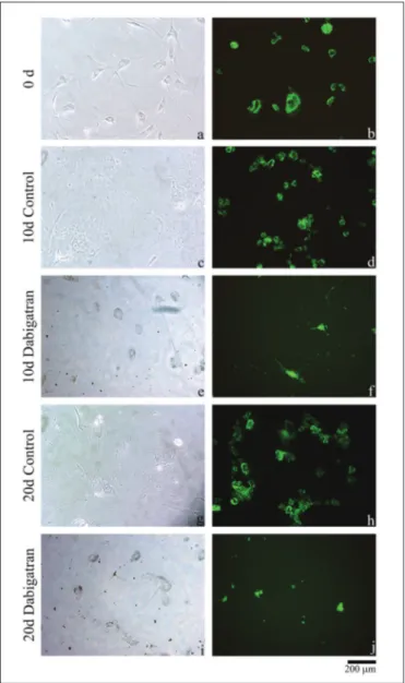
Morphological examination of cell cultures showed that cell proliferation in the control group increased with time. Contrastingly, cell proliferation was suppressed in dabigatran-supplemented cultures. Moreover, as seen in photographs of dabigatran-administered cultures, these cells lost their specific shapes and could be held in a more rounded and less attached form on the surface of culture flask (Figure 1). We wish to emphasize that dabigatran administration did not have an apoptotic effect on AF/NP cell cultures. No apoptotic cell death was observed in our AO/PI staining. Our MTT assays supported these results in the morphological analysis (Figure 2).
In the control group cultures, the number of viable cells increased with time, but proliferation was suppressed in cultures.
In our RT-PCR analysis, at the beginning of the experiment (Day 0), each gene’s expression in both cultures was accepted as a reference sample, and the gene expressions of these samples were assessed as 100% (in other words, RQ=1). On the 10th and 20th day, each gene’s expression in both
groups was normalized by comparison with the expression of the endogenous gene (β-actin) in the same culture, and fold changes were calculated and compared to those of the reference samples. Accordingly, in the control group, the gene expression of CHAD increased by 3.2-fold on day 10 and 8.6-fold on day 20. COMP gene expression was constant on day 10 and increased 2.6-fold on day 20. MMP-19 gene expression increased 1.7-fold on day 10 and 3.0-fold on day 20. Interestingly, the expression of MMP-13 decreased to 60% (RQ=0.4) on day 10 and 80% (RQ=0.2) on day 20. In dabigatran-treated cultures, no CHAD or MMP-13 expression was observed on either day 10 or 20 (Table I, Figure 3). In the study group, the expressions of COMP and MMP-19 decreased, and MMP-19 expression completely halted on day 20; the expressions of these genes were higher in the control
Figure 1: Inverted light microscope images of the
non-drug-administered control group samples “a”, on day 0; “c”, on day 10; and “g”, on day 20. It is observed that cells adhere to the floor, continue to proliferate as time progresses, and are healthy. It is seen that the proliferation decreases, and the matrix structure deteriorates, in the “e” and “” images. It is seen that the proliferation is suppressed following the administration of dabigatran, in the “f” and “j” images.
Figure 2: It is observed that the
obtained results were statistically significant (p<0.05) both in terms of application dose (F=148,99; p=0.00) and elapsed-time after application (F=56.98; p=0.00).
Figure 3: RQ values obtained with
qRT-PCR.
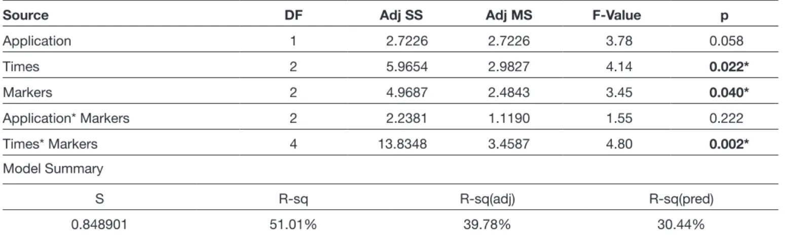
Table I: PCR Analysis: The Comparison of the PCR Analysis and the Changes in the Expression of CHAD, COMP, 13, and
MMP-19 Genes
Source DF Adj SS Adj MS F-Value p
Application 1 2.7226 2.7226 3.78 0.058 Times 2 5.9654 2.9827 4.14 0.022* Markers 2 4.9687 2.4843 3.45 0.040* Application* Markers 2 2.2381 1.1190 1.55 0.222 Times* Markers 4 13.8348 3.4587 4.80 0.002* Model Summary S R-sq R-sq(adj) R-sq(pred) 0.848901 51.01% 39.78% 30.44%
Bastiaans et al. reported in their study that the vitreous bodies of patients with proliferative vitreoretinopathy (PVR), a vitreoretinal disorder, contained increased thrombin activity that induced pro-inflammatory and proinflammatory programs in retinal pigment epithelial (RPE) cells (3). Thus, they examined the capacity of dabigatran, a direct thrombin inhibitor, to inhibit thrombin activity in vitreous fluids based on the hypothesis that the inhibition of intravitreal thrombin activity may represent a therapeutic option for PVR (3). They administered (i) thrombin, (ii) vitreous with no thrombin activity, and (iii) vitreous with increased thrombin activity in both the presence and absence of dabigatran to ARPE-19 cells, a commercial cell line. Subsequently, they determined CCL2, CXCL8, GM-CSF, IL6 and PDGFB mRNA expression levels through RQ-PCR. In addition, they detected the protein levels of 27 cytokines, chemokines, and growth factors in culture supernatants using a multiplex approach (3). The capacity of vitreous fluids obtained from patients after oral dabigatran uptake was tested in in vitro thrombin activity analysis. They reported that thrombin and vitreous fluids containing thrombin activity induced CCL2, CXCL8, GM-CSF, IL-6, and PDGF-BB expression in ARPE-19 cells but that it was inhibited by dabigatran. They also expressed that dabigatran entering the vitreous after repeated oral intake inhibited thrombin activity in the in vitro activity assay. They stressed that their findings provided evidence that the PVR activation pathway could possibly be inhibited by dabigatran (3).
Vianello and colleagues reported in their study that thrombin activates its G-coupled seven transmembrane protease-activated receptor (PAR-1) by cleaving the receptor’s N-terminal end and that PAR-1 expression and activation in many human cancers is associated with tumor progression and metastatization (29). After expressing that this presented compelling evidence for the effectiveness of an appropriate antithrombin agent for the adjuvant treatment of patients with cancer, they aimed to investigate whether dabigatran might affect the mechanisms favoring tumor growth by interfering with thrombin-induced PAR-1 activation (29). They reported that the exposure of tumor cells to thrombin significantly increased cell proliferation and correlated with the downregulation of p27 and the concomitant induction of cyclin D1 (29). They pointed out that dabigatran was constantly effective in antagonizing thrombin-induced proliferation and that it restored the baseline pattern of cell cycle protein expression (29). They also indicated that the chemoattractant effect of thrombin on tumor cells was lost in the presence of dabigatran (29). Next, they underlined that dabigatran could antagonize the adverse effects indicated for all cancers, thus impairing tumor growth and progression (29).
Castle et al. reported in their study (6) that cancer-like cells (CSCs) are tumor-inducers, resistant to chemotherapy, and play a role in metastasis. They hypothesized that NOACs could inhibit CSC activity, targeting specific factors in the thrombin pathway (6). In addition, they applied i) thrombin and ii) dabigatran to breast cancer cell lines. During analysis, they cultured MB-231, MCF-7, SKBR3, and MDA-MB-157 cell lines, which represented the spectrum of breast cancer subtypes, with i) 0.1 NIH units/mL of human thrombin such as sudden changes in patient color and appearance,
difficulty swallowing, difficulty with digestion, difficulty breathing, or abnormal laboratory test results concerning liver function, have been reported. In addition, a risk of hematoma formation, bloody coughing, bloody sputum, falling platelet and hemoglobin numbers, epistaxis, gastrointestinal bleeding, hematoma formation in penis, vagina, or urinary bladder may be associated with dabigatran use. Although many side effects or adverse events accompanying dabigatran use have been reported in the literature and in the drug’s prospectus, no study has yet examined its effects on intervertebral disc tissue or on NP and AF cells. Therefore, this study aimed to evaluate the effects of dabigatran on healthy, intact primary cell cultures isolated from intact intervertebral disc tissue at the pharmaco-molecular level.
Experimental setups using animal tissues have been used in cytotoxicity studies. However, the literature indicates that the sensitivity of human and animal tissues is different; therefore, the results obtained in these studies may differ from results obtained with human samples and may be misleading (1,2,13,14,17-20,25,26). In many studies where animal tissues are not used, commercial cell lines are used. However, commercial cell lines contain only a single cell type and cannot mimic the complex interactions of cells with their microenvironments or their ECM structures. Furthermore, since the genetic structure of the cells in the cell line has been modified, they do not have carry the genotypic and/or phenotypic characteristics that they have in the human body. Therefore, the results of the studies where commercial cell lines are used may be misleading (1,2,13,14,17-20,25,26). Dhar et al. aimed to evaluate the effects of factor Xa (FXa) and thrombin in vitro on stellate cells and their inhibition in
vivo using a rodent model of hepatic fibrosis in their study
and reported that the role of FXa in hepatic fibrosis was not elucidated (10). They used the HSC-LX2 commercial cell line during in vitro experiments. They added FXa and/or thrombin to the cell cultures and analyzed αSMA gene expression. Next, they administered thioacetamide (TAA) with rivaroxaban or dabigatran to 15 C57BL/6 J mice for 8 weeks during in vivo experiments. TAA was administered to live mammal subjects in the control group. Fibrosis was scored using digital image analysis, and hepatic tissue hydroxyproline was estimated (10). They reported that procollagen, transforming growth factor beta, and αSMA gene expressions were more upregulated in stellate cells treated with both FXa and thrombin than in groups treated with only FXa or thrombin alone. They also reported that the mean fibrosis score, fibrosis percentage area, and hepatic hydroxyproline content decreased significantly more in the FXa inhibitor-treated mice than in the control mice. These researchers emphasized that FXa inhibition was more effective than thrombin inhibition in reducing the fibrosis percentage area and the hepatic hydroxyproline content. They concluded that FXa increased stellate cell contractility and activation, that early inhibition of coagulation might be achieved through FXa inhibition, and that TAA-induced murine liver fibrosis might be significantly reduced (10).
It has been reported that dabigatran, which is commonly preferred by clinics because it is a safe and effective alternative to warfarin and does not require dose adjustment according to coagulation tests, may lead to severe bleeding in many tissues and organs, including intracranial bleeding (23). However, this drug represses proliferation in intervertebral disc tissue cells and disrupts the ECM structure.
█
CONCLUSION
Before prescribing dabigatran, it is necessary to consider the clinical profiles of patients, and the pharmaceuticals included in patient narratives or that are to be given alongside new prescriptions of this drug. Though this study used an in vitro experimental setup in which tissues of cases of the same race were applied, it should not be overlooked that dabigatran is toxic through the suppression of cell proliferation.
█
REFERENCES
1. Akyuva Y, Kaplan N, Yilmaz I, Ozbek H, Sirin DY, Karaarslan N, Guler O, Ates O: Delivering growth factors through a polymeric scaffold to cell cultures containing both nucleus pulposus and annulus fibrosus. Turk Neurosurg 29(2):180-193, 2019 2. Akyuva Y, Karaarslan N, Yilmaz I, Ozbek H, Sirin DY, Gurbuz
MS, Kaya YE, Kaplan N, Ates O: How scaffolds, which are polymeric drug delivery systems allowing controlled release, can be tested in human primary nucleus pulposus and annulus fibrosus cell cultures? Merit Res J Med Med Sci 5:477-487, 2017
3. Bastiaans J, Mulder VC, van Meurs JC, Smits-TeNijenhuis M, van Holten-Neelen C, van Hagen PM, Dik WA: Dabigatran inhibits intravitreal thrombin activity. Acta Ophthalmol 96(5):452-458, 2018
4. Beyer-Westendorf J: What have we learned from real-world NOAC studies in venous thromboembolism treatment? Thromb Res 163:83-91, 2018
5. Briggs MD, Rasmussen IM, Weber JL, Yuen J, Reinker K, Garber AP, Rimoin DL, Cohn DH: Genetic linkage of mild pseudoachondroplasia (PSACH) to markers in the pericentromeric region of chromosome 19. Genomics 18:656-660, 1993
6. Castle J, Farnie G, Kirwan CC: PO-11 - Thrombin and cancer stem-like cells: In vitro support for breast cancer anticoagulation. Thromb Res 140:80, 2016
7. Chen FH, Herndon ME, Patel N, Hecht JT, Tuan RS, Lawler J: Interaction of cartilage oligomeric matrix protein/ thrombospondin 5 with aggrecan. J Biol Chem 282:24591-24598, 2007
8. Chu DK, Hillis CM, Leong DP, Anand SS, Siegal DM: Benefits and risks of antithrombotic therapy in essential thrombocythemia: A systematic review. Ann Intern Med 167:170-180, 2017
9. Connolly SJ, Ezekowitz MD, Yusuf S, Eikelboom J, Oldgren J, Parekh A, Pogue J, Reilly PA, et al: Dabigatran versus warfarin in patients with atrial fibrillation. N Engl J Med 361:1139-11351, 2009
and ii) 0.5 μM of dabigatran. They indicated that thrombin increased MFE in the high PAR-1 expressing MDA-MB-231 and MDA-MB-157 cell lines. (6). Further, they emphasized that dabigatran abolished the stimulatory effect of thrombin on MFE in MDA-MB-231 and MDA-MB-157 cells treated with thrombin. They concluded that the clinical use of novel oral anticoagulants might be possible in targeting this critical cancer cell subpopulation (6). This study, conducted in vitro on AF/NP primer cell cultures, showed that the doses of dabigatran administered did not cause cell death but markedly suppressed cell proliferation and led to cell senescence. Although an absence of cell death suggests that the use of dabigatran is safe in clinical practice, clinicians should note that the dosage and duration of use of this drug may significantly slow the recovery process. It is an advantage that the drug did not reveal any short-term acute toxicity. However, the fact that it caused the long-term change in gene expression should be assessed in this context. CHAD and COMP gene expression increased time-dependently, which can be regarded as a healthy healing marker in our control group and is associated with the ECM and cell binding to it. The expression of these genes in the dabigatran-supplemented samples either disappeared or was reduced. Interestingly, it was observed that the expression of MMP-19 increased. The increase in its expression is regarded as an indication of the degeneration caused by inflammation in in vivo conditions. Although not morphologically observed, the increase in MMP-19 expression in the control group cultures established from intact AF/NP cells may indicate that the cells could have been affected by applications that may cause degeneration, such as trypsinization, which is performed during the culturing process. The direct growth factors in the culture medium may be another reason for this increase. We believe that the latter is likely the main reason for the increase, as MMP-13 expression decreased in the control groups. In cultures where dabigatran was applied, gene expressions were greatly reduced or completely disappeared due to repressed proliferation and cell senescence. Therefore, it should be emphasized that dabigatran may have a negative effect on the healing process in the clinic.
Since satisfactory results are not obtained with existing treatment modalities, it is hoped that treatment methods through tissue repair with the help of regenerative and reparative medicines can be developed. Through cellular studies carried out using molecular methods, it is believed that effective methods for the prevention and treatment of diseases can be developed.
Researchers have accelerated molecular studies to mechanically and biologically repair damaged tissues. Though it is believed that damaged tissues can be remedied using the findings of molecular investigations, these findings are obtained from studies carried out using in vitro experimental setups.
This study was carried out using an in vitro experimental setup, and the intervertebral disc cultures used in the human primary cell cultures were obtained from cases of the same race. These are this study’s limitations.
20. Karaarslan N, Yilmaz I, Sirin DY, Ozbek H, Kaplan N, Kaya YE, Akyuva Y, Gurbuz MS, Oznam K, Ates O: Does pregabalin used in the treatment of neuropathic pain damage intervertebral disc tissue? Exp Ther Med 16:1259-1265, 2018
21. Kennedy B, Gargoum FS, Kennedy L, Khan F, Curran DR, O’Connor TM: Emerging anticoagulants. Curr Med Chem 19: 3388-3416, 2012
22. Kwon WK, Moon HJ, Kwon TH, Park YK, Kim JH: The role of hypoxia in angiogenesis and extracellular matrix regulation of intervertebral disc cells during inflammatory reactions. Neurosurgery 81:867-875, 2017
23. Lau WCY, Li X, Wong ICK, Man KKC, Lip GYH, Leung WK, Siu CW, Chan EW: Bleeding-related hospital admissions and 30-day read missions in patients with non-valvularatrial fibrillation treated with dabigatran versus warfarin. J Thromb Haemost 15:1923-1933, 2017
24. Mao SH, Qian BP, Shi B, Zhu ZZ, Qiu Y: Quantitative evaluation of the relationship between COMP promoter methylation and the susceptibility and curve progression of adolescent idiopathic scoliosis. Eur Spine J 27: 272-277, 2018
25. Sirin DY, Kaplan N, Yilmaz I, Karaarslan N, Ozbek H, Akyuva Y, Kaya YE, Oznam K, Akkaya N, Guler O, Akkaya S, Mahirogullari M: The association between different molecular weights of hyaluronic acid and CHAD, HIF-1α, COL2A1 expression in chondrocyte cultures. Exp Ther Med 15:4205-4212,2018 26. Sirin DY, Karaarslan N: Evaluation of the effects of pregabalin
on chondrocyte proliferation and CHAD, HIF-1α, and COL2A1 gene expression. Arch Med Sci 14(6):1340–1347, 2018 27. Trivedi A, Zhang H, Ekeledo A, Lee S, Werb Z, Plant GW,
Noble-Haeusslein LJ: Deficiency in matrix metalloproteinase-2 results in long-term vascular instability and regression in the injured mouse spinal cord. Exp Neurol 284:50-62, 2016 28. Vadivelu S, Stewart TJ, Qu Y, Horn K, Liu S, Li Q, Silver J,
McDonald JW: NG2+ progenitors derived from embryonic stem cells penetrate glial scar and promote axonal outgrowth into white matter after spinal cord injury. Stem Cells Transl Med 4: 401-411, 2015
29. Vianello F, Sambado L, Goss A, Fabris F, Prandoni P: Dabigatran antagonizes growth, cell-cycle progression, migration, and endothelial tube formation induced by thrombin in breast and glioblastoma cell lines. Cancer Med 5:2886-2898, 2016 10. Dhar A, Sadiq F, Anstee QM, Levene AP, Goldin RD, Thursz
MR: Thrombin and factor Xa link the coagulation system with liver fibrosis. BMC Gastroenterol 18:60, 2018
11. Franchini M, Mannucci PM: Direct oral anticoagulants and venous thromboembolism. Eur Respir Rev 25: 295-302, 2016 12. Goode AP, Marshall SW, Kraus VB, Renner JB, Stürmer
T, Carey TS, Irwin DE, Jordan JM: Association between serum and urine biomarkers and lumbar spine individual radiographic features: The Johnston County Osteoarthritis Project. Osteoarthritis Cartilage 20:1286-1293, 2012
13. Gumustas SA, Yilmaz I, Isyar M, Sirin DY, Batmaz AG, Ugras AA, Oznam K, Ciftci Z, Mahirogullari M: Assessing the negative impact of phenyl alkanoic acid derivative, a frequently prescribed drug for the suppression of pain and inflammation, on the differentiation and proliferation of chondrocytes. J Orthop Surg Res 11:70, 2016
14. Guzelant AY, Isyar M, Yilmaz I, Sirin DY, Cakmak S, Mahirogullari M: Are chondrocytes damaged when rheumatologic inflammation is suppressed? Drug Chem Toxicol 40:13-23, 2017
15. Haleem-Smith H, Calderon R, Song Y, Tuan RS, Chen FH: Cartilage oligomeric matrix protein enhances matrix assembly during chondrogenesis of human mesenchymal stem cells. J Cell Biochem 113:1245-1252, 2012
16. Heidbuchel H, Verhamme P, Alings M, Antz M, Hacke W, Oldgren J, Sinnaeve P, Camm AJ, Kirchhof P: European Heart Rhythm Association pratical guide on the use of new oral anticoagulants in patients with non-valvular atrial fibrillation. Europace 15:625-651, 2013
17. Karaarslan N, Batmaz AG, Yilmaz I, Ozbek H, Caliskan T, Sirin DY, Kaplan N, Oznam K, Ates O: Do aryl acetic acid derivative analgesic and anti-inflammatory drugs affect proliferation and differentiation in primary cell cultures formed from human cartilage tissue? Exp Ther Med 16(3):1647-1654, 2018 18. Karaarslan N, Yilmaz I, Akgun FS, Caliskan T, Simsek AT,
Kaplan N, Kaya YE, Sirin DY, Ozbek H, Ates O: Which pharmacological agents may be used to establish healthy, proliferating human primer nucleus pulposus / annulus fibrosus cell cultures? Systematic evaluation of our experience in the light of literature. Merit Res J Med Med Sci 6:100-110, 2018 19. Karaarslan N, Yilmaz I, Ozbek H, Sirin Yasar D, Kaplan N,
Akyuva Y, Gonultas A, Ates O: Are specific gene expressions of extracellular matrix and nucleus pulposus affected by primary cell cultures prepared from intact or degenerative intervertebral disc tissues? Turk Neurosurg 29(1):43-52, 2019