140 Nigerian Journal of Clinical Practice • Jan-Feb 2015 • Vol 18 • Issue 1
Abstract
Anterior crown fractures are the most common type of injury in dental trauma. The ideal treatment is to reattach the fragments as quickly as possible following intraoral and radiographic examination, but sometimes delayed treatment appointments are necessary because of uninformed patients/parents or multidisciplinary cases included endodontically and periodontically. Delayed reattachment may lead to unesthetic results because of the dehydration of fragments. The purpose of this study was to present 1-year follow-ups of reattachment of dehydrated fragments using dentin bondings and flowable composites in two different cases. The color of the dehydrated fragments was natural in the control appointments and 1-year follow-ups show harmonious integration of color, form and texture after the reattachment of the original piece of tooth. Restoration of the tooth by reattaching the original fragment is the best way of treatment in esthetic, conservative and economic point of view.
Key words: Dehydrated, tooth fractures, tooth injuries
Date of Acceptance: 14-Jul-2014
Address for correspondence:
Dr. Funda Öztürk Bozkurt,
Department of Restorative Dentistry, School of Dentistry, Istanbul Medipol University, Atatürk Bulvari No: 27, Unkapani, 34083, Fatih-Istanbul, Turkey.
E-mail: [email protected]
Introduction
One of the problems frequently encountered in dental practice is trauma that can damage hard and soft tissues of the oral cavity. Anterior crown fractures are commonplace in children and adolescents and may affect up to 25% of this
patient population.[1] Anterior teeth, especially maxillary
incisors, are affected more than mandibular teeth because
of their position in the dental arch.[2] With the importance
to the person’s physical appearance, crown fractures of
anterior teeth requires immediate treatment solutions.[3,4]
Although newer formulations of composite resins offer us a large variety of products with optical properties such as translucency, opacity, different enamel and dentin colors, there is, however, no synthetic restorative material that
can replace the natural tooth structure better than itself.[3]
When it is present, reattaching the original fragment is the best way of treatment in esthetic, conservative and economic point of view.
The concept of reattachment began in 1964 by Chosack and
Eidelman.[5] In the following years, various techniques have
been described and due to the developments in adhesive technology reattachment treatment became very simple and
successful.[4] Clinical trials and long‑term follow‑up have
reported that reattachment using modern dentine bonding agents or adhesive luting systems may achieve functional and aesthetic success for up to 7 years.[1]
The ideal treatment is to reattach the fragments as quickly as possible following intraoral and radiographic examination, but sometimes delayed treatment appointments are necessary because of uninformed patients/parents or multidisciplinary cases included endodontically or periodontically. Delayed reattachment may lead to unaesthetic results because of the
dehydration of fragments.[1] The purpose of this study was
to present 1‑year follow‑ups of reattachment of dehydrated
Reattachment of dehydrated tooth fragments:
Two case reports
FÖ Bozkurt, B Demir1, E Erkan2
Departments of Restorative Dentistry, 1Periodontology and 2Endodontics, School of Dentistry,
Istanbul Medipol University, Istanbul, Turkey
Access this article online
Quick Response Code:
Website: www.njcponline.com
DOI: 10.4103/1119-3077.147004
PMID: *******
Bozkurt, et al.: Case reports of dehydrated fragment reattachment
141 Nigerian Journal of Clinical Practice • Jan-Feb 2015 • Vol 18 • Issue 1
fragments by using dentin bonding and flowable composites in two different cases.
Case Reports
Case 1
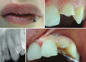
A 21‑year‑old female patient referred our faculty clinic with a complaint of broken tooth 2 days before [Figure 1]. In the extraoral examination, the injury of soft tissue was obvious with one suture in the lower lip. Intraoral examination revealed that the crown of maxillary left lateral incisor (22) was fractured and at the palatal region there was a mobile tiny piece of tooth fractured vertically without displacement. There was no pulp exposure, and the tooth was sensitive at cold pulp testing performed by chloroethyl refrigerant spray (IGS Aerosols GmbH, Wehr, Germany). There was no root fracture observed in the intraoral periapical radiograph. A written informed consent was obtained from the patient before the treatment. Past medical history was reviewed, and there was no remarkable report. The palatal gingiva was elevated by a flap surgery, and the mobile root fragment was removed. After checking the fracture lines,
etch and dry adhesive (S3 Bond Plus, Kuraray, Japan)
was applied to the tooth and the fragment with a brush by scrubbing for 20 s. The adhesive was air thinned and light‑cured (Elipar Free Light, 3M ESPE, AG, Germany) for 20 s. Flowable composite (Filtek™ Ultimate, 3M ESPE, USA) was placed over the fracture area on the tooth, and the fragment was placed over. The excess material was removed before light‑curing for 40 s. In order to reduce sensitivity and protect the pulp, light‑cured glass ionomer liner (Glass Liner, WP Dental, Germany) was placed over the exposed dentin of the crown. A second appointment was scheduled for the direct restoration of the tooth by composite resin since the patient didn’t have the broken fragment. However in the next appointment, the patient came with the broken piece which was recovered at the site of the injury and kept in dry conditions. A 2% chlorhexidine
digluconate (Cavity Cleanser, Bisco, USA) solution was used for disinfection of the fragment. The glass ionomer lining, which was placed for protection, was removed carefully with an explorer. The fragment was dehydrated but fitted perfectly to the remaining tooth and hence the treatment plan of choice was to reattach the fragment. The tooth fragment was bonded by the same manner as the root piece with the only exception of selective etching of enamel margins of the tooth and fragment for 10 s. The occlusion was checked and adjusted then finishing and polishing steps were completed by abrasive disks (Sof‑Lex™ Finishing and Polishing Systems, 3M ESPE, USA) and rubber cups
(Jiffy® Polishers, Ultradent, USA) [Figure 2a]. The patient
was told to avoid exerting heavy function with her tooth and to follow regular home care procedures relative to oral hygiene.
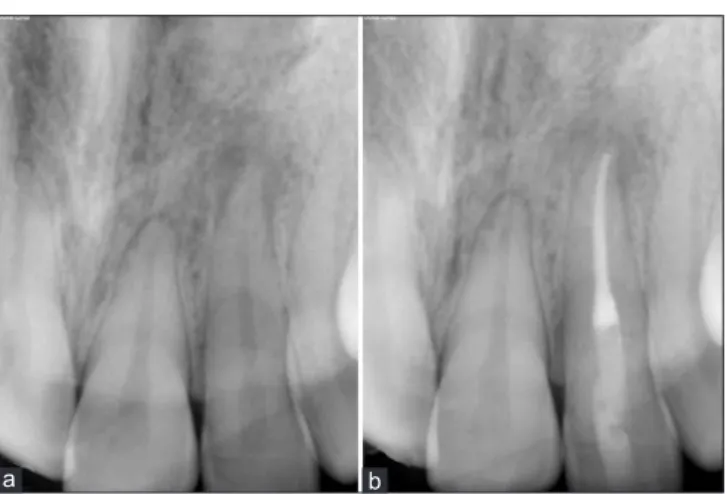
In the control appointment (2‑weeks later) the color of the reattached fragment was acceptable, and the form of the tooth was aesthetic with its original shape [Figure 2b]. In the 1‑year follow‑up, the tooth showed a marginal discoloration, which was easily removed by polishing [Figure 3a and b] but, unfortunately, there was a radiolucent area at the apex and the tooth was nonvital in cold pulp test, so an endodontic treatment was performed by using ProTaper Ni‑Ti Rotary System (Dentsply Malleiffer) and obturated with an AH 26 sealer (Dentsply; DeTrey, Konstanz, Germany) and gutta‑percha by lateral compaction technique[Figure 4a and b].
Case 2
A healthy 11‑year‑old female patient refereed for an aesthetic restoration of a fractured maxillary right central incisor (11) [Figure 5]. Dental history revealed that the tooth was fractured 1 month prior to presentation because of a bicycle accident. Her first dentist applied a root canal
Figure 1: Images of injury of a fractured maxillary left lateral incisor
Figure 2: Postoperative images (a) immediately after reattachment showing color difference because of dehydration (b) harmonious
integration of color in 2 weeks control b
Bozkurt, et al.: Case reports of dehydrated fragment reattachment
142 Nigerian Journal of Clinical Practice • Jan-Feb 2015 • Vol 18 • Issue 1
Figure 5: Preoperative images of a fractured maxillary right
central incisor Figure 6: Postoperative images (a) immediately after reattachment showing dehydration (b) acceptable color in 2 weeks control (c) 1-year follow-up c b a Figure 4: (a) Radiograph of 1-year follow-up showing radiolucency at the apex area (b) radiograph after endodontic
treatment b a Figure 3: One year follow-up (a) marginal discolouration (b) easily removed by polishing b a
treatment and suggested a temporary composite restoration until a crown restoration at the age of 18 for better clinical results. The parents hesitated for a final crown restoration and refereed our university clinics in order to find a remedy. As they were informed about the treatment alternatives of a fractured tooth, her mother gave us the dehydrated fragment she kept with her ever since the accident. The fragment was placed in 2% chlorhexidine digluconate solution for disinfection and tried in intraorally to check for proper positioning and fit with the fractured coronal structure. A written informed consent was signed by the patient’s mother as the guardian before the treatment. Following clinical and radiographic examination, the endodontic treatment was found to be successful but a horizontally located mesiodense in the apex area was noticed. Since the lamina dura surrounding roots of both teeth could be detected separately, the mesiodense was followed‑up closely for future recalls. The fragment and the tooth were acid etched for 30 s with 37% phosphoric acid gel, rinsed for 30 s and gently air dried. A one‑bottle etch and rinse adhesive (Adper Single Bond 2, 3M ESPE, USA) was applied with a brush for 20 s and carefully air thinned. Flowable composite was placed over the fracture area on the tooth, and the fragment was placed over. The excess material was removed before light‑curing for 40 s from labial and
palatal surfaces. After occlusion adjustment finishing and polishing steps were completed by the same manner as in case 1. The patient was told to avoid exerting heavy function with her tooth and to follow regular home care procedures relative to oral hygiene.
In the control appointment (2 weeks later), the color of the fragment was found to be acceptable by rehydrating [Figure 6a] and the broken part of the left central incisor was restored by a nanohybrid resin (Filtek™ Z550, 3M ESPE, USA) [Figure 6b]. The final 1‑year postoperative results reflected the harmonious integration of color, form and texture after the reattachment of the original piece of tooth [Figure 6c].
Discussion
Reattachment of fractured fragment is an excellent
approach for the treatment of coronal fractures.[1‑4] The
prognosis for re‑attached crown fragments is good and continues to improve with the increasing bond strengths
Bozkurt, et al.: Case reports of dehydrated fragment reattachment
143 Nigerian Journal of Clinical Practice • Jan-Feb 2015 • Vol 18 • Issue 1
Sometimes the fractured fragment is dehydrated because of being kept in the tissue. In these two cases, the fragments were intact, but dehydrated for being kept in dry conditions for more than 2 weeks. The fragments were unesthetic because of dehydration in the first trial but both the patients preferred to use their own tooth piece instead of a direct composite restoration.
Reis et al. reported different fracture strength for several
reattaching techniques[7] but in these two cases there was
no special preparation for the tooth or the fragment. All traumatic dental injuries need to be followed‑up over
time.[8,9] Endodontic treatment of teeth with uncomplicated
crown fractures is usually unnecessary unless pulp necrosis and infection develop subsequent to the injury, but the pulps in traumatized teeth can become necrotic and infected even a number of years subsequent to an injury.
[9] Here in the first case, the radiographic examination
in 1‑year recall showed a radiolucent area at the apex and vitally test revealed that the tooth was nonvital, so the endodontic treatment of the tooth was performed 1‑year after injury. The reattachment of dehydrated tooth fragments by bonding technique is a simple and satisfactory treatment for crown fractures.
References
1. Murchison DF, Burke FJ, Worthington RB. Incisal edge reattachment: Indications for use and clinical technique. Br Dent J 1999;186:614‑9.
2. Castro JC, Poi WR, Manfrin TM, Zina LG. Analysis of the crown fractures and crown‑root fractures due to dental trauma assisted by the Integrated Clinic from 1992 to 2002. Dent Traumatol 2005;21:121‑6.
3. Terry DA. Adhesive reattachment of a tooth fragment: The biological restoration. Pract Proced Aesthet Dent 2003;15:403‑9.
4. Macedo GV, Diaz PI, De O Fernandes CA, Ritter AV. Reattachment of anterior teeth fragments: A conservative approach. J Esthet Restor Dent 2008;20:5‑18.
5. Chosack A, Eidelman E. Rehabilitation of a fractured incisor using the patient’s natural crown: Case report. J Dent Child 1964;31:19‑21.
6. Farik B, Munksgaard EC, Andreasen JO, Kreiborg S. Fractured teeth bonded with dentin adhesives with and without unfilled resin. Dent Traumatol 2002;18:66‑9.
7. Reis A, Francci C, Loguercio AD, Carrilho MR, Rodriques Filho LE. Re‑attachment of anterior fractured teeth: Fracture strength using different techniques. Oper Dent 2001;26:287‑94.
8. Moule AJ, Moule CA. The endodontic management of traumatized permanent anterior teeth: A review. Aust Dent J 2007;52:S122‑37.
9. International Association of Dental Traumatology. Available from: http://www. iadt‑dentaltrauma.org. [Last accessed on 2013 Aug 15].
How to cite this article: Bozkurt FO, Demir B, Erkan E. Reattachment
of dehydrated tooth fragments: Two case reports. Niger J Clin Pract 2015;18:140-3.
Source of Support: Nil, Conflict of Interest: None declared.
View publication stats View publication stats