Clinical and Pathological Features of Rare Ovarian Tumors in Turkey
Asian Pac J Cancer Prev, 14 (11), 6493-6499
Introduction
Non-epithelial malignant ovarian tumors (germ cell and sex cord-stromal tumors), clear cell carcinoma, Brenner tumors, transitional cell tumors, and carcinoid tumors of the ovary are defined as rare ovarian tumors (ROTs). Of these tumors, germ cell tumors (GCTs) and sex cord-stromal tumors (SCSTs) comprise approximately 15% of all ovarian malignancies, and they have a variety of
1Department of Medical Oncology, Medical Faculty, Istanbul Medipol University, 5Dr. Lutfi Kirdar Kartal Education and Research Hospital, Istanbul, 2Erciyes University, Medical Faculty, Kayseri, 3Dr. Abdurrahman Yurtarslan Oncology Education and Research Hospital, 6Ankara Numune Education and Research Hospital, 10Gazi University, Medical Faculty, Ankara, 4Dokuz Eylul University, Medical Faculty, Izmir, 7Dicle University, Medical Faculty, Diyarbakir, 8Selcuk University, Meram Medical Faculty, Konya, 9Medical Faculty, Gaziantep University, Gaziantep, 11Medical Faculty, Inonu University, Malatya, Turkey *For correspondence:
Abstract
Background: Non-epithelial malignant ovarian tumors and clear cell carcinomas, Brenner tumors, transitional cell tumors, and carcinoid tumors of the ovary are rare ovarian tumors (ROTs). In this study, our aim was to determine the clinicopathological features of ROT patients and prognostic factors associated with survival. Materials and Methods: A total of 167 patients with ROT who underwent initial surgery were retrospectively analyzed. Prognostic factors that may influence the survival of patients were evaluated by univariate and multivariate analyses. Results: Of 167 patients, 75 (44.9%) were diagnosed with germ-cell tumors (GCT) and 68 (40.7%) with sex cord-stromal tumors (SCST); the remaining 24 had other rare ovarian histologies. Significant differences were found between ROT groups with respect to age at diagnosis, tumor localization, initial surgery type, tumor size, tumor grade, and FIGO stage. Three-year progression-free survival (PFS) rates and median PFS intervals for patients with other ROT were worse than those of patients with GCT and SCST (41.8% vs 79.6% vs 77.1% and 30.2 vs 72 vs 150 months, respectively; p=0.01). Moreover, the 3-year overall survival (OS) rates and median OS times for patients with both GCT and SCST were better as compared to patients with other ROT, but these differences were not statistically significant (87.7% vs 88.8% vs 73.9% and 170 vs 122 vs 91 months, respectively; p=0.20). In the univariate analysis, tumor localization (p<0.001), FIGO stage (p<0.001), and tumor grade (p=0.04) were significant prognostic factors for PFS. For OS, the univariate analysis indicated that tumor localization (p=0.01), FIGO stage (p=0.001), and recurrence (p<0.001) were important prognostic indicators. Multivariate analysis showed that FIGO stage for PFS (p=0.001, HR: 0.11) and the presence of recurrence (p=0.02, HR: 0.54) for OS were independent prognostic factors. Conclusions: ROTs should be evaluated separately from epithelial ovarian cancers because of their different biological features and natural history. Due to the rarity of these tumors, determination of relevant prognostic factors as a group may help as a guide for more appropriate adjuvant or recurrent therapies for ROTs.
Keywords: Rare ovarian tumors - overall survival - progression-free survival - prognostic factors
RESEARCH ARTICLE
Clinical and Pathologic Features of Patients with Rare Ovarian
Tumors: Multi-Center Review of 167 Patients by the Anatolian
Society of Medical Oncology
Ahmet Bilici
1*, Mevlude Inanc
2, Arife Ulas
3, Tulay Akman
4, Mesut Seker
5, Nalan
Akgul Babacan
6, Ali Inal
7, Oznur Bal
3, Lokman Koral
8, Alper Sevinc
9, Gulnihal
Tufan
10, Emin Tamer Elkiran
11, Bala Basak Oven Ustaalioglu
5, Tugba Yavuzsen
4,
Necati Alkis
3, Metin Ozkan
2, Mahmut Gumus
5histopathological subgroups (Quirk and Natarajan, 2005; Koulouris and Penson, 2009; Colombo et al., 2012). These two tumor forms also have curative potential with very different biological behaviors and treatment strategies. GCTs are commonly curable tumors, even in advanced stages. SCSTs, mostly granulosa cell tumors, are generally associated with a favorable prognosis, although they are much less chemosensitive and grow much more slowly (Young, 2005; Ayhan et al., 2009). Both GCTs and SCSTs
are histologically similar to other more common testicular tumors; therefore, the treatments for GCTs and SCSTs originated from testicular cancer research (Colombo et al., 2007; Matei et al., 2013).
The natural history of ROTs is poorly understood, and a consensus has not been reached with respect to the prognostic factors of ROTs. In ROTs, the clarification of prognostic factors that have an impact on survival is important for accurate diagnosis and treatment strategies, such as surgery, adjuvant treatment, and effective treatment of relapse (Garcia et al., 1999; Koulouris and Penson, 2009; Colombo et al., 2012). Because these tumors are rarely encountered in clinical practice, randomized clinical trials to evaluate the effects of proposed treatment regimens are limited in the literature. On the other hand, in SCSTs, especially granulosa cell tumors, the stage of disease and age at the time of diagnosis, the presence of residual tumor after the initial surgery, and the number of mitoses and nuclear atypia have been found to be important prognostic factors (Ayhan et al., 2009; Li et al., 2009; Suri et al., 2013).
In the present study, we aimed to determine the clinicopathological features of patients with ROTs in Turkey. In addition, the effect of these factors on survival and treatment outcomes of patients were also evaluated in the entire cohort.
Materials and Methods
A total of 167 patients with ROTs who had undergone initial surgery and follow up at 11 medical oncology centers in Turkey between 1993 and 2011 were included in the study. The primary tumor was staged according to the International Federation of Gynecology and Obstetrics (FIGO) staging classification. Eligibility was limited to patients with rare ovarian histologies including germ-cell tumors and sex-cord stromal tumors as a non-epithelial tumors and other relatively rare histologies such as clear cell carcinoma, transitional cell carcinoma, carcinoid tumor of the ovary or Brenner tumors that were histologically confirmed and who underwent primary surgery and had a postoperative survival expectancy longer than 3 months. After initial diagnosis by primary pathologist, all pathological slides were re-evaluated to confirm the histopathological subtypes by a pathologist who was an expert in matters of gynecological oncologic pathology in all centers. Patients who had insufficient disease information were excluded from data analysis. Data were retrospectively obtained from patients’ charts with respect to age, gender, presentation of symptoms at the time of diagnosis, surgery type, tumor location, histopathology, tumor stage, tumor size, histological grade, the presence of residual tumor after initial surgery, preoperative or postoperative tumor markers, type of adjuvant chemotherapy, responses to treatment, and survival after written informed consent was obtained from patients or their relatives.
Patients with stage III-IV disease underwent complete staging surgery including total abdominal hysterectomy (TAH), bilateral salpingo-oophorectomy (BSO), and omentectomy, including multiple biopsies of the pelvic
and abdominal peritoneum, retroperitoneal lymph node sampling, and peritoneal cytologic sampling. For patients who desired preservation of fertility and were found to be clinically appropriate, a fertility-sparing surgery including unilateral salpingo-oophorectomy (USO) or cystectomy was carried out with careful inspection of the abdominal cavity, and the biopsies obtained from suspected areas and the peritoneal cavity washing fluids were sent for analysis. In some patients, incomplete surgery including TAH and BSO or USO was performed due to the inappropriate disease.
Patients with a measurable disease response to the chemotherapy were evaluated by CT imaging and serial AFP and β-HCG levels in GCTs and CA 125 levels in other ROTs except for SCSTs. A complete response (CR) was defined as the complete disappearance of all clinically measurable disease and normalization of marker levels for at least 4 weeks. Partial response (PR) represented a decrease of at least 30% of the tumor volume, and progressive disease (PD) was defined as a greater than 20% increase in tumor volume, the presence of any new sites of disease, an increase in tumor markers (>25% increase of abnormal nadir) after the initial decrease, or an increase above normal values after returning to normal in less than 30 days. Recurrence was defined as new evidence of cancer after initial complete resection or after the CR of all detectable disease with tumor markers normalized for at least 30 days.
Adjuvant chemotherapy
Among patients with GCT who received postoperative chemotherapy (n=65), 60 (80%) patients were treated with the BEP regimen, which consists of bleomycin, etoposide and cisplatin with a median of 4 cycles (range: 2-5 cycles). Two patients were treated with the EP regimen, which consists of etoposide and cisplatin. Two patients were treated with the VIP regimen, which consists of etoposide, ifosfamide and cisplatin. The remaining patient with choriocarcinoma was treated with weekly methotrexate. Thirty-two patients (47.1%) with SCST received postoperative chemotherapy with a median of four cycles. Ten patients were treated with paclitaxel and carboplatin. Sixteen patients received the BEP regimen. The remaining six patients were treated with docetaxel and carboplatin (1 patient), EP regimen (2 patients), doxorubicin and cisplatin (2 patients), and the VIP regimen (1 patient). On the other hand, 22 patients (91.7%) with other ROTs were treated with postoperative chemotherapy, with a median of 6 cycles. Of these patients, 21 patients received paclitaxel and carboplatin regimen, while one patient was treated with doxorubicin and cisplatin regimen.
Statistical analysis
All data were analyzed using SPSS 17.0 (SPSS Inc., Chicago, IL, USA) software. The clinicopathological factors of the patients with ROTs were compared using a chi-squared test and Fisher’s exact test. Pre- and postoperative tumor markers were compared with a Wilcoxon test. The survival analyses and curves were established using the Kaplan-Meier method and compared with the log-rank test. The progression-free survival
Clinical and Pathological Features of Rare Ovarian Tumors in Turkey (PFS) was defined as the time from curative surgery to
recurrence, or to the date of death or loss of follow-up. Overall survival (OS) was described as the time from diagnosis to the date of the patient’s death or loss of follow-up. Post-recurrence survival (PRS) was also defined as the time from recurrence to the date of the patient’s death or loss of follow-up. Univariate and multivariate analyses were performed using the Cox proportional hazards model to evaluate the importance of clinical and pathological factors as prognostic factors. Multivariate p-values were used to characterize the independence of these factors. A 95% confidence interval (CI) was used to quantify the relationship between survival time and each independent factor. All p-values were two-sided in tests, and p-values less than 0.05 were considered statistically significant.
Results
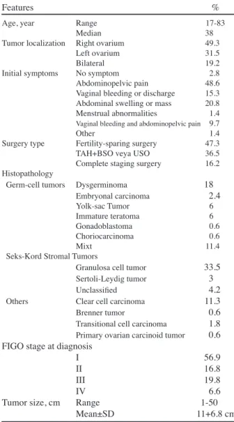
The median age of patients was 38 years (range: 17-83 years). The majority of patients were aged 50 years or younger (68.9%). The most frequent symptoms at the time of diagnosis were abdominopelvic pain (48.6%), abdominal swelling or mass (20.8%), and vaginal bleeding or discharge (15.3%). Complaints of irregular menses or amenorrhea occurred in only 1.45% of patients. The tumor was mostly localized in the right ovary (49.3%), while tumors were bilateral in only 19.3% of patients. After initial surgery, 75 patients (44.9%) were diagnosed with GCT, 68 patients (40.7%) were diagnosed with SCST, and the remaining 24 patients had other rare ovarian histologies (clear cell carcinoma, transitional cell carcinoma, carcinoid tumor of the ovary, and Brenner tumor). At the time of diagnosis, the majority of patients (56.9%) were classified as stage I, 16.8% of patients as stage II, 19.8% of patients as stage III, and 6.6% of patients as stage IV according to the FIGO staging system. The mean tumor size was 11±6.8 cm (range; 1-50 cm). Complete staging surgeries were performed in 27 patients (16.2%), while 79 patients underwent fertilty-sparing surgery. TAH and BSO or USO were carried out for the remaining 61 patients (36.5%). Histologic classifications and patient characteristics of the entire group are shown in Table 1.
Significant differences were present between ROT groups with respect to age at diagnosis, tumor site, initial surgery type, tumor size, tumor grade, and FIGO stage. Patients with GCTs were mostly localized in the left ovary, while the right ovary was the most common tumor site for patients with SCSTs and other ROTs (p=0.02). Patients with GCTs were more likely to be 50 years old or younger compared to patients with SCST and other ROCs (p<0.001). Fertility-sparing surgery was more frequently performed for GCTs; however, patients with other ROTs commonly underwent complete staging surgery (p<0.001). Furthermore, GCTs tended to be large in diameter and well or moderately differentiated compared to patients with SCST or other ROTs (p=0.04 and 0.01, respectively). Patients with other ROTs had advanced-stage disease compared to those with GCT and SCSTs (p=0.001). Table 2 shows the clinicopathological factors in ROT groups. For patients with GCTs, the preoperative mean AFP
Table 1. Patient Characteristics in Entire Cohort with
Rare Ovarian Cancers
Features %
Age, year Range 17-83
Median 38
Tumor localization Right ovarium 49.3 Left ovarium 31.5
Bilateral 19.2
Initial symptoms No symptom 2.8 Abdominopelvic pain 48.6 Vaginal bleeding or discharge 15.3 Abdominal swelling or mass 20.8 Menstrual abnormalities 1.4
Vaginal bleeding and abdominopelvic pain 9.7
Other 1.4
Surgery type Fertility-sparing surgery 47.3 TAH+BSO veya USO 36.5 Complete staging surgery 16.2 Histopathology
Germ-cell tumors Dysgerminoma 18
Embryonal carcinoma 2.4 Yolk-sac Tumor 6 Immature teratoma 6 Gonadoblastoma 0.6 Choriocarcinoma 0.6 Mixt 11.4
Seks-Kord Stromal Tumors
Granulosa cell tumor 33.5
Sertoli-Leydig tumor 3
Unclassified 4.2
Others Clear cell carcinoma 11.3
Brenner tumor 0.6
Transitional cell carcinoma 1.8
Primary ovarian carcinoid tumor 0.6 FIGO stage at diagnosis
I 56.9
II 16.8
III 19.8
IV 6.6
Tumor size, cm Range 1-50 Mean±SD 11+6.8 cm
*TAH: total abdominal histerectomy, BSO: bilateral salpingo-oophorectomy; USO: unilateral salpingo-oophorectomy, SD: standard deviation
Table 2. Clinicopathological Factors in Patients with
Rare Ovarian Tumors
Factors Germ- Sex-cord Other p cell Stromal
n=75 n=68 n=24 % % %
Age (year) <50 97.3 48.5 37.5 <0.001* >50 2.7 51.5 62.5 Tumor site Right Ovarium 31.6 56.7 54.2 0.02*
Left Ovarium 52.6 33.3 12.5 Bilateral 15.8 10 33.3 Initial surgery type
Fertility-sparing surgery 73.3 33.8 4.2 <0.001* TAH+BSO or USO 21.3 55.9 29.2 Complete staging surgery 5.4 10.3 66.7 Tumor diameter (cm)
<15 64.4 85.7 75 0.04* >15 35.6 14.3 25 Tumor grade I-II 82.9 78.7 47.1 0.01*
III 17.1 21.3 52.9 FIGO stage I-II 65.3 89.7 58.3 0.001*
III-IV 34.7 10.3 41.7 Recurrence Absent 77.3 69.1 62.5 0.3
Present 22.7 30.9 37.5
*TAH: total abdominal histerectomy, BSO: bilateral salpingo-oophorectomy; USO: unilateral salpingo-oophorectomy
and β-HCG levels were 4832.2±368 ng/mL (range: 0-213358 ng/mL) and 3025.2±246 U/L (range: 0-91028), respectively. However, postoperative mean AFP and β-HCG levels were 78.6±16.9 ng/mL (range: 0.6-1000) and 11.3±4.9 U/L (range: 0.1-51), respectively. Preoperative levels of both APF and β-HCG were significantly higher compared to postoperative levels (p=0.001 and 0.005, respectively). The preoperative mean CA 125 levels were significantly elevated compared with the postoperative values (637±470.2 vs 28±7.4 U/mL, respectively, p<0.001) in patients with other ROTs. At the median follow-up period of 44.4 months (range: 4-228 months), the 3-year PFS rates and median PFS interval for patients with other ROT were worse than those of patients with GCT and SCST (41.8% vs 79.6% vs 77.1% and 30.2 vs 72 vs 150 months, respectively; p=0.01, Figure 1). Moreover, the 3-year OS rates and median OS times for patients with both GCT and SCST were better compared to patients with other ROT, but these differences were not statistically significant (87.7% vs 88.8% vs 73.9% and 170 vs 122 vs 91 months, respectively; p=0.20, Figure 2). After postoperative chemotherapy, the recurrence rate for GCTs was 22.7% (n=17), while the rates of recurrence were 30.9% (n=21) for SCSTs and 37.5% (n=9) for other ROCs, but these differences were not significant (p=0.30, Table 2). The most common sites of recurrence were the abdominopelvic cavity and liver, respectively. Twelve patients with recurrence underwent secondary debulking surgery. Furthermore, thirty-eight of the 47 patients (80.8%) who had recurrent disease were treated with second-line chemotherapy. Objective response
rates (complete plus partial response) were 47.4%, while the rates of stable disease and progressive disease were 13.2% and 39.4%, respectively. The 3-year PRS rates and median PRS times for patients with both GCT and SCST were significantly better compared to patients with other ROT (69% vs 61% vs 35% and 132 vs 66 vs 24 months, respectively; p=0.001).
In the univariate analysis for entire cohorts, tumor localization (p<0.001), FIGO stage at diagnosis (p<0.001), and tumor grade (p=0.04) were significant prognostic factors for PFS. For OS, the univariate analysis indicated that tumor localization (p=0.01), FIGO stage at diagnosis (p=0.001), and the presence of recurrence (p<0.001) were important prognostic indicators. When survival analysis was performed according to the FIGO stage at diagnosis, median PFS times of patients with stage I-GCTs and SCSTs were better than those of patients with stage I-other ROTs (160.3 vs 113.6 vs 38 months, respectively, p=0.02,
Figure 1. Progression-free survival curve according
to the histopathological subgroups of rare ovarian tumors. 250 200 150 100 50 0 T ime (months ) 1,0 0,8 0,6 0,4 0,2 0,0 Pr ob ab ilit y S ur viv ing fo r P ro gr es sio n-fre e ( %) p=0.01
G erm-c ell tumors
S ex -c ord s tromal tumors O ther rare tumors
Figure 2. Comparison of overall survival for patients
with rare ovarian tumors with respect to the histopathological subgroups. 250 200 150 100 50 0 T ime (months ) 1,0 0,8 0,6 0,4 0,2 0,0 Pr ob ab ilit y S ur viv ing (% ) p=0.203
O ther rare tumors
G erm-c ell tumors S ex -c ord s tromal tumors
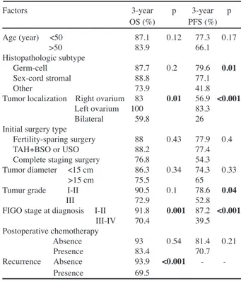
Table 3. Univariate Analysis of Patients with ROCs
for Both Overall Survival (OS) and Progression-free Survival (PFS) According to Clinicopathological Factors
Factors 3-year p 3-year p OS (%) PFS (%) Age (year) <50 87.1 0.12 77.3 0.17 >50 83.9 66.1 Histopathologic subtype Germ-cell 87.7 0.2 79.6 0.01 Sex-cord stromal 88.8 77.1 Other 73.9 41.8 Tumor localization Right ovarium 83 0.01 56.9 <0.001
Left ovarium 100 83.3 Bilateral 59.8 26 Initial surgery type
Fertility-sparing surgery 88 0.43 77.9 0.4 TAH+BSO or USO 88.2 77.4 Complete staging surgery 76.8 54.3 Tumor diameter <15 cm 86.3 0.34 74.3 0.33 >15 cm 75.5 65 Tumur grade I-II 90.5 0.1 78.6 0.04 III 72.9 52.8 FIGO stage at diagnosis I-II 91.8 0.001 87.2 <0.001
III-IV 70.4 39.5 Postoperative chemotherapy Absence 93 0.54 81.4 0.21 Presence 83.4 70.7 Recurrence Absence 93.9 <0.001 Presence 69.5
*ROCs: rare ovarian cancers
Figure 3. Progression-free survival times of patients
with stage I germ-cell tumors and sex-cord stromal tumors were better than those of patients with stage I other rare ovarian tumors (p=0.02).
250 200 150 100 50 0 T ime (months ) 1,0 0,8 0,6 0,4 0,2 0,0 Pr ob ab ilit y S ur viv ing af ter re lap se (% ) p=0.01
G erm-c ell tumors
O ther rare tumors
Clinical and Pathological Features of Rare Ovarian Tumors in Turkey Figure 3). Stage II-SCSTs had worse median PFS interval
compared to patients with stage II-GCTs (81 vs 122.4, respectively, p=0.04). In stage II group, there were no other ROTs. The median PFS times were similar in all stage III patients (37 months for GCTs vs 24.6 months for SCSTs vs 28.4 months for other ROTs, p=0.86). After PFS analysis was carried out with respect to stage IV patients, all of the three pathological groups had worse PFS interval (2.8 vs not reached vs 5 months, respectively, p=0.09). Thereafter, OS analysis was performed according to the tumor-stage specific. The median OS times for patients with stage I-GCTs and stage I-SCSTs were better than that of patients with stage I-other ROTs, but this difference was not significant (177.1 vs 199.3 vs 124.8 months, respectively, p=0.60). In stage II-GCTs, median OS time was 170 months, while stage II-SCTs had 122 months of median OS time (p=0.56). The median OS intervals were not reached in all of the stage III patients in pathological subgroups, but not significant (p=0.20). Moreover, the median OS times were similar (8 months for GCTs vs not reached for SCSTs vs 6.7 months for other ROTs, p=0.21) in stage IV histological types.
Multivariate analysis showed that the FIGO stage at the time of diagnosis was an independent prognostic factor for PFS (p=0.001, HR: 0.11). Thereafter, the multivariate analysis was carried out for OS; only the presence of recurrence was an independent prognostic indicator (p=0.02, HR: 0.54). The results of univariate and multivariate analysis for PFS and OS are summarized in Table 3 and 4.
Thereafter, histopathological type-specific survival analysis was separately performed. Univariate analysis for patients with GCTs indicated that tumor localization (p<0.001), initial surgery type (p<0.001), FIGO stage at diagnosis (p<0.001) and tumor grade (p=0.001) were significant prognostic factors for PFS. For OS, initial surgery type (p=0.006), FIGO stage at diagnosis (p=0.003) tumor grade (p=0.008) and the presence of recurrence (p<0.001) were found to be important prognostic indicators in the univariate analysis. In the patients with SCSTs, univariate analysis showed that initial surgery type (p=0.031) and FIGO stage at diagnosis (p=0.05)
for PFS were significant prognostic factors, however, no significant factor was detected for OS. Moreover, when univariate anaysis was carried out for patients with other ROTs, it demonstrated that tumor localization (p=0.004) and FIGO stage at diagnosis (p=0.05) for PFS and only the presence of recurrence (p=0.02) were significant factors. No significant prognostic indicator was found when the type-specific multivatiate analysis was carried out.
Discussion
ROTs comprise GCTs, SCSTs, Brenner tumors, clear cell carcinoma, transitional cell tumors, and carcinoid tumors of the ovary. They have different biological behaviors with a variety of histopathological subtypes; in addition, they have curative potential with different treatment approaches compared with much more commonly encountered epithelian ovarian cancers (Quirk and Natarajan, 2005; Young, 2005; Ayhan et al., 2009; Koulouris and Penson, 2009; Colombo et al., 2012; Penson, 2013; Takeuchi et al., 2013). The cure rates of early stage GCTs approach 100%, and even in the advanced stage of the disease, surgery and the BEP regimen result in cure rates of approximately 75% (Gershenson, 2007; Parkinson et al., 2011; Matei et al., 2013). SCSTs of the ovary are considered low grade malignancies with a relatively more favorable prognosis and are generally treated more often with repeat surgeries compared to epithelial ovarian cancers (Young, 2005; Ayhan et al., 2009). Their natural history is unclear, and the prognostic factors that impact survival remain to be clarified.
In our study, tumors that are mostly localized in the left ovary had GCT histology, but SCST and other ROTs were mostly localized in the right ovary. In addition, GCTs also occurred in younger patients and tended to be larger in size and well or moderately differentiated compared to SCST and other ROTs. Median PFS time for patients with GCTs or SCSTs was better than that of other ROTs, but OS was similar for the three ROT groups. However, PFS and OS in tumors that were localized in the right ovary or bilaterally were worse compared to patients with left ovarian ROTs. Multivariate analysis showed that FIGO stage at the time of diagnosis was an independent prognostic factor for PFS, while only the presence of recurrence was found to be an independent prognostic indicator for OS. Ray-Coquard et al indicated that both PFS and OS were similar in all ROT groups, a finding that agrees with our results (Ray-Coquard et al., 2010). In addition, the authors found that age, tumor size, and FIGO stage for PFS were independent prognostic factors. Thus, our results were compatible with the literature with respect to FIGO stage, but they were different according to the age and tumor size (Ray-Coquard et al., 2010). This may be related to the heterogeneous patient group of our study.
Surgery is the first treatment of choice, because it provides accurate information about the initial extent of disease; thereafter, it guides to the adjuvant treatment (Karimi Zarchi et al., 2011; Colombo et al., 2012, Matei et al., 2013). Although the extent of the initial surgery is still controversial, higher relapse rates have been reported by fertility-sparing surgery, especially in SCSTs (Pautier
Table 4. The Results of Multivariate Analysis for Both
Progression-free and Overall Survival
Factor X2 p HR 95%CI
Progression-free survival
Tumor localization 0.18 0.66 1.16 0.57-2.37 FIGO stage at diagnosis 11.4 0.001 0.11 0.03-0.40 Histopathological subtype (GCT vs SCST vs other ROC)
0.36 0.85 0.92 0.40-2.13 Tumor size 0.13 0.96 0.97 0.29-3.20 Tumor grade 0.26 0.6 0.7 0.18-2.69 Overall survival Initial symptoms 0.19 0.65 0.73 0.19-2.80 Tumor localization 0.24 0.62 0.8 0.34-1.89 Histopathological subtype (GCT vs SCST vs other ROC)
0.11 0.73 0.83 0.28-2.42 FIGO stage at diagnosis 0.12 0.72 1.37 0.23-8.25 Tumor size 1.61 0.2 3.56 0.50-12.2 Tumor grade 0.56 0.45 1.86 0.36-9.48 The presence of recurrence 5.04 0.02 0.54 0.05-1.69
et al., 1997). The rate of complete staging surgery has been reported to be 25% in the literature (Ray-Coquard et al., 2010). However, complete staging surgery could be performed for 16.2% of patients in our study. In GCTs, fertility-sparing surgery was more frequently carried out, while patients with other ROTs commonly underwent complete staging surgery. These differences were significant. This situation may be attributed to the fact that patients with other ROT had more aggressive histologies, such as clear cell and transitional cell carcinomas, and GCTs or SCSTs were more suitable for fertility-sparing surgery because patients with these diagnoses were typically younger. On the other hand, survival rates did not differ according to the initial surgical procedure. In a study performed by Cicin et al., the rate of complete staging surgery has been reported to be 15.7% in 70 patients with malignant GCTs (Cicin et al., 2009).
Limited and non-uniform studies are present in the literature, including those with small patient sizes and with respect to ROTs due to different histological subtypes and multiple treatment strategies. Therefore, making decisions about standard medical treatment approaches can be difficult due to the results of limited studies (Colombo et al., 2007; 2012; Ray-Coquard et al., 2010; Matei et al., 2013). In addition, the prognostic factors for these rare tumors need to be clarified. Thus, trials with respect to the ROTs to date tend to consist of too few patients to detect prognostic factors clearly. Of the ROTs, some prognostic factors have been documented in SCSTs, especially granulosa cell tumors (Jamieson et al., 2008; Ayhan et al., 2009; Li et al., 2009; Fotopoulou et al., 2010; Suri et al., 2013). Ayhan et al. (2009) analyzed 80 patients with granulosa cell tumor (Ayhan et al., 2009). In their study, abnormal uterine bleeding was the most common symptom with which patients initial presented. The authors indicated that advanced stage, advanced age, the presence of residual tumor after initial surgery, and the requirement of adjuvant treatment were found to be important prognostic factors by univariate analysis. However, multivariate analysis showed that only the initial disease stage was an important prognostic indicator for survival (Ayhan et al., 2009).
Another study of patients with granulose cell tumors found that nuclear atypia, FIGO stage, increased mitosis, and the presence of tumor rupture before or during operations were independent prognostic factors by multivariate analysis for survival (Li et al., 2009). In a study performed by Zhang et al. (2007) the authors showed that early FIGO stage and patients’ age ≤50 years were important prognostic factors for granulose cell tumors (Zhang et al., 2007). In our study, an analysis of histological subgroups could not be performed separately because of the small sample size of subgroups. However, tumor localization, FIGO stage and tumor grade for PFS, tumor localization, FIGO stage, and the recurrence of OS were important prognostic factors in univariate analysis. In the present study, clinicopathological features of three different ROTs groups were compared. Patients with GCTs were mostly localized in the left ovary, while the right ovary was the most common tumor site for SCSTs and other ROTs. Patients with GCTs were more like to
be 50 years old or younger compared to patients with SCST and other ROCs. Fertility-sparing surgery was more frequently performed for GCTs; however, patients with other ROTs commonly underwent complete staging surgery. Furthermore, GCTs tend to be larger in diameter and well or moderately differentiated compared to SCSTs or other ROTs. Patients with other ROTs had advanced-staged disease compared to patients with GCTs and SCSTs. The rate of bilateral involvement has been reported to be 3.8-8.5% (Ayhan et al., 2009; Cicin et al., 2009). We found that the rate of bilateral tumor localization was 19.2%, which was higher than the values found in other studies in the literature. In addition, tumor localization was found to be prognostic indicator for both PFS and OS in the univariate analysis, but not in the multivariate analysis. The 3-year PFS and OS rates were worse for bilateral tumors compared to patients with both left and right tumors. This may be related to the fact that our study population included many different histologies and the high rate bilaterality of tumors compared with literature (Ayhan et al., 2009; Cicin et al., 2009).
There have been no randomized studies assessing the significance of postoperative adjuvant therapy in high-risk patients until now due to the low incidence of ROTs. Therefore, the impact of adjuvant treatment on survival is still not clear (Cronjé et al., 1999; Schumer et al., 2003; Colombo et al., 2012; Matei et al., 2013). In our study, 71.3% of patients who had advanced-stage or high-risk disease received adjuvant treatment after the initial surgery. However, the significant survival advantage conferred by adjuvant treatment for wither PFS or OS could not be found. The recurrence rate was 28.1% for the entire cohort in the current study. In addition, the most frequent site of recurrence was the abdominal cavity in our study, which agreed with similar studies in the literature (Ayhan et al., 2009; Cicin et al., 2009). Eleven of the patients with recurrence had not received adjuvant chemotherapy, while the remaining 36 patients with recurrent disease were treated with adjuvant treatment. In fact, the majority of mortalities (75%) were observed in patients who received adjuvant chemotherapy. Therefore, it might be thought that there was no any effect of adjuvant treatment on recurrent disease or mortality for this group. The unfavorable results of adjuvant treatment groups might be due to the fact that this group comprised advanced stage or high-risk patients. On the other hand, the presence of adjuvant treatment was not found to be an independent prognostic factor in our study. Our findings were concordance with a study by Ayhan et al. (2009). The 3-year PRS rates and median PRS times for patients with both GCT and SCST were significantly better than those of other ROT (69% vs 61% vs 35% and 132 vs 66 vs 24 months, respectively; p=0.001). In other words, these patients with rare ovarian histologies, especially GCTs and SCSTs can be cured despite the recurrence. Therefore, regular follow-up examinations including tumor markers and radiologic imagings are needed.
There are a number of limitations of our study. The retrospective nature of our study was an important limitation and may have influenced our results. Therefore, some patients had an incomplete follow-up period. The
Clinical and Pathological Features of Rare Ovarian Tumors in Turkey 0 25.0 50.0 75.0 100.0 Newl y di agnosed wi thout tr eatment Newl y di agnosed wi th tr eatment Persi stence or recurr ence Remi ssi on None Chemother ap y Radi other ap y Concurr ent chemor adi ati on 10.3 0 12.8 30.0 25.0 20.3 10.1 6.3 51.7 75.0 51.1 30.0 31.3 54.2 46.8 56.3 27.6 25.0 33.1 30.0 31.3 23.7 38.0 31.3 0 25.0 50.0 75.0 100.0 Newl y di agnosed wi thout tr eatment Newl y di agnosed wi th tr eatment Persi stence or recurr ence Remi ssi on None Chemother ap y Radi other ap y Concurr ent chemor adi ati on 10.3 0 12.8 30.0 25.0 20.3 10.1 6.3 51.7 75.0 51.1 30.0 31.3 54.2 46.8 56.3 27.6 25.0 33.1 30.0 31.3 23.7 38.0 31.3 0 25.0 50.0 75.0 100.0 Newl y di agnosed wi thout tr eatment Newl y di agnosed wi th tr eatment Persi stence or recurr ence Remi ssi on None Chemother ap y Radi other ap y Concurr ent chemor adi ati on 10.3 0 12.8 30.0 25.0 20.3 10.1 6.3 51.7 75.0 51.1 30.0 31.3 54.2 46.8 56.3 27.6 25.0 33.1 30.0 31.3 23.7 38.0 31.3 0 25.0 50.0 75.0 100.0 Newl y di agnosed wi thout tr eatment Newl y di agnosed wi th tr eatment Persi stence or recurr ence Remi ssi on None Chemother ap y Radi other ap y Concurr ent chemor adi ati on 10.3 0 12.8 30.0 25.0 20.3 10.1 6.3 51.7 75.0 51.1 30.0 31.3 54.2 46.8 56.3 27.6 25.0 33.1 30.0 31.3 23.7 38.0 31.3 0 25.0 50.0 75.0 100.0 Newl y di agnosed wi thout tr eatment Newl y di agnosed wi th tr eatment Persi stence or recurr ence Remi ssi on None Chemother ap y Radi other ap y Concurr ent chemor adi ati on 10.3 0 12.8 30.0 25.0 20.3 10.1 6.3 51.7 75.0 51.1 30.0 31.3 54.2 46.8 56.3 27.6 25.0 33.1 30.0 31.3 23.7 38.0 31.3 0 25.0 50.0 75.0 100.0 Newl y di agnosed wi thout tr eatment Newl y di agnosed wi th tr eatment Persi stence or recurr ence Remi ssi on None Chemother ap y Radi other ap y Concurr ent chemor adi ati on 10.3 0 12.8 30.0 25.0 20.3 10.1 6.3 51.7 75.0 51.1 30.0 31.3 54.2 46.8 56.3 27.6 25.0 33.1 30.0 31.3 23.7 38.0 31.3 0 25.0 50.0 75.0 100.0 Newl y di agnosed wi thout tr eatment Newl y di agnosed wi th tr eatment Persi stence or recurr ence Remi ssi on None Chemother ap y Radi other ap y Concurr ent chemor adi ati on 10.3 0 12.8 30.0 25.0 20.3 10.1 6.3 51.7 75.0 51.1 30.0 31.3 54.2 46.8 56.3 27.6 25.0 33.1 30.0 31.3 23.7 38.0 31.3
other limitations of this study were the relatively small sample size in determining the relevant prognostic factors for ROTs. In addition, our heterogeneous study population from many treatment centers might also have influenced our findings. Although our results should be confirmed by prospective studies including large sample sizes, we believe that they contribute to the literature because there were no randomized studies assessing prognostic factors in ROTs.
In conclusion, our study indicates that tumor localization, FIGO stage, and tumor grade for PFS and tumor localization, and FIGO stage and the presence of recurrence for OS were important prognostic factors. Moreover, FIGO stage at the time of diagnosis was an independent prognostic factor for PFS, while only its recurrence was found to be an independent prognostic indicator for OS. Due to the rarity of these tumors of the ovary, multi-center, prospective, randomized, controlled trials including a large number of patients are needed; then, relevant prognostic factors, which may help as a guide for more appropriate adjuvant or recurrent therapies for ROTs with advanced stage or high-risk, may be accurately clarified.
References
Ayhan A, Salman MC, Velipasaoglu M, et al (2009). Prognostic factors in adult granulosa cell tumors of the ovary: a retrospective analysis of 80 cases. J Gynecol Oncol, 20, 158-63.
Cicin I, Eralp Y, Saip P, et al (2009). Malignant ovarian germ cell tumors: a single-institution experience. Am J Clin Oncol,
32, 191-6.
Colombo N, Parma G, Zanagnolo V, et al (2007). Management of ovarian stromal cell tumors. J Clin Oncol, 25, 2944-51. Colombo N, Peiretti M, Garbi A, et al (2012). Non-epithelial
ovarian cancer: ESMO Clinical Practice Guidelines for diagnosis, treatment and follow-up. Ann Oncol, 7, 20-6. Cronjé HS, Niemand I, Bam RH, et al (1999). Review of the
granulosa-theca cell tumors from the emil Novak ovarian tumor registry. Am J Obstet Gynecol, 180, 323-7.
Fotopoulou C, Savvatis K, Braicu EI, et al (2010). Adult granulosa cell tumors of the ovary: tumor dissemination pattern at primary and recurrent situation, surgical outcome.
Gynecol Oncol, 119, 285-90.
Garcia AA, Morrow CP (1999). Stromal tumors of the ovary. In: Raghavan D, Brecher M, Johnson DH, Meropol NJ, Moots PL, Thigpen JT, editos. Textbook of Uncommon Cancer. New-York: John Wiley & Sons, 661-9.
Gershenson DM (2007). Management of ovarian germ cell tumors. J Clin Oncol, 25, 2938-43.
Jamieson S, Fuller PJ (2008). Management of granulosa cell tumour of the ovary. Curr Opin Oncol, 20, 560-4.
Karimi Zarchi M, Mousavi A, Gilani MM, et al (2011). Fertility sparing treatments in young patients with gynecological cancers: Iranian experience and literature review. Asian Pac
J Cancer Prev, 12, 1887-92.
Koulouris CR, Penson RT (2009). Ovarian stromal and germ cell tumors. Semin Oncol, 36, 126-36.
Li W, Wu X, Fang C, et al (2009). Prognostic factors in adult granulosa cell tumor of the ovary. Saudi Med J, 30, 247-52. Matei D, Brown J, Frazier L (2013). Updates in the management of ovarian germ cell tumors. Am Soc Clin Oncol Educ Book,
2013, 210-6.
Parkinson CA, Hatcher HM, Ajithkumar TV (2011). Management of malignant ovarian germ cell tumors. Obstet Gynecol Surv,
66, 507-14.
Pautier P, Lhommé C, Culine S, et al (1997). Adult granulosa-cell tumor of the ovary: a retrospective study of 45 cases. Int J
Gynecol Cancer, 7, 58-65.
Penson RT, Dizon DS, Birrer MJ (2013). Clear cell cancer of the ovary. Curr Opin Oncol, 25, 553-7.
Quirk JT, Natarajan N (2005). Ovarian cancer incidence in the United States, 1992-1999. Gynecol Oncol, 97, 519-23. Ray-Coquard I, Weber B, Lotz JP, et al (2010). Management of
rare ovarian cancers: the experience of the French website “Observatory for rare malignant tumours of the ovaries” by the GINECO group: interim analysis of the first 100 patients.
Gynecol Oncol, 119, 53-9.
Schumer ST, Cannistra SA (2003). Granulosa cell tumor of the ovary. J Clin Oncol, 21, 1180-9.
Suri A, Carter EB, Horowitz N, Denslow S, Gehrig PA (2013). Factors associated with an increased risk of recurrence in women with ovarian granulosa cell tumors. Gynecol Oncol,
131, 321-4.
Takeuchi T, Ohishi Y, Imamura H, et al (2013). Ovarian transitional cell carcinoma represents a poorly differentiated form of high-grade serous or endometrioid adenocarcinoma.
Am J Surg Pathol, 37, 1091-9.
Young RH (2005). Sex cord-stromal tumors of the ovary and testis: their similarities and differences with consideration of selected problems. Mod Pathol, 18, 81-98.
Zhang M, Cheung MK, Shin JY, et al (2007). Prognostic factors responsible for survival in sex cord stromal tumors of the ovary--an analysis of 376 women. Gynecol Oncol, 104, 396-400.