Balloon Laryngoplasty for Pediatric Subglottic Stenosis:
A 5-year Experience
Serap Şahin Önder1 , Aslı Şahin Yılmaz1 , Özgül Gergin2 , Begüm Yılmaz1
1Department of Otolaryngology, University of Health Sciences, Ümraniye Training and Research Hospital, İstanbul, Turkey 2Department of Otolaryngology, Medipol University Hospital, İstanbul, Turkey
Original Investigation
Corresponding Author:
Serap Şahin Önder; [email protected] Received Date: 19.07.2020
Accepted Date: 25.08.2020
Content of this journal is licensed under a Creative Commons Attribution 4.0 International License. Available online at www.turkarchotolaryngol.net
DOI: 10.5152/tao.2020.5795
Abstract Objective: The objective of this study was to present
our experience and evaluate our results of endoscopic balloon laryngoplasty (BL) in children with subglot-tic stenosis (SGS) at a pediatric tertiary center over a 5-year period.
Methods: This study reviewed 41 pediatric patients
diagnosed with acquired SGS who had undergone BL as the primary course of treatment. Cases were analyzed for details including patient demographics, SGS grade and length, timing and the type of surgery, presence of tracheostomy, comorbidities, postoper-ative management, complications and outcomes of balloon dilatation.
Results: Forty-one children (22 girls and 19 boys)
who had undergone BL at a mean age of 26 months (range, 1 month to 14 years) were included in the
study. Nineteen (46.3%) were diagnosed with acute SGS (12 thin stenosis, 7 thick stenosis) and 22 (53.7%) with chronic SGS (9 thin stenosis, 13 thick stenosis). The success rate of BL was 100% in patients with acute and chronic thin membranous stenosis. The effectiveness of BL was significantly higher in pa-tients with acute thick stenosis than in papa-tients with chronic thick stenosis (p=0.016).
Conclusion: This study confirms that BL in patients
presenting with acquired SGS with thin membranous stenosis, regardless of whether acute or chronic, can have a good prognosis. However, the results are less promising in cases of chronic thick stenosis.
Keywords: Subglottic stenosis, balloon laryngoplasty,
airway, pediatric, pediatric otolaryngology
ORCID iDs of the authors:
S.Ş.Ö. 0000-0002-3576-0953; A.Ş.Y. 0000-0002-7846-9453; Ö.G. 0000-0002-4544-863X; B.Y. 0000-0002-1345-2503.
Cite this article as: Şahin Önder S,
Şahin-Yılmaz A, Gergin Ö, Yılmaz B. Balloon Laryngoplasty for Pediatric Subglottic Stenosis: A 5-year Experience. Turk Arch Otorhinolaryngol 2020; 58(4): 208-13.
Introduction
Subglottic stenosis (SGS) encompasses a range of potentially life-threatening conditions and is characterized by the narrowing of the airway be-low the vocal folds. SGS has an incidence report-ing of 1 to 8% after endotracheal intubation (1). Advancements in neonatal care coupled with the introduction of prolonged intubation methods in neonatal and pediatric intensive care units have markedly reduced mortality rates in critically ill children over the last 60 years. Consequently, there is an increase in the incidence of SGS due to the application of mechanical ventilation (2, 3). The treatment of SGS should be patient-centric with the utmost importance placed on all practic-es that aid in delivering succpractic-essful surgeripractic-es. This involves a number of factors including the type of the stenosis, the surgeon’s experience as well as the preferred course of treatment, expectations of the patient and the family, the quality of post-opera-tive care the institution can provide, and the skill level of the caregivers (anesthesiologist, pulmonol-ogist and gastroenterolpulmonol-ogist) (4).
The surgical techniques deployed fall into two groups, as endoscopic and open procedures.
La-ryngotracheal reconstruction, cricotracheal resec-tion and cervical slide tracheoplasty feature promi-nently in the rankings for the frequently used open airway reconstruction procedures (3).However, the last 5-20 years, has seen the increased use of endoscopic approaches, especially after the intro-duction of the balloon dilator (4). Although bal-loon laryngoplasty (BL) has been used since the 1980s, it remains a relatively new technique in the treatment of SGS and is now heralded as an ef-fective and less invasive treatment for appropriate stenosis cases (5).
The objective of this study was to present our ex-perience and evaluate our results of endoscopic BL in children with SGS at a pediatric tertiary center over a 5-year period.
Methods
This is a retrospective review of all patients who presented with SGS, were diagnosed and referred to our tertiary care hospital between 2015 and 2020. Patient charts were analyzed and reviewed for data points, including patient demograph-ics, grade of subglottic stenosis (Cotton-Myers classification (6)), timing (acute or chronic SGS; Avelino et al. (7)), craniocaudal extension of SGS
(thin or thick; McCaffrey classification (8)), type of surgery, presence of tracheotomy, comorbidities, postoperative manage-ment, complications, as well as the following outcomes: change in endoscopic grading of SGS, postoperative symptoms, rate of decannulation, tracheotomy status. Informed consent was taken from the parents. Local Ethics Committee of Ümraniye Train-ing and Research Hospital approved the study (Approval Date: March 19, 2020; Approval Number: 7507).
This series included 41 pediatric patients with documented SGS who had undergone endoscopic BL as a primary modality of treat-ment. Patients who had other coexisting tracheal pathologic ditions, grade 4 SGS, SGS with glotto-subglottic stenosis, con-genital SGS, previous intervention for their SGS, comorbidities requiring a high likelihood of prolonged ventilation in the future or lost to follow-up after treatment were excluded. The diagnosis and description of subglottic stenosis was performed by an expert pediatric otolaryngologist at the time of the direct laryngobron-choscopy (DLB) with spontaneous ventilation technique. The Myer-Cotton grade was used to assess the severity of SGS (6). The timings of SGS were classified as follows.
1) Acute SGS was defined as a diagnosis of SGS within 30 days of extubation,
2) Chronic SGS was defined as a diagnosis of SGS 30 days after extubation (7).
The lengths of the scar tissue were classified as follows.
1) Thin SGS, which has a craniocaudal extension of <1 cm (McCaffrey classification grade 1)
2) Thick SGS, which has a craniocaudal extension of more than 1 cm and features either soft or hard scar contraction (McCaffrey classification grades 2 and 3) (8).
Prior to BL, dynamic upper airway examination was performed under spontaneous respiration using a flexible bronchoscope (3.1 mm or 3.8 mm Olympus Medical Systems; Hamburg, Ger-many) in order to exclude the presence of possible additional airway diseases including laryngomalacia, vocal cord paralysis or tracheomalacia. DLB was performed under general anesthesia using a 0º rigid endoscope (Storz Corporation; Tuttlingen, Ger-many) with a diameter between 2.7 and 4 mm. After establish-ing the diagnosis, high-pressure balloon (Acclarent Technology; Boston, United States) was used and inflation pressure of 8-12 atm was maintained for 1 to 2 minutes or until the oxygen sat-uration level dropped below 90%. The balloon size was selected according to the theoretical ideal subglottic diameter for the pa-tient’s age (9). The procedure was performed three times each session. A pledget soaked with betamethasone dipropionate and gentamycin sulfate (Belogent; Farmatek, İstanbul, Turkey) was then applied to the dilated segment. Adjunctive procedure, such as radial divisions of stenosis was performed using cold knife in thick SGS featuring hard scar contraction. No patient was intubated after dilatation; instead, monitored in the pediatric intensive care unit for 24 to 48 hours. Follow-up endoscopy was performed after an interval of three weeks in all cases, and BL was performed when required. All patients received systemic steroids (Prednisolone; Actavis, İstanbul, Turkey) (1-2 mg/kg/d) for two days, proton pump inhibitors (Ulcuran; Avis, İstanbul, Turkey) for one month postoperatively. Follow-up DLB was performed three weeks after the initial DLB and the procedure was repeated until complete healing. After complete resolution of SGS, the children underwent a course of awake flexible fi-beroptic laryngoscopy (FFL) every three months for a year. If the results were not satisfactory, BL was performed three times at 3-week intervals. If this failed to yield positive results, open surgery was performed. All DLB and awake FFL examinations were recorded (Figures 1, 2, 3, 4). Only when patients had been weaned from tracheotomy or had become symptom free for the duration of the study were deemed successful BL outcomes. The Main Points
• SGS is not a rare complication for the newborns or infants with prolonged intubation for various reasons in neonatal and pediatric intensive care units.
• SGS is a life-threatening condition. • BL is a less invasive treatment for SGS.
• BL can have a good prognosis in patients with acquired SGS with thin stenosis regardless of whether acute or chronic.
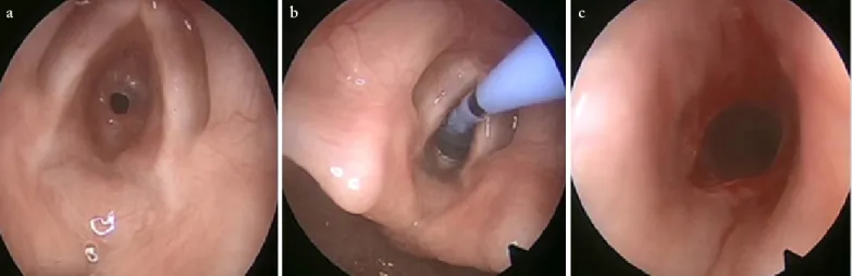
Figure 1. a-c. Acute grade III thin SGS in a 3-month old child. (a) pre-balloon laryngoplasty, (b) with balloon laryngoplasty, (c) post-balloon laryngoplasty
effectiveness of BL was calculated according to the changes in the Cotton-Myer grade with measurements taken before and after balloon dilatation.
Statistical Analysis
IBM Statistical Package for the Social Sciences version 20.0 (IBM SPSS Corp.; Armonk, NY, USA) was used for statisti-cal analysis. The changes in the Cotton-Myer grades before and
after the BL were compared using the Mann-Whitney U Test. P-values of ≤0.05 were regarded as significant.
Results
Forty-one children (22 girls and 19 boys) who had undergone BL, at a mean age of 26 months (range, one month to 14 years) were included in the study. Table 1 summarizes the breakdown of the patients by demographics, comorbidities, and details of
Figure 2. a-c. Acute grade II thick subglottic and tracheal stenosis in an 11-year-old child. (a) subglottic view of the lumen, (b) with balloon laryngoplasty, (c) post-balloon laryngoplasty
a b c
Figure 3. a-c. Chronic grade II thin SGS in a 7-month old child. (a) subglottic view of the lumen, (b) with balloon laryngoplasty, (c) post-balloon laryngoplasty
a b c
Figure 4. a-d. Chronic grade III thick SGS in a 22-month old child (a) subglottic view of the lumen, (b) radial divisions of stenosis using cold knife, (c) with balloon laryngoplasty, (d) post-balloon laryngoplasty
the etiology of stenosis. Nineteen (46.3%) children were diag-nosed with acute SGS (12 thin stenosis, seven thick stenosis) and 22 (53.7%) with chronic SGS (nine thin stenosis, 13 thick stenosis). Thirteen patients had undergone a tracheotomy before
laryngeal stenosis assessment. The mean period of follow-up was 15±8.6 months. The etiology pointed to all patients having developed SGS due to prolonged intubation. Other associated medical comorbidities were shown in Table 1.
The details of the children regarding their SGS grade and oth-er features woth-ere shown in Table 2. In patients with acute and chronic thin stenosis, the number of BLs ranged from one to two times. Moreover, the mean numbers of BLs in acute and chronic thin SGS groups were 1.3±0.7 and 1.2±0.4, respectively. However, in acute and chronic thick SGS groups this range was from two to three times. The mean numbers of BLs in acute and chronic thick SGS groups were 2.4±0.7 and 2.3±0.7, respective-ly. In all patients, BL was performed at 3-week intervals. The success rate of BL was 100% in patients with acute and chronic thin stenosis. In the acute thick SGS group (n=7) six children had undergone tracheotomies before BL and successful decan-nulation was achieved in two patients after balloon laryngoplas-ty. Three patients required additional open airway surgeries and the success rate of BL was four out of seven (57.1%). In the chronic thick SGS group (n=13) tracheotomy was performed in seven children before BL and one case was successfully de-cannulated after balloon laryngoplasty. Nine patients required additional open airway surgeries. In this group, the success rate of BL was four out of 13 (30.7%) (Table 2).
There was no significant statistical difference between the effec-tiveness of BL in patients with acute and chronic thin stenosis (p=0.56). BL was significantly more effective in patients with thin SGS (acute or chronic) than in patients with thick SGS (acute or chronic) (p=0.000). Moreover, the effectiveness of BL
Table 1. Demographics, comorbidities, etiology and follow up
Patients (n=41)
Age range 1 month-14 years
(mean: 26 months) Gender Male 19 (46.3%) Female 22 (53.7%) Comorbidity Prematurity 11 (26.8%) Only CHD 5 (12.1%) Down syndrome + CHD 3 (7.3%) CEA 1 (2.4%) Type 1 DM 1 (2.4%) None 20 (48.7%) Etiology of SGS Prolonged intubation 41 (100%)
Mean follow up period 15±8.6 months
CHD: congenital heart disease; CEA: congenital esophageal atresia; DM: diabetes mellitus; SGS: subglottic stenosis
Table 2. Comparison of outcomes between acute and chronic SGS
Type of SGS Acute (n=19) Chronic (n=22)
Length of stenosis Thin stenosis (n=12) Thick stenosis (n=7) Thin stenosis (n=9) Thick stenosis (n= 13)
Pre-op grade* Grade I - - 11.1% (n=1) 7.6% (n=1) Grade II 8.3% (n=1) 57.1% (n=4) 33.3% (n=3) 53.8% (n=7) Grade III 91.7% (n=11) 42.9% (n=3) 55.5% (n=5) 38.4% (n=5) Post-op grade* Grade 0 100% (n=12) 28.5% (n=2) 100% (n=9) Grade I - 42.8%(n=3) - 23.1% (n=3) Grade II - 14.2% (n=1) - 53.8% (n=7) Grade III - 14.2% (n=1) - 23.1% (n=3) Number of BL 1.3±0.7 2.4±0.7 1.2±0.4 2.3±0.75 Success rate of BL 100% 57.1% 100% 30.7%
Presence of tracheotomy N/A 85.7% (n=6) None 53.8% (n=7)
Decannulation rate after BL N/A 33.3% (n=2) None 14.2% (n=1)
*Cotton-Myer Classification
was significantly higher in patients with acute thick stenosis than in patients with chronic thick stenosis (p=0.016).
There were no significant clinical complications related to the procedures.
Discussion
Airway narrowing in children is mostly located in the subglottis. The most expected pathogenesis of acquired SGS commences with subglottic mucosal pressure necrosis due to endotracheal intubation, then mucosal ulceration, followed by perichondritis, and finally mature scar tissue formation (10).This condition ne-cessitates immediate treatment on symptom presentation. The purpose of BL is to arrest the progress of mature scar formation during the evolution of acquired SGS.
The lack of prospective studies evaluating the effectiveness of bal-loon dilatation has come to mean that balbal-loon dilatation is pre-dominantly featured in discussions. Designing controlled studies comparing BL to other more conventional therapies is challeng-ing due to patients havchalleng-ing a wide range of comorbidities and lack of uniformity amongst airway patients (e.g., age, weight, gender, ethnicity, stenosis diameter, length, histology, neurologic func-tions, glottal status and comorbidities) (11). Consequently, case series are an important mechanism for experience sharing. The presented case series included 41 pediatric patients with acquired SGS who had undergone BL. The subglottic steno-sis cases were divided into four subgroups: acute thin stenosteno-sis, acute thick stenosis, chronic thin stenosis, and chronic thick stenosis. Our results revealed that the effectiveness of BL may vary depending on the type of the subglottic stenosis. This study adhered to the guidelines established by the previous reporting by McCaffrey (8) for the purposes of this case series, however, it should be stressed that SGS measurements can be subjective. The management of SGS in children is a frustrating and distress-ing process both for the patients and their families. In acute cases, it is possible that several extubation attempts are unsuccessful or extubation is successful but the child ends up with severe respira-tory distress. In chronic cases, the child has already had a trache-otomy or is receiving treatment for recurring laryngitis or asthma. According to several studies, the success rate of BL for acute SGS was significantly higher compared to chronic SGS (2,7). Avelino et al. (7) demonstrated a success rate of 100% for acute SGS and 39% for chronic SGS and determined that success rates could be linked to the SGS grade and the immature scar tissue. Lee et al. (2) suggested that patients with acquired SGS, once diagnosed, should promptly undergo balloon dilatation as it increases treat-ment success rates, and decannulation as well, to reduce the need of tracheotomies. However, Simpson et al. (12) warned that in patients with chronic acquired SGS, the prognosis could be worse if circumferential stenosis and scar contraction are present. In the presented study, balloon dilatation demonstrated a 100% success rate in patients with SGS with thin and soft membrane, regard-less of whether the scar tissue was acute or chronic. This success
rate was 57.1% in patients with acute thick stenosis and 30.4% in patients with chronic thick stenosis. These results reaffirmed that when the stenosis features thin and soft membranous scar tis-sue, regardless of whether acute or chronic, the success of balloon dilatation is virtually certain. If the stenosis is fibrotic and thick in character, prompt execution of balloon dilatation is paramount to ensure success, as the stenosis expands easier during the acute period than in the chronic period. Therefore, early diagnosis and treatment of acquired SGS is vital to prevent recurrence. Children who have undergone prolonged endotracheal intubation and de-velop stridor and respiratory distress after extubation need extra consideration and care.
On reviewing the literature, we found that one systematic re-view, which included 7 studies and 150 patients, estimated the overall success rate of BL as 65.3%. The authors claimed that failures may be linked to more severe grades of stenosis (13). In our series, the success rate was calculated by ignoring the types of stenosis and scar tissue. The presented study demonstated that where the grade of SGS is severe, the success of balloon dilatation is high in the patients with acute and chronic thin SGS. Moreover, this outcome shows that the nature of stenosis is more important than its grade.
There are varying numbers of dilatations reported in the litera-ture. Some authors describe performing up to seven dilatations until achieving success, but most report that up to three dilata-tions were necessary for successful treatments (14). In our study, serial DLB was performed as previously described and deemed successful if there were any responses in the form of grading, extension, or clinical picture. If there was no improvement in the patient’s condition after three consecutive sessions, we moved to open surgical procedures. For each patient with acute or chron-ic thin SGS, the number of BLs performed ranged from one to two times. In patients with acute and chronic thick SGS, however, this number ranged from two to three times. Unfor-tunately, the outcomes of three patients in the acute thick SGS group and nine patients in the chronic thick SGS group were not satisfactory.
Special attention should be devoted to balloon dilatation in chil-dren who have not undergone tracheotomy. Close collaboration with the anesthesiology team is vital in pediatric airway surger-ies (15). Reported complications related to balloon dilatation of the lower airway include granuloma formation, mucosal lac-eration, tracheitis, bleeding, pneumothorax, pneumomediastini-tis, atelectasis, and death (15). Furthermore, a rapid, complete luminal obstruction secondary to edema in response to balloon dilatation was reported by Gungor (16). This complication can cause an inability to intubate. All surgeons need to be alert about the presence of this complication should it arise, the occurrence of which would necessitate an emergency tracheotomy. The downside risks posed by BL can be managed by reviewing the location, the size of the stenosis, and by analyzing the patient’s underlying health condition (15). In our study, none of the pa-tients experienced any major complications from BL.
Conclusion
This study highlights the evidence that BL in pediatric pa-tients with acquired thin SGS, regardless of whether of acute or chronic, can have a good prognosis. BL may be considered as a first-course treatment for thin acquired SGS. However, further research is advocated for its use in the management of SGS es-pecially with thick and mature stenosis.
Ethics Committee Approval: Ethics committee approval was received
for this study from the Local Ethics Committee of Ümraniye Train-ing and Research Hospital (Approval Date: March 19, 2020; Approval Number: 7507).
Informed Consent: Informed consent was obtained from the parents
of the patients who participated in this study.
Peer-review: Externally peer-reviewed.
Author Contributions: Concept - S.Ş.Ö., Ö.G.; Design - S.Ş.Ö.,
Ö.G.; Supervision - A.Ş.Y., Ö.G.; Resources: Ö.G.; Materials - S.Ş.Ö., B.Y.; Data Collection and/or Processing - S.Ş.Ö., B.Y.; Analysis and/or Interpretation - S.Ş.Ö., B.Y.; Literature Search - S.Ş.Ö., A.Ş.Y., B.Y.; Writing - S.Ş.Ö., A.Ş.Y.; Critical Reviews - S.Ş.Ö., A.Ş.Y.
Conflict of Interest: The authors have no conflicts of interest to declare. Financial Disclosure: The authors declared that this study has received
no financial support.
References
1. Morrissey MS, Bailey CM. Diagnosis and management of subglottic stenosis after neonatal ventilation. Arch Dis Child 1990; 65: 1103-4. 2. Lee JC, Kim MS, Kim DJ, Park DH, Lee IW, Roh HJ, et al.
Sub-glottic stenosis in children: our experience at a pediatric tertiary center for 8 years in South Korea. Int J Pediatr Otorhinolaryngol 2019; 121: 64-7.
3. Marston AP, White DR. Subglottic stenosis. Clin Perinatol 2018; 45: 787-804.
tracheal stenosis. Expert Rev Respir Med 2018; 12: 709-17. 5. Avelino MG, Fernandes EJ. Balloon laryngoplasty for subglottic
stenosis caused by orotracheal intubation at a tertiary care pediat-ric hospital. Int Arch Otorhinolaryngol 2014; 18: 39-42.
6. Myer CM 3rd, O'Connor DM, Cotton RT. Proposed grading sys-tem for subglottic stenosis based on endotracheal tube sizes. Ann Otol Rhinol Laryngol 1994; 103: 319-23.
7. Avelino M, Maunsell R, Jubé Wastowski I. Predicting outcomes of balloon laryngoplasty in children with subglottic stenosis. Int J Pediatr Otorhinolaryngol 2015; 79: 532-6.
8. McCaffrey TV. Classification of laryngotracheal stenosis. Laryn-goscope 1992; 102: 1335-40.
9. Hautefort C, Teissier N, Viala P, Van Den Abbeele T. Balloon di-lation laryngoplasty for subglottic stenosis in children: eight years' experience. Arch Otolaryngol Head Neck Surg 2012; 138: 235-40. 10. Baker S, Kelchner L, Weinrich B, Lee L, Willging P, Cotton R, et al. Pediatric laryngotracheal stenosis and airway reconstruction: a review of voice outcomes, assessment, and treatment issues. J Voice 2006; 20: 631-41.
11. Bent JP, Shah MB, Nord R, Parikh SR. Balloon dilation for recur-rent stenosis after pediatric laryngotracheoplasty. Ann Otol Rhi-nol Laryngol 2010; 119: 619-27.
12. Simpson GT, Strong MS, Healy GB, Shapshay SM, Vaughan CW. Predictive factors of success or failure in the endoscopic manage-ment of laryngeal and tracheal stenosis. Ann Otol Rhinol Laryn-gol 1982; 91: 384-8.
13. Wentzel JL, Ahmad SM, Discolo CM, Gillespie MB, Dobbie AM, White DR. Balloon laryngoplasty for pediatric laryngeal ste-nosis: case series and systematic review. Laryngoscope 2014; 124: 1707-12.
14. Rutter MJ, Cohen AP, de Alarcon A. Endoscopic airway manage-ment in children. Curr Opin Otolaryngol Head Neck Surg 2008; 16: 525-9.
15. Lee KH, Ko GY, Song HY, Shim TS, Kim WS. Benign tracheo-bronchial stenoses: long-term clinical experience with balloon di-lation. J Vasc Interv Radiol 2002; 13: 909-14.
16. Gungor A. Balloon dilation of the pediatric airway: potential for disaster. Am J Otolaryngol 2012; 33: 147-9.