Accepted: 2014.01.28 Published: 2014.05.20
2495
4
2
38
Malignant pleural mesothelioma: A single-center
experience in Turkey
ABCDEFG 1
Ahmet Sezer
ABCDEFG 2
Ahmet Taner Sümbül
ABCDEFG 1
Hüseyin Abalı
G 1
Hüseyin Mertsoylu
AD 1
Özgür Özyılkan
Corresponding Author: Ahmet Taner Sümbül, e-mail: [email protected] Source of support: Departmental sources
Background: Malignant pleural mesothelioma is a rare lethal malignancy caused by asbestos exposure. It is more frequent-ly seen in certain regions in Turkey. In this retrospective study, we aimed to anafrequent-lyse demographic, clinical, and pathological data and treatment-related features in 54 patients.
Material/Methods: The study included 54 patients diagnosed with malignant mesothelioma that were followed and treated.
Results: Of the 54 patients, 34 (55.6%) were male. The median age in men and women were 60.3 (38.2–77.2) and 65.8 (37.7–77.5) years, respectively. In 35 (64.8%), exposure to asbestosis was present. Epithelial type was found in 27 (50.0%), followed by mixed type in 7 (13.0%) patients, and in 20 (37.0%) patients the subtype could not be determined. The disease was staged as IV in 37 (68.5%) patients. In 28 patients (51.9%), it was right-sid-ed and in 1 (1.9%) it was bilateral. The most frequent metastatic sites (in decreasing order) were lungs, me-diastinum, diaphragm, liver, and thoracal wall. Of the 54 patients, 36 (66.6%) received 1st-line chemotherapy
and 20 (37%) 2nd-line chemotherapy. Eighteen patients (33.3%) received radiotherapy; 11 (20.3%) with
palli-ative intention and 7 (12.9%) with curpalli-ative intention. Median overall survival (OS) was 12.03 months (95% CI 7.2–16.8). OS was not affected by sex (p=0.32), smoking history (p=0.51), alcohol consumption (p=0.36), fami-ly history (p=0.67), pleural effusion presence (p=0.80), operation (p=0.14), clinical stage (p=0.072), symptom at presentation (p=0.66), having mixed type histology (p=0.079), asbestos exposure (p=0.06), and type of 1st-line
chemotherapy (p=0.161). On the contrary, it may be positively affected by good ECOG PS (0-1) (p<0.01), age below 65 (p=0.03), left-sided disease (p=0.01), receiving chemotherapy (p<0.01), having unilateral pleural ef-fusion (p=0.018), and type of 2nd-line chemotherapy (p=0.025).
Conclusions: OS of our patients was better than that found in the literature, seeming to be positively affected by early stag-es, better ECOG PS, age below 65 years, left side involvement, and having second-line chemotherapy with cis-platin-gemcitabine or 3M. Overall treatment success seems to be comparable to what is currently expected.
MeSH Keywords: Risk Factors • Overall Survival • Risk Factors • Mesothelioma • Survival Rate Full-text PDF: http://www.medscimonit.com/download/index/idArt/890020 Authors’ Contribution: Study Design A Data Collection B Statistical Analysis C Data Interpretation D Manuscript Preparation E Literature Search F Funds Collection G
1 Department of Medical Oncology, Başkent University, Medical Faculty, Adana, Turkey
2 Department of Medical Oncology, Mustafa Kemal University, Medical Faculty, Hatay, Turkey
Background
Malignant pleural mesothelioma (MPM) is an insidious can-cer that emerges in pleura, peritoneal cavities, tunica vagi-nalis, or on pericardial surfaces. Of the total cases, 80% are pleural. In 70% of the cases, a relationship with exposure to asbestos is detected. Other possible etiologic factors are radiotherapy, viral oncogenes, and genetic factors [1–3]. Mesothelioma is mainly seen in adults. It generally occurs in the 5th and 6th decades of the life and 70–80% of the
pa-tients are male. Most of these papa-tients have a history of long-term asbestos exposure during childhood [4]. The incidence of MPM in people without asbestos exposure is expected to be 1 in 1 million persons, whereas in people with exposure this ratio may be up to 10 in 100 persons [5]. Median over-all survival ranges from 6 to 18 months. In patients with lo-calized disease, survival can be increased by multimodality therapy [6]. Although extrapleural pneumonectomy is advo-cated by some groups, pleurectomy and decortication is most widely utilized [7]. In advanced stages, combination chemo-therapy of cisplatin and pemetrexed is the current standard [8]. Radiotherapy can be applied as a part of multimodali-ty therapy for rather early stages and as a palliative tool at later stages [9].
The incidence of mesothelioma is decreasing in developed countries due to reduced exposure in working and living ar-eas, but in underdeveloped and developing countries it is in-creasing because of increased industrial and environmental ex-posure [5,10]. In Western countries mesothelioma occurs as a result of environmental exposure in people living near asbes-tos mines and through occupational exposure, but in coun-tries such as Turkey, Cyprus, Greece, and Afghanistan it results from secondary contact with soil (white soil) mixed with asbes-tos for domestic use [11–15]. In Turkey, the problem is main-ly environmental and is an important public health problem in central and southeastern Anatolia. A study conducted by the Turkish Ministry of Health, Directorate of Cancer Control Department, in 8 Turkish cities of during 2004–2006, report-ed that MPM was not among the 10 most common types of cancer in men and women, and its incidence in both sexes was below 1% [16].
As MPM is a rare disease, each study performed in large se-ries of patients and published in the literature will advance the therapeutic approach to this disease. We think that sharing our experience about this disease, which is more commonly encountered in the neighboring cities compared to other re-gions of our country, both by revealing its clinico-pathologi-cal data and evaluating our therapeutic approaches and their outcomes, will make a substantial contribution to the med-ical literature.
Material and Methods
Prior to study initiation, approval was obtained from the Başkent University ethics committee (Date and number: 20/09/2010-KA10/133). In this retrospective study, we aimed to enroll pa-tients treated at Adana Başkent Research and Training Hospital. The charts of 54 patients followed between January 2000 and June 2010 at Başkent University Medical Oncology Department were enrolled in this study. Key clinical parameters, disease, and therapies had been recorded in the electronic health re-cords, including data on sex, age, smoking and drinking hab-its, exposure to asbestosis, clinic stages, ECOG PS, disease ex-tension, and treatment protocols).
The patients were generally diagnosed using VATS and pleu-ral biopsy (25 with pleupleu-ral biopsy, 7 with thoracotomy, 20 with VATS, and 2 with pleural fluid cytology). All patients had histo-logical diagnosis, including hematoxylin-eosin and immunohis-tochemistry for keratin, calretinin, EMA, mesothelin, CEA, and TTF-1. CT was used 25% of patients, MRI for 20% of patients, and PET CT for 55% of patients for initial staging. Operation decisions are taken in oncology tumor boards for patients suit-able for operation and operation notes are also considered in our study. Only 5 of the patients with extrapulmonary pleurec-tomy had lymph node sampling; all other patients’ lymph node status was considered and compared with PET CT.
All patients were followed and screened with computerized tomography at 3-month intervals, in line with general follow-up protocols of the medical oncology department.
Statistical analysis
Statistical analysis was performed by using SPSS 15.0 soft-ware. For all data, frequency, mean, and standard deviation were calculated. Survival was analyzed by Kaplan-Meier meth-od and factors that might have an effect on the survival were examined using the log-rank Test. Statistical data were con-sidered to be significant at p<0.05.
Results
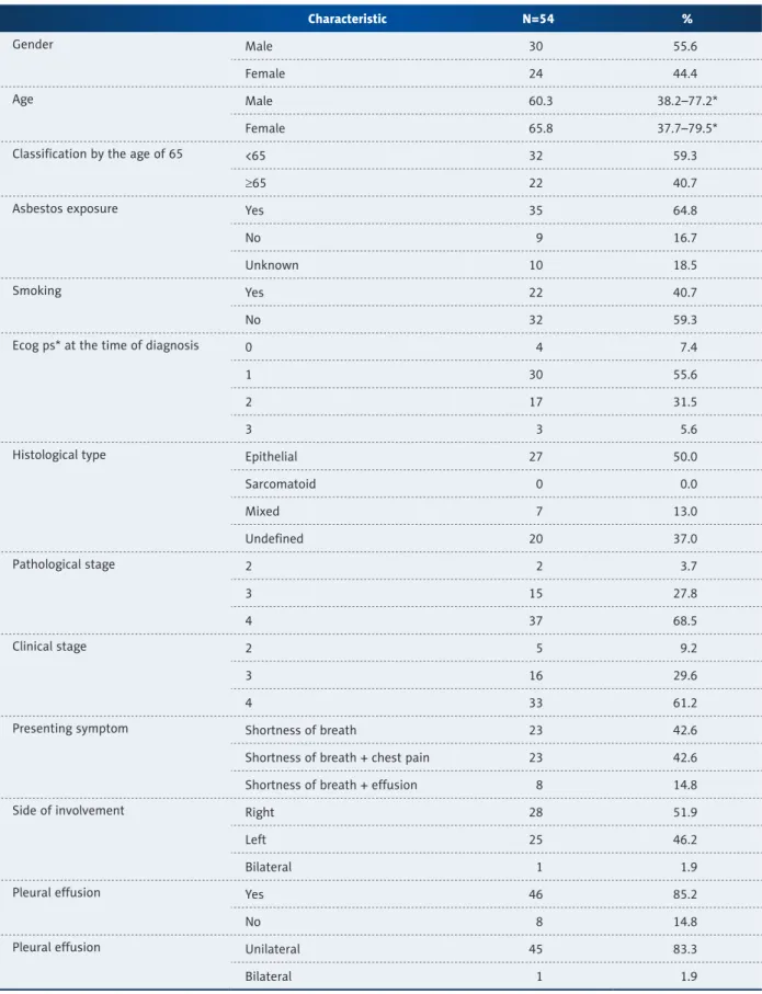
A total of 54 patients, of whom 30 (55.6%) were male, were enrolled in the study. Demographic and clinical characteristics features of patients are given in Table 1.
At the time of diagnosis, 22 patients were found to have non-pleural metastasis. Of the patients, 22 had palliative or cura-tive operations (16 patients had pleurectomy/decortication, 5 patients had extrapleural pneumonectomy, and 1 patient had pleurodesis). No mortality was seen in patients with sur-gical operation. Five of them had postoperative complication
Characteristic N=54 %
Gender Male 30 55.6
Female 24 44.4
Age Male 60.3 38.2–77.2*
Female 65.8 37.7–79.5*
Classification by the age of 65 <65 32 59.3
≥65 22 40.7
Asbestos exposure Yes 35 64.8
No 9 16.7
Unknown 10 18.5
Smoking Yes 22 40.7
No 32 59.3
Ecog ps* at the time of diagnosis 0 4 7.4
1 30 55.6
2 17 31.5
3 3 5.6
Histological type Epithelial 27 50.0
Sarcomatoid 0 0.0 Mixed 7 13.0 Undefined 20 37.0 Pathological stage 2 2 3.7 3 15 27.8 4 37 68.5 Clinical stage 2 5 9.2 3 16 29.6 4 33 61.2
Presenting symptom Shortness of breath 23 42.6
Shortness of breath + chest pain 23 42.6
Shortness of breath + effusion 8 14.8
Side of involvement Right 28 51.9
Left 25 46.2
Bilateral 1 1.9
Pleural effusion Yes 46 85.2
No 8 14.8
Pleural effusion Unilateral 45 83.3
Bilateral 1 1.9
Table 1. Demographic and clinical characteristics of the patients.
(e.g., empyema, bleeding). Mean hospitalization of patients who had surgery was 8.1 days.
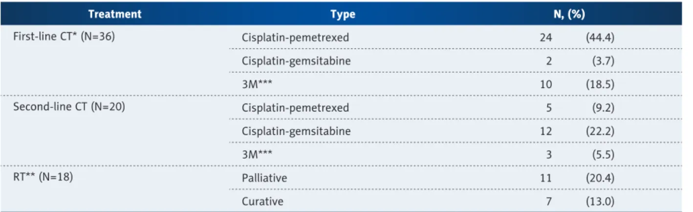
Only 1 patient was treated with neoadjuvant therapy. In the first-line treatment, 36 patients received a total of 176 cy-cles of therapy (median 5.1) and the most common adverse effects were anemia (13.8%, grade I–II), neutropenia (13.8%, grade I–III), nausea and vomiting (10.2%, grade II), and throm-bosis and asthenia (2%). In the second-line treatment, 20 pa-tients received a total of 75 cycles of therapy (median 3.75) and common adverse effects were neutropenia (25%, grade I–IV), thrombocytopenia (10%, grade II–III), anemia (grade I–II), and neuropathy (5%). The therapies given to the patients are shown in Table 2.
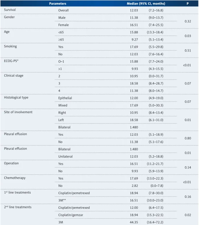
Overall survival data and their relation with other factors are shown in Table 3. Kaplan-Meier curve for overall survival is given in Figure 1. An insignificant correlation was observed between sex and overall survival (12.03, 95% CI: 7.24–16.83) (Figure 2). A statistically significant difference was found between overall survival and age above or below 65, ECOG PS status 0–1 or >1, second-line treatment with gemcitabine or cisplatpemetrexed, 1-sided or 2-sided pleural effusion with, site of in-volvement left or right side, and taking chemotherapy or not. Median progression-free survival was 7.33±0.87 (95% CI: 5.61–9.05) months. No statistically significant correlation was found between sex and receiving chemotherapy and progres-sion-free survival (Table 4).
Discussion
MPM is a rare, insidious type of cancer. Progress in its treat-ment has been slow. This disease is also an important health problem in Turkey. Asbestos exposure is still a problem in
developing countries such as Turkey. In this study, at one of the most comprehensive oncology centers of the region, we studied the patients who were treated at our hospital. Of 54 patients enrolled to our study, 30 were male and 24 were female. We found that, in line with the published liter-ature, the disease seems to be more common in men, proba-bly because of higher levels of occupational and environmen-tal exposure to asbestos, as in other countries [14,17,18]. In our study, the M/F ratio was 1.25. Among our patients, mean age was calculated to be slightly lower in men compared to women, which may be because men are exposed to asbestos most intensely and at an earlier age [13,19].
The disease initiation period after asbestos exposure is not ex-actly known, but the mean period was calculated as 32 years in 1 study [20]. Of the patients, 35 (64.8%) were definitively exposed to asbestos for at least a part of their lives and only 9 (18.5%) had no history of exposure to asbestos. Thirty-six patients were born in the regions of Turkey in which the ex-posure to asbestos is high and, although they immigrated to other regions after spending an average of 20–30 (mean 23.4 years) years in high-risk regions, they could not escape from malignant mesothelioma after the 6th decade of life. This is
especially important for showing that in some places of our country, there is a high level of asbestos exposure (men who work in marble and stone quarries where there are high lev-els of asbestos exposure and women who paint their house annually with asbestos-containing lime) [21]. When we eval-uated our patients for other additional factors, we found that 40% smoked and only 1.9% consumed alcohol. Previous ad-ministration of radiotherapy, which is an etiological risk factor for malignant mesothelioma, was not reported in any patient. The fact that nearly all of our female patients are house-wives suggests that, in our country, in this group, painting the house with asbestos-containing lime paint rather than in-dustrial or occupational exposure is the leading type risk [22].
Treatment Type N, (%) First-line CT* (N=36) Cisplatin-pemetrexed 24 (44.4) Cisplatin-gemsitabine 2 (3.7) 3M*** 10 (18.5) Second-line CT (N=20) Cisplatin-pemetrexed 5 (9.2) Cisplatin-gemsitabine 12 (22.2) 3M*** 3 (5.5) RT** (N=18) Palliative 11 (20.4) Curative 7 (13.0)
Table 2. Treatment modalities initiated to the patients.
Interestingly, only 1 of the patients had a familial history of malignant mesothelioma.
In MPM, age (below vs. above 65 years) is an important fac-tor affecting prognosis [23]. Aggressive treatment is suggested
for patients under age 65, even if there are additional risk fac-tors [24]. In our patient group, overall survival was significant-ly higher in the patients below age 65 compared to those over 65 (15.8 and 9.2 months, p=0.037). Another factor that shows a good prognosis is the performance status of the patient at the
Parameters Median (95% CI, months) P
Survival Overall 12.03 (7.2–16.8) Gender Male 11.38 (9.0–13.7) 0.32 Female 16.51 (7.4–25.5) Age <65 15.88 (13.3–18.4) 0.03 ≥65 9.27 (5.1–13.4) Smoking Yes 17.69 (5.5–29.8) 0.51 No 12.03 (7.6–16.4) ECOG-PS* O–1 15.88 (7.7–24.0) <0.01 >1 9.93 (4.3–15.5) Clinical stage 2 10.95 (0.0–31.7) 0.07 3 18.58 (8.4–28.7) 4 11.38 (8.0–14.7)
Histological type Epithelial 12.00 (4.9–19.0)
0.07
Mixed 17.69 (5.0–30.3)
Site of involvement Right 10.95 (8.4–13.4)
0.01
Left 18.58 (6.1–31.0)
Bilateral 1.480
Pleural effusion Yes 12.03 (5.1–18.9)
0.80
No 11.38 (5.1–17.6)
Pleural effusion Bilateral 1.480
0.01 Unilateral 12.03 (5.2–18.8) Operation Yes 16.51 (11.2–21.7) 0.14 No 9.93 (5.9–13.9) Chemotherapy Yes 17.69 (13.0–22.3) <0.01 No 2.82 (0.0–7.8) 1st line treatments Cisplatin/pemetrexed 18.94 (7.8–30.0) 0.16 3M** 16.51 (10.0–23.0)
2nd line treatments Cisplatin/pemetrexed 12.00 (6.4–17.5)
0.02
Cisplatin/gemzar 18.94 (15.3–22.5)
3M 44.35 (16.4–72.2)
Table 3. Overall survival data and their relation with other clinical factors.
time of diagnosis [23]. As expected in the patients with a perfor-mance status of 0–1 at the time of diagnosis, overall survival is better. In our study, 34 patients (62.9%) had a performance sta-tus of 0–1 at the time of diagnosis and a better overall survival was calculated compared to the patients with ECOG performance >1 (p<0.01). Life expectancy is longer in the patients with earli-er clinical and pathological stage [23]. In our study, the patients with stage III disease had longer lifespan compared to those with stage II or stage IV diseases (18.5 months, 10.9 months, and 11.3 months, respectively). This difference may be explained by the fact that the patients have other risk factors (e.g., histology, age, ECOG PS). Reports in the general literature suggest aggressive therapies will not contribute to overall survival, patients with ep-ithelial histology have a better prognosis, and aggressive thera-py may not be suitable in patients with non-epithelial histology [24,25]. In our patient group, the patients with mixed histology had surprisingly prolonged survival compared to those with ep-ithelial histology (17.6 months and 12.0 months, respectively, p=0.079). This result, which differs from results in the general lit-erature, may be due to the lack of histopathologic subgroup de-scription in 20 patients in our series, which might have changed the statistical results. Difficulties in the diagnosis of malignant mesothelioma should be kept in mind [26].
At the time of diagnosis, the most commonly reported com-plaints were shortness of breath and chest pain [27]. Although the presence of chest pain at the time of admission was re-ported to be a good prognostic factor [23], no significant cor-relation could be detected between the presenting symptom and overall survival (p=0.661). In more than one-third of pa-tients, there is pleural effusion-related shortness of breath without chest pain [28]. In the patients with pleural fluid, cy-tological examination may demonstrate malignant cells in one-third of the cases [4]. In our case series, 85.2% of the patients were reported to have pleural effusion at the time of diagno-sis and all but 1 had unilateral effusion. Although the major-ity of the patients have pleural effusion, in our clinic, only 2 patients had the diagnosis using pleural cytology. Histological subgroup classification of mesothelioma may not be performed using other closed biopsy methods such as pleural cytology [29]. However, biopsy using VATS and open thoracotomy is more likely to lead to a diagnosis [30]. In our study, 44.4% of the patients were diagnosed by VATS and open thoracotomy surgery. Inadequate biopsy material is a problem in our study and inability to perform subgroup analysis in the majority of the patients may be caused by this factor.
Characteristic Median (95% CI; months) P
General 7.66 (4.7–10.5) Gender Male 7.33 (3.6–10.9) 0.314 Female 7.76 (2.7–12.7) Chemotherapy Yes 7.76 (3.6–11.8) 0.572 No 6.57 (1.3–11.8)
Table 4. Progression-free survival after firstline chemotherapy. Figure 1. Overall survival curve for all patients (n: 54).
1.0 0.8 0.6 0.4 0.2 0.0 Survival function Censored Survival function 0.00 20.00 40.00 OS 60.00 Cum sur vival
Figure 2. Overall survival curve according to gender (p=0.32). 1.0 0.8 0.6 0.4 0.2 0.0 Men Women Men − censored Women − censored Survival function 0.00 20.00 40.00 OS 60.00 Cum sur vival
For the treatment of MPM, 3 important surgical procedures were defined: surgical pleurodesis, P/D, and EPP with VATS [25,31,32]. In the surgical procedures, mortality is less than 5% [31]. In our study, 22 patients underwent surgery (9 pleu-rectomy, 1 pleurodesis, 5 EPP, and 7 P/D) and no mortality occurred. Despite the substantial potential for morbidity, sur-gery may be beneficial in the palpation of the major symp-toms such as resection pain and dyspnea [33]. Reports on the effect of P/D on overall survival are contradictory. Based on some data, VATS + P/D may be beneficial to ensure the sur-vival benefit in the patients who are not candidates for EPP [34]. Despite the high morbidity (60%) and mortality (4–9%) observed with EPP, when administered along with chemo-therapy and radiochemo-therapy, this procedure may allow use of higher doses of radiotherapy, as well as prolonging surviv-al in the presence of locsurviv-al recurrence and early disease [35]. However, EPP alone does not seem to prolong life expectan-cy [24]. In our study, there was not difference of overall sur-vival between the patients who underwent surgery and those who did not undergo surgery. In Turkey most patients are di-agnosed at an advanced stage, so there is scant opportuni-ty to initiate trimodal therapy. A study considering trimod-al therapy in stage 1–3 MPM patients revetrimod-aled that median overall survival is approximately 16.8 months after neoadju-vant treatment with cisplatin-pemetrexed and following EPP and radiotherapy [36].
Although local control of the patients is ensured using surgery and radiotherapy, many patients develop systemic metasta-sis. Chemotherapy leads to prolonged life expectancy and im-proved quality of life and relieves the symptoms. The response rates obtained with chemotherapy ranged between 0% and 45% [37]. A phase 2 study reported by Nowak et al. revealed that median overall survival was 11.2 months with cisplatin-gemcitabine therapy [38]. In our study, overall survival time was found to be significantly longer in the patients treated with chemotherapy compared to those who did not receive it
(17.6 months and 2.8 months, respectively, p<0.001). In the first-line treatment, we administered cisplatin-pemetrexed, cisplatin-gemcitabine and 3M (mitoxantrone, methotrexate, mitomycin-c) protocols and we did not find a difference in survival times among these 3 groups (p=0.161). In the sec-ond-line treatment, both the patients who received cisplatin-gemcitabine and those who received 3M protocols showed a significant difference of survival in second-line therapy com-pared with best supportive care (p=0.025). In the analyses of progression-free survival of the patients, the contribution of the chemotherapy did not show a significant difference be-tween sexes.
Conclusions and Recommendations
1. MPM patients have a poor life expectancy, with a mean sur-vival of approximately 1 year after diagnosis.
2. Because exposure to asbestos is the main etiological factor in 80% of the cases, primary prevention should be the first goal, especially in developing countries.
3. Selection of the potentially most appropriate surgical meth-od in the patients who were diagnosed allows at least con-trolling the symptoms.
4. In the patients with early-stage disease, triple therapy (sur-gery, radiotherapy, and chemotherapy) options may be tried. 5. In the patients with advanced-stage disease, in light of
many studies that showed significant survival differences by the best supportive therapy, we recommend giving at least first-line therapies to all patients with an appropriate performance status.
Conflict of interest
The authors of this manuscript have no conflicts of interest to disclose. The authors have full control of all the primary data and agree to allow the journal to review the data if requested.
References:
1. Tward JD, Wendland MM, Shrieve DC et al: The risk of secondary malignan-cies over 30 years after the treatment of non-hodgkin lymphoma. Cancer, 2006; 107: 108–15
2. De Luca A, Baldi A, Espesito V et al: The retinoblastoma gene family pRb/ p105, p107, pRb2/p130, and simian virus 40 large T-antigen in human me-sotheliomas. Nat Med, 1997; 3: 913–16
3. Sekido Y: Genomic abnormalities and signal transduction dysregulation in malignant mesothelioma cells. Cancer Sci, 2010; 101(1): 1–6
4. Jett J, Aubry M: Malignant pleural Mesothelioma. Clinical Respiratory Medicine. Mosby 2nd ed., 735–41
5. Antman KH: Natural history and epidemiology of malignant mesothelioma. Chest, 1993; 103: 373S–76
6. Herndon JE, Gren MR, Chahinian AP et al: Factors predictive of survival among 337 patients with mesothelioma treated between 1984 and 1994 by the Cancer and Leukemia Group B. Chest, 1998; 113: 723–31
7. Treasure T,Lang-Lazdunski L, Waller D et al: Extra-pleural pneumonecto-my versus no extra-pleural pneumonectopneumonecto-my for patients with malignant pleural mesothelioma: clinical outcomes of the Mesothelioma and Radical Surgery (MARS) randomised feasibility study. Lancet Oncol, 2011; 12(8): 763–72
8. Carteni G, Manegold C, Garcia GM et al: Malignant peritoneal mesothelio-ma-Results from the International Expanded Access Program using peme-trexed alone or in combination with a platinum agent. Lung Cancer, 2009; 64: 211–18
9. Flores RM, Krug LM, Rosenzweig KE et al: Induction chemotherapy, extra-pleural pneumonectomy, and postoperative high-dose radiotherapy for lo-cally advanced malignant pleural mesothelioma: a phase II trial. J Thorac Oncol, 2006; 1: 289–95
10. Hodgson JT, Mcelvenny DM, Darton AJ et al: The expected burden of meso-thelioma mortality in Great Britain from 2002 to 2050. Br J Cancer, 2005; 92: 587–93
11. Selçuk ZT, Coplu I, Emri S et al: Malignant pleural mesothelioma due to en-vironmental mineral fiber exposure in Turkey. Analysis of 135 cases. Chest, 1992; 102: 790–96
12. Coplu I, Dumortıer P, Demir AU: An epidemiological study in an Anatolian village in Turkey environmentally exposed to tremolite asbestos. J Environ Pathol Toxicol Oncol, 1996; 15: 177–82
13. Metintaş M, Ozdemir N, Hıllerdal G: Environmental asbestos exposure and malignant pleural mesohelioma. Respir Med, 1999; 93: 349–55 14. Senyiğit A, Bayram H, Babayiğit C: Malignant pleural mesothelioma caused
by environmental exposure to asbestos in the southeast of Turkey: CT find-ings in 117 patients. Respiration, 2000; 67: 615–22
15. Emri S, Semir AU: Malign pleural mesothelioma in Turkey, 2000–2002. Lung Cancer, 2004; 45S: S17–20
16. Eser S, Olcayto E, Karakılınç H et al: Sağlık Bakanlığı Kanserle Savaş Dairesi Başkanlığı (Epidemiyoloji ve Koruma Şube Müdürlüğü). Nüfus tabanlı kans-er kayıt mkans-erkezlkans-eri vkans-eri havuzu: sekiz il, 2004-2006 değkans-erlendirilmesi. http:
//www.kanser.gov.tr/folders/file/8iL-2006-SON.pdf-erişim tarihi 03.01.2011).
[in Turkish]
17. Schwartz DA: New developments in asbestos induced pleural disease. Chest, 1991; 99: 191–98
18. Adams VI, Unnı KK, Muhm JR et al: Diffuse malignant mesothelioma of pleura: Diagnosis and survival in 92 cases. Cancer, 1986; 58: 1540–51 19. Gennaro V, Ugolini D, Viarengo P et al: Incidence of pleural mesothelioma
in Liguria Region, Italy (1996–2002). Eur J Cancer, 2005; 41: 2709–14 20. Lanphear BP, Buncher CR: Latent period for malignant mesothelioma of
oc-cupational origin. J Occup Med, 1992; 34(7): 718–21
21. Yazıcıoglu S, Ilcayto R, Balcı K et al: Pleural calcification, pleural mesothe-liomas, and bronchial cancers caused by tremolite dust. Thorax, 1980; 35: 564–69
22. Senyigit A, Babayigit C, Gökirmak M et al: Incidence of Malignant Pleural Mesothelioma due to Environmental Asbestos Fiber Exposure in the Southeast of Turkey. Respiration, 2000; 67: 610–14
23. Curran D, Sahmoud T, Therasse P et al: Prognostic factors in patients with pleural mesothelioma: the European Organization for Research and Treatment of Cancer experience. J Clin Oncol, 1998; 16: 145–52 24. Balduyck B, Trousse D, Nakas A et al: Therapeutic surgery for
nonepithe-loid malignant pleural mesothelioma: is it really worthwhile? Ann Thorac Surg, 2010; 89: 907–11
25. Sugarbaker DJ, Jaklitsch MT0, Liptay MJ: Mesothelioma and radical multi-modality therapy: who benefits? Chest, 1995;107: 345S–50
26. Addis B, Roche H: Problems in mesothelioma diagnosis. Histopathology, 2009; 54(1): 55–68
27. Stewart DJ, Edwards JG, Symthe WR et al: Malignant pleural mesothelio-ma-an update. Int J Environ Health, 2004; 10: 26–39
28. Yates DH, Corrin B, Stidolph PN, Browne K: Malignant mesothelioma in South east England: clinicopathologic experience of 272 cases. Thorax, 1997; 52: 507–12
29. Bueno R, Reblando J, Glickman J et al: Pleural biopsy: a reliable method for determining the diagnosis but not subtype in mesothelioma. An Thorac Surg, 2004; 78: 1774–76
30. British Thoracic Society Standart of Care Committee. Statement on malig-nant mesothelioma in the United Kingdom. Thorax, 2001; 56: 250–65 31. Rice DC, Erasmus JJ, Stevens CW et al: Extended surgical staging for
poten-tially resectable malignant pleural mesothelioma. Ann Thorac Surg, 2005; 80: 1988–92; discussion 1992–93
32. Van Ruth S, Baas P, Zoetmulder FA: Surgical treatment of malignant pleu-ral mesothelioma: a review. Chest 2003;123(2): 551–61
33. Herndon JE, Gren MR, Chahinian AP et al: Factors predictive of survival among 337 patients with mesothelioma treated between 1984 and 1994 by the Cancer and Leukemia Group B. Chest, 1998; 113: 723–31 34. Halstead JC, Lim E, Venkateswaran RM et al: Improved survival with VATS
pleurectomy-decortication in advanced malignant mesothelioma. Eur J SurgOncol, 2005; 31: 314–20
35. Rusch VW, Rosenzweig K, Venkatraman E et al: A phase II trial of surgical resection and adjuvant high-dose hemithoracic radiation for malignant me-sothelioma. J Thorac Cardiovasc Surg, 2001; 122: 788–95
36. Krug ML, Pass JH, Rusch WV et al: Multicenter Phase II Trial of Neoadjuvant Pemetrexed Plus Cisplatin Followed by Extrapleural Pneumonectomy and Radiation for Malignant Pleural Mesothelioma. J Clin Oncol, 2009; 27: 3007–13
37. Green J, Dundar Y, Dodd S et al: Pemetrexed disodium in combination with cisplatin versus other cytotoxic agents or supportive care for the treatment of malignant pleural mesothelioma. Cochrane Database Syst Rev, 2007; CD005574
38. Nowak AK, Byrne MJ, Williamson R et al: A multicenter phase II study of cisplatin and gemcitabine in malignant mesothelioma. Br J Cancer, 2002; 87: 491–96