ACTA FACULTATIS
MEDICAE NAISSENSIS
DOI: 10.5937/afmnai2001034K
UDC: 615.245:618.11-006-07
O r i g i na l a r t ic l eInvestigation of Possible Effects of Exendin-4 during
Exposure to Mild Chronic Stress on
Dehydroepiandrosterone-Induced Polycystic Ovary
Syndrome in Rats
Burcu Köksal
1, Sedat Yildiz
2,Tugba Ozgöcer
3, Azibe Yildiz
2,
Nigar Vardi
2, Özlem Barutcu
2¹Lokman Hekim University, Faculty of Medicine, Ankara, Turkey
2Inonu University, Faculty of Medicine, Malatya, Turkey 3Harran University, Faculty of Medicine, Sanlıurfa, Turkey
S U MMA RY
The purpose of the study was to investigate the effects of Exendin-4 on polycystic ovary syndrome (PCOS) in rats in chronic mild stress medium. For establishing the PCOS model, dehydroepiandrosterone (DHEA) (6mg/100g) in 0.2ml sesame oil was injected subcutaneously to 21-day old rats (n = 67). In addition, 0.2ml sesame oil was injected subcutaneously to the rats in groups involving solution injection only. At the initial stage of the study, the rats were grouped as control, solution and PCOS, whereas stress and Exendin-4 groups were also added in the second stage of the study. In PCOS groups, Exendin-4 was applied intraperitoneally (10μg/kg/day) in mild chronic stress medium for four weeks. The results revealed that weight, fasting blood glucose, fasting blood insulin and HOMA-IR levels in the rats with PCOS were significantly higher than in the other groups; also, corticosterone levels of stress groups were significantly higher than in the other groups. In addition, harmful effects of PCOS on ovarian tissues were observed in histological examinations. However, after Exendin-4 application in PCOS groups, weight, fasting blood glucose, fasting blood insulin, HOMA-IR and LH/FSH levels were decreased, whereas Exendin-4 application in PCOS group treated with stress was not as effective as the application of Exendin-4 on rats with PCOS. Exendin-4 application also increased the number of healthy follicles in PCOS group, whereas there was no change in the number of healthy follicles in PCOS+Stress group. Key words: DHEA, PCOS, insulin resistance, chronic stress, Sprague Dawley, Exenatide
Corresponding author: Burcu Köksal
INTR ODU CTIO N
Polycystic ovary syndrome (PCOS) is a com-mon reproductive endocrinopathy affecting 5%-10% of reproductive-aged women (1). PCOS is associated with hyperandrogenism, enlarged ovaries, irregu-larities in menstrual cycles, hirsutism, cysts in ovaries and acnes (2). Moreover, the patients with PCOS might have type II diabetes, obesity, cardiovascular diseases, degenerated fertility and endometrial cancer (3, 4). In PCOS, changes in gonadotropin release lead to the development of abnormal follicles; moreover, disproportionate increases in LH/FSH ratio are seen in 94% of women with PCOS (5). As an effect of increased LH/FSH ratio, ovulation does not occur and corpus luteum is not formed (6). As the LH/FSH ratio, adrenal androgens are also increased during PCOS, and being converted to testosterones, they lead to hirsutism in women with PCOS (7). Another problem associated with PCOS is insulin resistance caused by beta cell degenerations (8). In the treatment of PCOS, different interventions involving diet, exercise, anti-estrogens, anti-androgens, ovarian drilling and agents increasing insulin sensitivity are used (9-13). Indeed, PCOS is a very complicated syndrome (13) and the majority of the studies focus on compensating the metabolic and reproductive malfunctions in PCOS in the treatment. Insulin resistance is the major metabolic problem and it is seen 70% of women with PCOS (14). Currently, GLP-1 agonists are used for decreasing insulin resistance (15). GLP-1 is an incretine hormone decreasing appetite and food intake by stimulating insulin release, repressing glucagon synthesis and inhibiting gastric emptying (16). Exendine-4 (Exe-4) is a synthetic mammalian incretin hormone analogue and has 53% of similarity with GLP-1 (17). Exe-4 binds to GLP-1 receptors with high affinity, and initiates glucose-dependent insulin secretion and increase in mass of β-cells (18). Moreover, it suppresses appetite and glucagon secretion by retarding gastric emptying (18). Effects of Exe-4 on obesity and diabetes has been thoroughly examined, but its effects on ovaries and endometrium have not been studied enough (19). At the same time, its effects on insulin levels, ovaries and endometrium in PCOS during stress condition have not been studied and discussed enough in spite of the existence of stress situation in PCOS. Stress situation is defined as impairment of homeostasis by a stressor (20). Stress
situation lead to the secretion of corticotropin-releasing hormone (CRH) by activating hypo-thalamus-hypophysis-adrenal (HPA) axis, and thus increased CRH causes anovulation, impairment of corpus luteum and infertility (21, 22). It is clear that stress is the reason of different reproductive function impairments. When importance of stress on reproduc-tive functions in PCOS is considered, effects of Exe-4 on insulin resistance and PCOS in chronic mild stress medium have to be studied well to contribute to the cure of women with PCOS. Therefore, the aim of the study was to investigate the effects of Exe on insulin resistance and PCOS in rats in chronic mild stress medium.
MA TERIA LS A ND METHODS
The study was initiated by taking permissions about ethic situations from Inonu University Ethic Committee of Medicine Faculty (Protocol number: 2015/A-07). Then, the determination of the number of rats in every group was done by setting the alpha level as 0.05, power as 0.80 and effect size as 1,5σ-2σ. Based on the standard table for sample size, 10 rats per group (7 groups) were determined to be sufficient (23). Sprague Dawley Rats were bought from Inonu University Research Center for Experimental Animals. Seventy 21-day-old female rats were taken and they were weighted. They were randomly put into the cages so as to place 5 rats to every cage. Then, their tails were painted by using different colors. The rats were weighted every week during the study. They were kept in the fixed room
temperature (220C ± 2) during 12 hours of dark and
daylight periods. All of the rats were fed with tap water and were pellet feed. In the study, seven groups were classified as three groups before stress applications, although seven groups were consid-ered in analysis after stress applications.
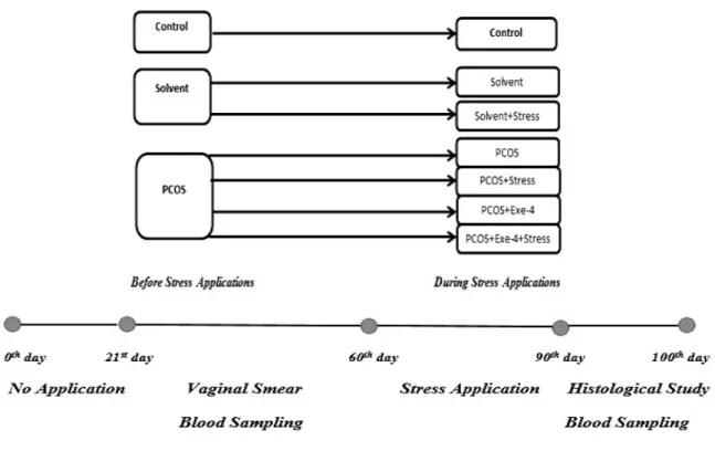
Three groups were determined as control (n = 10), PCOS (n = 39) and solvent (n = 18) before stress applications. In the control group, there was no intervention, while DHEA (Acros 154980100) was subcutaneously applied as 6 mg/100g weight/0.2ml of sesame oil in PCOS group until vaginal opening was observed. Hence, PCOS model was established. In the solvent group, 0.2ml of sesame oil was subcu-taneously applied until vaginal opening was ob-served. The groups of the study before and during the stress applications are presented below. (Figure 1)
Figure 1. The groups of the study before and during stress applications
Before Stress Applications
Blood sample collection
Estrous cycle observation was done by taking vaginal smear samples after the observation of vaginal opening. The rats in diestrus stage were not given any food one night before sample collection. Then, blood samples from tails of the rats were taken for examining LH, FSH, fasting blood glucose level and serum glucose levels. For taking blood samples, the rats were anesthetized by low concentration ether. Then, the tails were cleaned by warm water and ethanol, and blood samples were taken from tail vein by 1 cc insulin injector. Fasting blood glucose levels (mg/dl) were determined by On-Call-Plus blood glucometer. The remained blood samples were centrifuged with 3000 rpm for 10 min to separate serum and blood cells. Serum samples were put into
Ependorf tubes and were kept in -200C to examine
insulin, FSH and LH.
Measurement of blood serum insulin levels
Fasting serum insulin levels of the rats were determined by rat insulin ELISA kit (No: YHB0584Ra, Yehuda Biotechnology, China) and kit protocol. The
samples were examined by spectrophotometer with 450 nm wavelength and the results were reported as mlU/L. Then, HOMA-IR value was calculated with the following formula:
HOMA-IR= Fasting insulin levels (ml U/L) x Fasting glucose levels (mmol/L)/22.5.
Measurement of blood serum LH and FSH levels
Plate with 96 microwells (Nunc Roskilde, Denmark) was used to determine the levels of serum LH and FSH. The samples in microwell and standards were incubated to bind antibodies, and then the samples were transferred to coated plates for competitive binding process. Moreover, the plates were washed and secondary antibodies conjugated with streptavidin peroxidase were put into the wells. Tetramethylbenzidin was added to substrates and changes in color were observed at 450nm by plate reader (Biotek Synergy HT, Winooski, VT, USA). Primer LH and FSH antibodies of rats were taken from Dr. A. Parlow (National Institute of Diabetes and Digestive and Kidney Diseases National Hormone and Peptide Program, National Institutes of Health, USA). Secondary antibodies were bought from Sigma
for LH and FSH, 1ng/ml for LH and 2ng/ml for FSH, respectively.
Vaginal smear method
Checking the formation of PCOS model, vaginal smear was taken from the rats having vaginal opening during 10 days. Observation time of the
vaginal openings was observed on the 60th day of the
rats. First, 0.5 ml of the physiological saline solution
was taken to Pastor Pipet and then the rat was hold in motionless position by another researcher. The tip of the pipet was inserted into the vagina (0,5 – 1 cm) and physiologic saline solution was injected into the vagina. Without pulling the tip of the pipet, the liquid in the vagina was drawn back and was put on the microscope slide. Without any staining, the sample liquid was examined by light microscope for estrus stages. (Figure 2).
Figure 2. Application of vaginal smear method
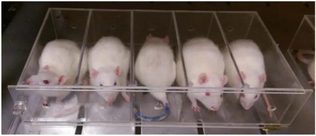
Figure 3. Mobility limiting cages for mild stress
During Stress Applications
Chronic mild stress method
After formation of the PCOS model, the stress process was applied to the experimental groups. The stress model was based on mobility limiting process. For the process, the boxes (6 x 7 x 18 cm) with 5 com-partments were used. In the comcom-partments, 2 holes in 1 cm diameter were involved to make breath-ing easy. The rats were placed inside for 1 hour in a day during 30 days without giving water and food. The
temperature was held stable at 230C. Scratching hairs,
urination, defecation and frequent breathing were accepted as stress indicators (24, 25). After the 1-hour period, the rats were put into their cages and the stress cages were cleaned by water. The stress cages
are presented inFigure 3.
After stress applications
After 30 days of Exe-4 and the stress appli-cations, vaginal smears were taken from the rats during 10 days and estrus cycles were observed. Then, food for the rats was picked up 1 night before blood collecting and blood samples were taken from
the tails for determining the fasting glucose levels. Fasting blood glucose levels (mg/dl) were determined by On-Call-Plus blood glucometer. For anesthesia of the rats, ksilasin/ketamine was injected as 90mg/kg i.p. (intraperitonally) /10mg/kg i.p. In the following stage, blood samples were taken from heart in the amount of 5ml by an injector and the samples were transferred to yellow-cap tubes involving a separator gel. Then blood cells and serum part of the samples were separated by centrifugation (10min., 3000rpm). Serum samples were transferred into Eppendorf tubes
and were kept at -200C. At the same time, the tissue
samples of ovaries were put into bottles involving 10% formaldehyde. The amount of the formaldehyde was ten times higher than the tissue volume.
Measurement of corticosterone in serum
Rat Coticosterone ELISA kit (Kat. No: 201111300, Sunredbio, China) and kit protocol was used to determine the corticosterone level in serum. The samples were observed by spectrophotometer at 450 nm wavelength and the records were taken as ng/ml.
Histological studies
The tissue samples of ovaries in 10% formaldehyde were fixed and then they were washed by tap water. After the dehydration and polishing processes, the samples were buried in paraffin. Paraffin blocks of ovaries were sliced to four sections having 100-120μm spaces. Then, hematoxilen–eosin (H-E) staining was applied to the samples after deparaffinization and rehydration. The stained samples of the tissues were investigated by Leica DFC-280 and Leica Q Win Image Analysis System (Leica Micros Imaging Solutions Ltd., Cambridge,
UK). In the histological investigations of ovaries, follicle types and corpus luteum were calculated. However, Langerhans islets were investigated in terms of sinusoidal dilatation, intracellular vacuol-ization and picnotic nucleus. Histological changes were recorded as 0 for none, 1 for few, 2 for medium, 3 for severe.
Statistical analyses
For checking the data normality, Shapiro Wilk Test was used and there was no violation of this assumption for parametric tests. For the assumption of homogeneity of variances, Levene test was used. For between-group comparisons, One-way ANOVA was applied while Benferoni test, as a follow-up test, was used if homogeneity of variances was equal and Games-Howell test was used in violence of the assumption of homogeneity of variances. Statistical analyses of histological data about ovaries were done by reporting the mean and standard deviations, and Mann-Whitney U test was used for between-group comparisons. All of the analyses were done at 0.05 level of type I error rate.
Findings
The results of this study will be represented under three different titles: findings before stress application, findings after stress application and histological findings.
Findings before stress application
The rats in seven groups were considered as three groups before stress application. They were control (n = 10), solvent (n=18) and PCOS (n = 39) groups. Mean weights of the groups per week before
Table 1. Mean weights of the groups per week before stress applications
Groups Weeks (Mean±SD)
1 2 3 4 5 6 7
Control (n=10) 30.0 ± 2.1a 43.1 ± 6.4a 54.7 ± 5.5a 65.2 ± 3.7a 86.2 ± 5.9a 98.1 ± 6.4a 121.1 ± 7.2a Solvent (n=18) 31.8 ± 3.1a 51.5 ± 5.6b 68.1 ± 6.44b 79.0 ± 5.4b 105.0 ± 8.0b 121.4 ± 7.8b 140.2 ± 7.9b PCOS (n=39) 30.7 ± 2.8a 51.8 ± 4.7b 66.7 ± 6.7b 81.3 ± 6.7b 104.4 ± 7.6b 120.3 ± 8.9b 144.5 ± 9.4b
p 0.21 0.00 0.00 0.00 0.00 0.00 0.00
stress applications are represented in Table 1.
As seen in the Table 1, the rats in PCOS and solvent groups gained more weight than the control group rats, although there was no significant difference between the groups in terms of weight at the beginning of the study. As the other variables of the study, LH, FSH, and HOMA-IR values before stress applications are given in Table 2.
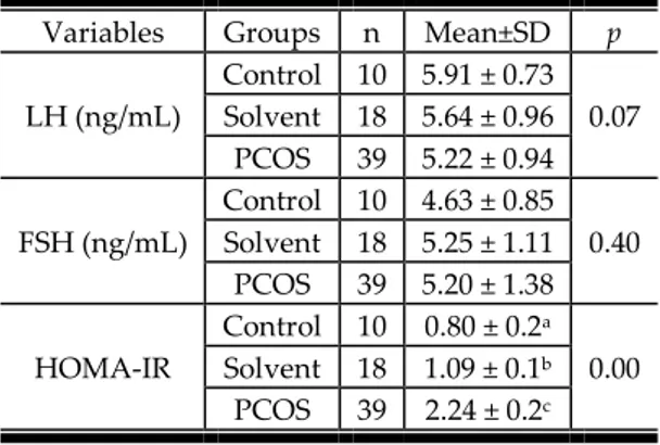
Table 2. LH, FSH, and HOMA-IR values before stress applications
Variables Groups n Mean±SD p
LH (ng/mL) Control 10 5.91 ± 0.73 0.07 Solvent 18 5.64 ± 0.96 PCOS 39 5.22 ± 0.94 FSH (ng/mL) Control 10 4.63 ± 0.85 0.40 Solvent 18 5.25 ± 1.11 PCOS 39 5.20 ± 1.38 HOMA-IR Control 10 0.80 ± 0.2a 0.00 Solvent 18 1.09 ± 0.1b PCOS 39 2.24 ± 0.2c
Table 2 shows that LH and FSH levels of the rats were not significantly different between the groups, while HOMA-IR level of PCOS group were significantly higher than the solvent and control groups. Moreover, HOMA-IR level of the solvent group was also higher than the control group level. Another important variable in PCOS is duration of estrus cycle. Table 3 shows the duration of completion of the estrus cycles in the groups before stress applications, Table 3.
Table 3. Duration of completing the estrus cycles in the groups before stress applications
Obser- vation Day Control (n = 10) % Solvent (n = 18) % (n=39) PCOS % 1stDay 0 0 0 2ndDay 0 0 0 3rdDay 0 0 0 4thDay 0 1 5.5 0 5thDay 7 70 7 38.9 1 2.6 6thDay 10 100 14 77.8 3 7.7 7thDay 0 17 94.4 5 12.8 8thDay 0 18 100 6 15.4 9thDay 0 0 7 18 10thDay 0 0 39 100
As represented in Table 3, the majority of the rats in PCOS group showed a delay in completing the estrus cycle, while control group rats completed their cycles in 5 and 6 days.
Findings after stress application
After the seven weeks, four-week stress application procedure was executed. The results on weight change during stress application period are given in Table 4.
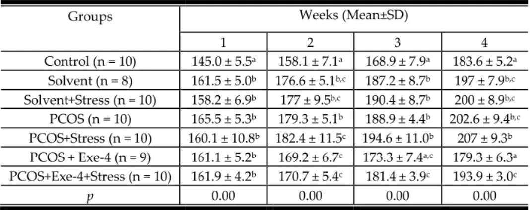
As seen in Table 4, the weight increases in the PCOS groups were significantly higher than in the control group. However, solvent groups also gained significantly high increases in body weight. As the other variables, LH/FSH, corticosterone and HOMA-IR levels were also analyzed after the stress application. The results can be seen in Table 5.
Table 5 shows that LH/ FSH ratio in PCOS group is significantly higher than in the control, solvent and the other PCOS groups. Exe-4 appli-cations also produced a lower ratio of LH/FSH than in the other groups even if it was applied under the stress condition. Corticosterone levels of the stress groups were also found to be significantly higher than in the other groups. HOMA-IR levels of PCOS and PCOS+Stress groups were significantly higher than in the other groups. However, the groups involving exe-4 application had lower levels of HOMA-IR than PCOS+Stress group. The duration of completing estrus cycle after stress application was also recorded. The results are seen in Table 6.
As illustrated in Table 6, delays in estrus cycles of the rats in PCOS groups were clearly seen. In addition to evidence from delay in estrus cycle, an increase in HOMA-IR levels and increase in LH/FSH ratio, histological examinations were also needed in investigation of the effect of exe-4 during stress conditions. Under the following title, histological findings are represented.
Histological findings
Histopathological examinations of the ovaries showed different patterns in different groups. Structure of ovaries in the solvent group seemed normal as well as the ovaries in the control group. In these ovaries,germinal epitheliums involved cubic, prismatic and squamous epitheliums. In the cortex regions, follicles and corpus luteums in different de-
Table 4. Mean weights of the groups per week after stress applications
Groups Weeks (Mean±SD)
1 2 3 4 Control (n = 10) 145.0 ± 5.5a 158.1 ± 7.1a 168.9 ± 7.9a 183.6 ± 5.2a Solvent (n = 8) 161.5 ± 5.0b 176.6 ± 5.1b,c 187.2 ± 8.7b 197 ± 7.9b,c Solvent+Stress (n = 10) 158.2 ± 6.9b 177 ± 9.5b,c 190.4 ± 8.7b 200 ± 8.9b,c PCOS (n = 10) 165.5 ± 5.3b 179.3 ± 5.1b 188.9 ± 4.4b 202.6 ± 9.4b,c PCOS+Stress (n = 10) 160.1 ± 10.8b 182.4 ± 11.5c 194.6 ± 11.0b 207 ± 9.3b PCOS + Exe-4 (n = 9) 161.1 ± 5.2b 169.2 ± 6.7c 173.3 ± 7.4a,c 179.3 ± 6.3a PCOS+Exe-4+Stress (n = 10) 161.9 ± 4.2b 170.7 ± 5.4c 181.4 ± 3.9c 193.9 ± 3.0c
p 0.00 0.00 0.00 0.00
Note: The groups having different word (a and b) in the same column are different from each other (p < 0.001) Table 5. LH/FSH, corticosterone and HOMA-IR values after stress applications
Variables Groups (Mean±SD) Control (n = 10) Solvent (n = 8) Solvent +Stress (n = 10) PCOS (n = 10) PCOS +Stress (n = 10) PCOS +Exe-4 (n = 9) PCOS+ Exe-4+Stress (n = 10) LH/FSH 1.78 ± 0.9a 1.78 ± 0.1a 1.92 ± 0.2a 2.36 ± 0.3b 1.58 ± 0.3a,c 1.43 ± 0.1c 1.50 ± 0.2c Cortico-sterone (ng/mL) 52.62 ± 3.33 a 53.74 ± 1.19a 59.34 ± 3.11b 53.49 ± 3.01a 58.92 ± 1.90b 51.72 ± 3.47a 58.33 ± 1.44b HOMA-IR 1.18 ± 0.13a 1.32 ± 0.10a 1.29 ± 0.09a 2.08 ± 0.31b 2.51 ± 0.06c 1.37 ± 0.12a 1.87 ± 0.35b Note: The groups having different word (a and b, c) in the same row are different from each other (p < 0.001)
Table 6. Duration of completing an estrus cycles in the groups before stress applications Obser-vation Day Con-trol n=10 % Sol-vent n= 8 % Sol-vent+ Stress % PCOS % PCOS+
Stress % PCOS+ Exe-4 %
PCOS+ Exe-4+ Stress % 1stDay 0 0 0 0 0 0 0 2ndDay 0 0 0 0 0 0 0 3rdDay 0 0 0 0 0 0 0 4thDay 1 10 1 12.5 0 0 0 0 0 5thDay 7 70 6 75 2 20 2 20 0 0 0 6thDay 10 100 8 100 3 30 3 30 0 2 22.2 0 7thDay 0 0 6 60 0 1 10 3 33.3 4 40 8thDay 0 0 0 4 40 2 20 5 55.5 5 50 9thDay 0 0 7 70 0 0 4 40 6 66.7 7 70 10thDay 0 0 10 100 10 100 10 100 9 100 10 100
velopment stages were seen. The total number of healthy follicles in the control group was 24.0 ± 11.3, while they numbered 20.33 ± 8.08 in the solvent group. There was no significant difference between the control and solvent groups in terms of the number of the healthy follicles. A sample histological image of the control group tissue is presented in Figure 4.
When analyzing the samples of PCOS (0.94 ± 1.29) and PCOS+Stress (0.85 ± 1.25) group, the number of cystic follicles in these groups was significantly higher than those for the control group. At the same time, the total number of healthy follicles in these groups was also different from the control group. Their number of healthy follicles was significantly lower than in the
Figure 4. An image of the control group ovary. Tertiary follicles (thick arrow), secondary follicle (thin arrow), primary follicle (arrowhead) and corpus luteum (KL).
H-E, X40.
control group (PCOS = 17.3 ± 11.1, PCOS + Stress =18.8 ± 10.5). In the PCOS + Exe-4 group, the number of healthy follicles was significantly higher than in the PCOS groups. However, there was no statistically significant difference between PCOC and PCOS + Exe-4 groups in terms of the numbers of atretic and cystic follicles. Figure 4
For the solvent and control group, similar images were obtained in histological examinations. Images of sample ovary tissues of Solvent and Solvent + Stress groups are givenin Figure 5.
As seen in Figure 5, similar structures are seen in images of Solvent and Control groups. However, Solvent + Stress group image captured a cystic follicle. In PCOS and PCOS + Stress groups, atretic and cystic follicles are seen clearly. Figure 6 represents images of sample ovary tissues of PCOS and PCOS + Stress groups. Figure 6
Figure 5. A: An image of the solvent group ovary. Tertiary follicles (thick arrow), secondary follicle (thin errow), primary follicle (arrowhead) and corpus luteum (KL). B: An image of the Solvent+Stress group ovary.
Corpus luteum (KL),cystic follicle (KF) H-E, X40.
Figure 6. A: An image of a sample ovary tissue of PCOS group; atretic follicle (arrows) and cystic follicle(KF). B: An image of sample ovary PCOS+Stress group; atretic follicle (arrows) and cystic follicle (KF). H-E, X40.
Figure 7. A: An image of a sample ovary tissue of PCOS+Exe-4 group; atretic follicle (arrow) and cystic follicle (KF). B: An image of a sample ovary PCOS+ Stress+Exe-4 group; atretic follicle (arrows) and cystic follicle (KF). H-E, X40.
In PCOS groups, the number of cystic follicles is higher than in the other groups. As illustrated in Figure 6 and 7, atretic and cystic follicles were seen in spite of the 4 use. Especially, PCOS + Stress + Exe-4 group was not different from PCOS group in terms of atretic and cystic follicles. Figure 7 shows the images of sample ovary tissues of PCOS + Exe-4 and PCOS+Stress+Exe-4 groups. Figure 7
DIS CU S S ION A ND SU GGES TIONS
The findings of this study about establishing a PCOS model showed that the PCOS model, as expected, indicated an increase in weight and HOMA-IR (insulin resistance) and a delay in completing the estrus cycle after DHEA application. Sun et al. (26) used a similar way to induce PCOS and they found that DHEA-application increased the weight of the rats. The increase in weight might be related to increase in androgens since increased levels of androgens lead to hypertrophy in adipocytes by changing lipid and carbohydrate metabolisms (27). As another point in this study, weight increase was also observed in the solvent groups. This situation might be associated with lipid nature of the solvent used in this study. Hala and Magbolah (28) also observed that sesame oil led to the increase in weight. Another change regarding the PCOS model related to the increase in HOMA-IR, which was expected. HOMA-IR is a sign of insulin resistance and the application of DHEA should increase HOMA-IR level. In this study, HOMA-IR levels of PCOS group were increased over 2.2 as a sign of insulin resistance (29, 30). Sun el at. (26) used DHEA for establishing PCOS model and their
find-ings also supported the idea that HOMA-IR levels were increased in rats with PCOS. Another change found in this study is about delay in estrus cycles of PCOS group. The findings showed that DHEA application led to delay in estrus cycles in 82.1% of the rats in PCOS group. The rats in solvent and control groups completed the cycles in 5-6 days, while the rats of PCOS group completed the cycles in 7-10 days. Sun et al. (26) also reported that the rats in the control group completed the cycles in 4-5 days. Iwasa et al. (31) applied DHEA for 12 weeks and their results showed that DHEA disarranged menstrual cycles of the rats. DHEA application led to a delay of menstrual cycles in the rats in their study.
After the four-week stress application, the effects of Exe-4 in PCOS on weight, LH/FSH ratio, HOMA-IR levels, corticosterone levels and menstru-al cycles during mild stress were examined. The application of Exe-4 in PCOS-Exe-4 group held the mean weight of the group similar to the mean weight of the rats in the control groups. This is an evidence of the effect of Exe-4 on weight loss. Similarly, the mean weight of the PCOS-Exe-4 group was less than the mean weight of PCOS+Stress + Exe-4. It can be said that effect of Exe-4 on weight loss is mediated by stress condition. The findings on the effect of Exe-4 of weight loss were supported by previous studies. Szayna et al. (32) applied Exe-4 as 10μg/kg/day during 13 days and they reported significant weight loss in the rats. Exe-4 was re-ported to be an effective agent for increasing insulin sensitivity and so was an effective agent for food intake (33). In spite of its effects in PCOS groups, stress application influenced its effect on weight loss. Torres and Nowson (34) reported that chronic stress
had an effect on HPA axis and the stress increased corticosterone levels and food intake in rats. Actu-ally, when the findings about corticosterone levels were examined, it was seen that the groups in-volving stress application had higher levels of corticosterone than the other groups. This finding is in line with the findings of previous studies (34-36). Stress response was related to HPA axis and sympatoadrenal system (37). In HPA axis, cortisol levels are increased after stress condition and the increase causes insulin resistance and release of free fatty acids (38). From the results of this study, it is seen that the stress and Exe-4 applications functions on a common pathway involving insulin metabo-lism. Hence application of Exe-4 in PCOS should be done by taking into account stress level of the rat.
The result about the effects of Exe-4 in PCOS on LH/FSH ratio showed that LH/FSH ratio for PCOS + Exe-4 was found to be decreased, while the level of PCOS group was higher than those for the other groups. The finding about PCOS+ Exe-4 might be explained as the effect of Exe-4 on decreasing insulin resistance, since the decrease in insulin resistance was found to be associated with decreases in LH/FSH ratio (26). Exe-4 applications in PCOS+ Exe-4 group also decreased HOMA-IR levels. However, Exe-4 application in PCOS+EXE-4+Stress group did not decrease the HOMA-IR levels to the levels of the control and solvent groups. This is an indication of stress effect on Exe-4 application. In-terestingly, stress application decreased the effects of Exe-4 on insulin resistance in rats with PCOS. This finding might be related to glucocorticoid release during stress, since chronic stress changes glucose metabolism by leading to glucocorticoid release (39). Stress hormones increase the catabolism of glycogen, triglycerides and proteins, hence they increase insulin resistance (40).
Another point of this study was to examine the changes in duration of estrus cycles after stress application. Stress application in PCOS groups caused a delay in the duration of completing the estrus cycles. The longest duration was recorded for PCOS+Stress group members. The application of Exe-4 did not prevent a delay in duration of estrus cycles. Hence the application of stress condition with Exe-4 negatively mediated the effects of Exe-4 on estrus cycles. Previous studies showed that stress application increases the activities of HPA axis and thereby it increases glucocorticoid synthesis. These changes damage the reproductive functions of
ova-ries (41-43). The metabolic pathways of these changes should be investigated by considering the HPA axis and ovaries.
After the stress application, it was found that the number of cystic follicles was higher in PCOS and PCOS+ Stress groups than the control group,while the number of healthy follicles was lower in PCOS and PCOS + Stress groups than in the control group. Lee et al. (44) reported that 20-day DHEA application increased estradiol and estrone levels, and these agents led to cyctic follicles in ovaries. Similarly, Singh et al. (45) also found that DHEA application caused cyctic follicles by increasing testosterone and androgen levels. In this study, Exe-4 application increased the number of healthy follicles in PCOS + Exe-4 group. However, these were no significant difference between the Solvent, PCOS, PCOS+Exe-4, Solvent-Stress, PCOS + Stress and PCOS + Exe-4 + Stress groups in terms of the numbers of healthy and cystic follicles. There-fore, it can be claimed that stress application pre-vented the effects of Exe-4 on ovaries in the rats with PCOS.
Findings of this study have merits for balancing stress-dependent metabolic changes in PCOS. As seen in the findings in using Exe-4 for balancing insulin and weight in PCOS, there should be attention about stress condition and duration of using Exe-4 should also be determined by considering stress condition. In addition to using Exe-4, stress control ways should also be taught for increasing the effects of Exe-4 in PCOS. Moreover, determination of stress level of patients with PCOS is needed before deciding to use Exe-4. In spite of the strong sides of this study, sample size should be increased in the following studies. Also, different types of exenatides should also be tested in stress conditions. In this study,as a limitation, one type of stress (chronic mild stress) was considered, the other types of stress should also be applied to rats during Exe-4 application to test its effects. At the same time, two groups involving “solvent+PCOS” and “solvent + PCOS+stress” are not involved in this study. This is a limitation for the study, and further studies should consider this limitation.
Acknowledgments
Portions of this work were presented at the TFBD 42. National Physiology Congress (Düzce, Turkey, 2016). This research did not receive any specific funding.
References
1. Lecke SB, Morsch DM, Spritzer PM. Association between adipose tissue expression and serum levels of leptin and adiponectin in women with polycystic ovary syndrome. Genet Mol Res 2013; 1: 4292-6.
https://doi.org/10.4238/2013.February.28.16
2. Fauser BC, Tarlatzis BC, Rebar RW, et al. Consensus on women's health aspects of polycystic ovary syndrome (PCOS). Hum Reprod 2012; 1: 14-24.
https://doi.org/10.1093/humrep/der396
3. Talbott EO, Zborowski JV, Rager JR, et al. Polycystic ovarian syndrome (PCOS): a significant contributor to the overall burden of type 2 diabetes in women. J Womens Health 2007; 1: 191-7.
https://doi.org/10.1089/jwh.2006.0098
4. Homburg R. Pregnancy complications in PCOS. Best Pract Res Clin Endocrinol Metab 2006; 30: 281-92.
https://doi.org/10.1016/j.beem.2006.03.009
5. Taylor AE, McCourt B, Martin MA, et al. Determinants of abnormal gonadotropin secretion in clinically defined women with polycystic ovary syndrome. J Clin Endocrinol Metab 1997; 82:2248-56.
https://doi.org/10.1210/jcem.82.7.4105
6. Cheung AP, Lu JK, Chang RJ. Pulsatile gonadotrophin secretion in women with polycystic ovary syndrome after gonadotrophinreleasing hormone agonist treatment. Hum Reprod 1997; 12: 1156-64.
https://doi.org/10.1093/humrep/12.6.1156
7. Memi G. Effect of mild excercise on chronic stress responses and role of hormones in this effect. Institute of Health Sciences, Department of
Physiology, Master Thesis, Marmara University, Istanbul, Turkey. 2015.
8. DeUgarte CM, Bartolucci AA, Azziz R. Prevalence of insulin resistance in the polycystic ovary syndrome using the homeostasis model assessment. Fertil Steril 2005: 83: 1454-60.
https://doi.org/10.1016/j.fertnstert.2004.11.070
9. Bremer AA. Polycystic ovary syndrome in the pediatric population. Metab Syndr Relat Disord 2010; 8: 375-94.
https://doi.org/10.1089/met.2010.0039
10. Mitwally MF, Casper RF. Aromatase inhibitors for the treatment of inferftility. Expert Opin Investig Drugs 2003;12: 353-71.
https://doi.org/10.1517/13543784.12.3.353
11. Baysal B. Polikistik over sendromu ve hirsutizm. İÜ Cerrahpaşa Tıp Fakültesi Sürekli Tıp Eğitimi Etkinlikleri Sempozyum Dizisi 2008; 63: 99-107. 12. Lebbi I, Temime RB, Fadhlaoui A, Feki A. Ovarian
drilling in PCOS: is it really useful Frontiers in surgery 2015; 2: 1-3.
https://doi.org/10.3389/fsurg.2015.00030
13. Sheehan MT. Polycystic ovarian syndrome: diagnosis and management. J. Clin Med Res 2004; 2: 13-27.
https://doi.org/10.3121/cmr.2.1.13
14. Dokras, A. Cardiovascular Risk in Women with PCOS. Steroids 2013;78(8): 773-6.
https://doi.org/10.1016/j.steroids.2013.04.009
15. Tura A, Göbl C, Pacini G. Effects of antidiabetic agents on pancreatic beta-cell function in gestational diabetes: is there enough evidence? Expert Opin Drug Metab Toxicol 2016; 1-5.
16. Drucker DJ, Nauck MA. The incretin system: glucagon-like peptide-1 receptor agonists and dipeptidyl peptidase-4 inhibitors in type 2 diabetes. The Lancet 2006; 368: 1696-05.
https://doi.org/10.1016/S0140-6736(06)69705-5
17. Kolterman OG, Buse JB, Fineman MS, et al. Synthetic exendin-4 (exenatide) significantly reduces postprandial and fasting plasma glucose in subjects with type 2 diabetes. J Clin Endocrinol Metab 2003; 88: 3082-89.
https://doi.org/10.1210/jc.2002-021545
18. Nielsen LL, Young AA, Parkes DG. Pharmacology of exenatide (synthetic exendin-4): a potential therapeutic for improved glycemic control of type 2 diabetes. Regulatory peptides 2004; 117: 77-88.
https://doi.org/10.1016/j.regpep.2003.10.028
19. Artunc-Ulkumen B, Pala HG, Pala EE, et al. Exenatide improves ovarian and endometrial injury and preserves ovarian reserve in streptozocin induced diabetic rats. Gynecol Endocrinol 2015; 4: 196-201.
https://doi.org/10.3109/09513590.2014.975686
20. Bienertová-Vasku J, Zlámal F, Necesánek I, et al. Calculating Stress: From Entropy to a Thermodynamic Concept of Health and Disease. PLoS One 2016; 11:1.
https://doi.org/10.1371/journal.pone.0146667
21. Kalantaridou SN, Makrigiannakis A, Zoumakis E, Chrousos GP. Stress and the female reproductive system. J Reprod Immunol 2004; 62: 61-8.
https://doi.org/10.1016/j.jri.2003.09.004
22. Chrousos GP, Torpy DJ, Gold PW. Interactions between the hypothalamic-pituitary-adrenal axis and the female reproductive system: Clinical implications. Ann Intern Med 1998; 129: 229-40.
https://doi.org/10.7326/0003-4819-129-3-199808010-00012
23. Hinkle, Dennis E, William Wiersma, and Stephen G. Jurs. Applied Statistics for the Behavioral Sciences. Boston, Mass: Houghton Mifflin, 2003. Print.
24. Smith C. Using rodent models to simulate stress of physiologically relevant severity: when, why
and how. In Xiaoxiao Q. (ed). Glucocorticoids - New Recognition of Our Familiar Friend. Intech Open Access Publisher, 2012:211-30.
https://doi.org/10.5772/52045
25. McDougall SJ, Paull JR, Widdop RE, Lawrence A J. Restraint stress differential cardiovascular responses in Wistar-Kyoto and spontaneously hypertensive rats. Hypertension 2000; 35: 126-9.
https://doi.org/10.1161/01.HYP.35.1.126
26. Sun L, Ji C, Jin L, et al. Effects of Exenatide on Metabolic Changes, Sexual Hormones, Inflammatory Cytokines, Adipokines, and Weight Change in a DHEA-Treated Rat Model. Reprod Sci 2016; 1-8.
https://doi.org/10.1177/1933719116635278
27. Conway GS, Dewailly D, Diamanti-Kandarakis E, et al. The polycystic ovary syndrome: an endocrinological perspective from the European Society of Endocrinology. Eur J Endocrinol 2014; 171: 489-98.
https://doi.org/10.1530/EJE-14-0253
28. Hala MA, Magbolah SH. Effect of feeding on diets supplemented by some vegetable oils on blood lipids and bone mineral content in osteoporotic rats. Life Science Journal 2013; 10: 1.
29. Gokcel A, Baltali M, Tarim E, et al. Detection of insülin resistance in Turkish adults: A hospital-based study. Diabetes ObesMetab 2003; 5: 126-30.
https://doi.org/10.1046/j.1463-1326.2003.00253.x
30. İlk, B. The Relationship Between Hepatosteatosis And Insulin Directors In Normal Glucose Tolerance Obeses, Proficiency Thesis, Taksim Education and Research Hospital, İstanbul, Turkey, 2006
31. Iwasa T, Matsuzaki T, Tungalagsuvd A, et al. Effects of chronic DHEA treatment on central and peripheral reproductive parameters, the onset of vaginal opening and the estrous cycle in female rats. Gynecol Endocrinol 2016; 28: 1-4.
32. Szayna M, Doyle ME, Betkey JA, et al. Exendin-4 decelerates food intake, weight gain, and fat deposition in Zucker rats. Endocrinology 2000; 141: 1936-41.
https://doi.org/10.1210/endo.141.6.7490
33. DeFronzo RA, Ratner RE, Han J, et al. Effects of exenatide (exendin-4) on glycemic control and weight over 30 weeks in metformin-treated patients with type 2 diabetes. Diabetes Care 2005; 28: 1092- 00.
https://doi.org/10.2337/diacare.28.5.1092
34. Torres SJ, Nowson CA. Relationship between stress, eating behavior, and obesity. Nutrition 2007; 23: 887-94.
https://doi.org/10.1016/j.nut.2007.08.008
35. Marin MT, Cruz FC, Planeta CS. Chronic restraint or variable stresses differently affect the behavior, corticosterone secretion and body weight in rats. Physiol Behav 2007; 90: 29-35.
https://doi.org/10.1016/j.physbeh.2006.08.021
36. Shah ZA, Sharma P, Vohora SB. Ginkgo biloba normalises stress-elevated alterations in brain catecholamines, serotonin and plasma corticosterone levels. Eur Neuropsychopharmacol 2003; 13: 321-5.
https://doi.org/10.1016/S0924-977X(03)00005-1
37. Sternberg EM. Neural-immune interactions in health and disease. J Clin Invest 1997; 100: 2641-7.
https://doi.org/10.1172/JCI119807
38. Rosmond R. Stress induced disturbances of the HPA axis: a pathway to type 2 diabetes Med Sci Monit Basic Res 2003, 9: 35-9.
39. Raison CL, Miller AH. When not enough is too much: The role of insufficient glucocorticoid
signaling in the pathophysiology of stress-related disorders. Am J Psychiatry 2003; 160: 1554-65.
https://doi.org/10.1176/appi.ajp.160.9.1554
40. Black PH, Garbutt LD. Stress, inflammation and cardiovascular disease. J Psychosom Res 2002; 52: 1-23.
https://doi.org/10.1016/S0022-3999(01)00302-6
41. Kyrou I, Tsıgos C. Stress, visceral obesıty, and gonadal function. Ανηρ Anır 2008;188.
https://doi.org/10.14310/horm.2002.1209
42. Ralph CR, Lehman MN, Goodman RL, Tilbrook AJ. Impact of psychosocial stress on gonadotrophins and sexual behaviour in females: role for cortisol? Reproduction 2016; 152: 1-14.
https://doi.org/10.1530/REP-15-0604
43. Kala M, Nivsarkar M. Role of cortisol and superoxide dismutase in psychological stress induced anovulation. Gen Comp Endocrinol 2016; 225: 117-24.
https://doi.org/10.1016/j.ygcen.2015.09.010
44. Lee MT, Anderson E, Lee GY. Changes in ovarian morphology and serum hormones in the rat after treatment with dehydroepiandrosterone. Anat Rec1991; 231: 185-92.
https://doi.org/10.1002/ar.1092310206
45. Singh P, Srivastava RK, Krishna A. Effects of gonadotropin-releasing hormone agonist and antagonist on ovarian activity in a mouse model for polycystic ovary. J Steroid Biochem Mol Biol 2016; 1-10.
Ispitivanje mogućih efekata eksendina-4 u toku izlaganja
blagom hroničnom stresu na sindrom policističnih jajnika
izazvan dehidroepiandrosteronom kod pacova
Burcu Köksal1, Sedat Yildiz2, Tugba Ozgöcer3, Azibe Yildiz2, Nigar Vardi2,
Özlem Barutcu2
1Univerzitet Lokman Hekim, Medicinski fakultet, Ankara, Turska 2Univerzitet Inonu, Medicinski fakultet, Malatya, Turska 3 Univerzitet Harran, Medicinski fakultet, Sanlıurfa, Turska
SA ŽE TA K
Cilj rada bilo je ispitivanje efekata eksendina-4 na sindrom policističnih jajnika (PCOS – eng.) kod pacova u uslovima hroničnog blagog stresa. Zbog formiranja modela policističnih jajnika, dehidroepiandrosteron (DHEA) (6 mg / 100 g), sa 0,2 ml susamovog ulja, ubrizgan je potkožno pacovima starim 21 dan (n = 67). Takođe, 0,2 ml susamovog ulja ubrizgano je potkožno i pacovima kojima je data samo injekcija rastvora. Na početku studije pacovi su grupisani u kontrolnu grupu, grupu kojoj je dat samo rastvor, i grupu sa sindromom policističnih jajnika, dok su grupe izložene stresu i eksendinu-4 dodate u drugoj fazi studije. U grupi sa sindromom policističnih jajnika, eksendin-4 dat je pacovima intraperitonalno (10 μg/kg/dan) u uslovima blagog stresa u trajanju od četiri nedelje. Rezultati su pokazali da su težina kao i jutarnje vrednosti glukoze i insulina i nivoi HOMA-IR bili značajno povišeni kod pacova sa sindromom policističnih jajnika nego kod pacova ostalih grupa; takođe, nivoi kortikosterona kod pacova u grupi izloženoj stresu bili su značajno povišeni u poređenju sa pacovima ostalih grupa. Pored toga, štetni efekti sindroma policističnih jajnika na tkivo jajnika evidentirani su prilikom histološkog pregleda. Međutim, nakon primene eksendina-4 na pacovima u grupi sa sindromom policističnih jajnika, težina, jutarnje vrednosti šećera i insulina, kao i nivoi HOMA-IR i LH/FSH bili su smanjeni, dok primena eksendina-4 na pacovima u grupi sa sindromom policističnih jajnika, koji su izlagani stresu, nije bila efektna kao primena eksendina-4 kod pacova sa policističnim jajnicima. Primena eksendina-4 takođe je povećala broj zdravih folikula kod pacova u grupi sa sindromom policističnih jajnika, dok nije bilo nikakvih promena u broju zdravih folikula kod pacova u grupi sa sindromom policističnih jajnika izlaganoj stresu.