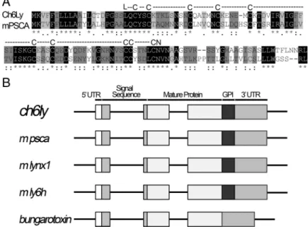
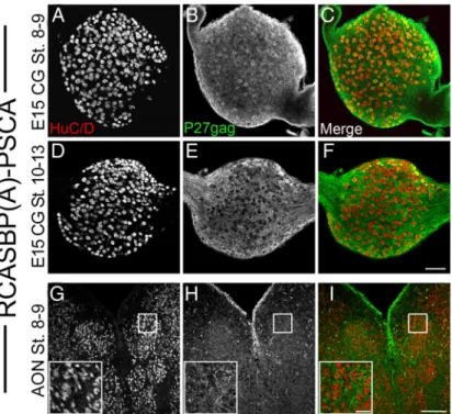
Development/Plasticity/Repair
Prostate Stem Cell Antigen Is an Endogenous lynx1-Like
Prototoxin That Antagonizes
␣7-Containing Nicotinic
Receptors and Prevents Programmed Cell Death of
Parasympathetic Neurons
Martin Hruska,
1Julie Keefe,
1David Wert,
1Ayse Begum Tekinay,
2Jonathan J. Hulce,
1Ines Iban˜ez-Tallon,
3and Rae Nishi
11Department of Anatomy and Neurobiology, University of Vermont College of Medicine, Burlington, Vermont 05405,2Laboratory of Molecular Biology, Howard Hughes Medical Institute, Rockefeller University, New York, New York 10021, and3Max-Delbru¨ck-Center, 13125 Berlin, Germany
Vertebrate
␣-bungarotoxin-like molecules of the Ly-6 superfamily have been implicated as balancers of activity and survival in the adult
nervous system. To determine whether a member of this family could be involved in the development of the avian ciliary ganglion, we
identified 6 Gallus genes by their homology in structure to mouse
lynx1 and lynx2. One of these genes, an ortholog of prostate stem cell
antigen (
psca), is barely detectable at embryonic day (E) 8, before neuronal cell loss in the ciliary ganglion, but increases
⬎100-fold as the
number of neurons begins to decline between E9 and E14. PSCA is highly expressed in chicken and mouse telencephalon and peripheral
ganglia and correlates with expression of
␣7-containing nicotinic acetylcholine receptors (␣7-nAChRs). Misexpressing PSCA before cell
death in the ciliary ganglion blocks
␣7-nAChR activation by nicotine and rescues the choroid subpopulation from dying. Thus, PSCA, a
molecule previously identified as a marker of prostate cancer, is a member of the Ly-6 neurotoxin-like family in the nervous system, and
is likely to play a role as a modulator of
␣7 signaling-induced cell death during development.
Introduction
Nicotinic signaling has been implicated in controlling
pro-grammed cell death during the development of the nervous
sys-tem. By far the most effective means of rescuing spinal cord
motor neurons from dying is to treat embryos with nicotinic
antagonists that block neuromuscular transmission such as
D-tubocurarine (dTC) or
␣-bungarotoxin (␣btx) (Pittman and
Oppenheim, 1979). In contrast, in the avian ciliary ganglion, up
to 90% of the neurons in the avian ciliary ganglion are rescued by
antagonists of
␣7-containing nicotinic acetylcholine receptors
(nAChRs) such as
␣btx and ␣-methyllycaconitine (MLA) (Bunker
and Nishi, 2002; Hruska et al., 2007); however, nonselective
nAChR antagonists that completely block ganglionic
transmis-sion exacerbate cell death (Wright, 1981; Meriney et al., 1987;
Maderdrut et al., 1988). In cell culture, nicotine promotes
sur-vival of ciliary ganglion neurons, but it does so in the absence of
trophic support (Pugh and Margiotta, 2000). Thus, the
contribu-tion of nicotinic signaling to neuronal survival during
develop-ment is complex.
Endogenous prototoxins that are homologous to snake
venom neurotoxins modulate signaling through nAChRs. These
molecules are members of the Ly6/neurotoxin (lynx) family
characterized by their cysteine-rich motif that predicts their
folding into the typical three-fingered loop structure of
␣-bungarotoxin (Miwa et al., 1999; Chimienti et al., 2003).
Mem-bers of this family that are GPI-linked and expressed in the
ner-vous system are lynx1, lynx2, Ly6H (Horie et al., 1998; Miwa et
al., 1999; Dessaud et al., 2006); others are secreted and expressed
in non-neuronal cells, e.g., SLURP-1 and SLURP-2 (Chimienti et
al., 2003; Tsuji et al., 2003). Mouse lynx1 binds to and alters the
kinetics of nAChRs (Iban
˜ez-Tallon et al., 2002). Furthermore,
cortical neurons of mice lacking lynx1 have an enhanced
sensi-tivity to nicotine that potentiates excitotoxicity, whereas aging
lynx1-null mice exhibit exacerbated neurodegeneration in the
brain that is enhanced by nicotine and attenuated by loss of
nAChRs (Miwa et al., 2006).
In the present study we determined whether the expression of
endogenous lynx-like molecules could play a role during the
de-velopment of the avian ciliary ganglion. The ganglion contains
two types of neurons, ciliary and choroid, and both receive
cho-linergic innervation that is detectable at embryonic day (E) 5.5
and complete by E8 (Landmesser and Pilar, 1972). Over half of
the ciliary and choroid neurons are lost between E8 and E14
(Landmesser and Pilar, 1974). By E8, all neurons express
homo-Received May 13, 2009; revised Oct. 1, 2009; accepted Oct. 9, 2009.This project was funded by Grant DA17784 from the National Institutes of Health to R.N. The real-time PCR and calcium imaging were made possible by the Neuroscience Centers of Biomedical Research Excellence (Grant 5 P20 RR016435). We are grateful to Dr. Nathanial Heintz at Rockefeller University for providing us with anti-mouse lynx1 antibodies that started this project and for his insights. We also thank Priscilla Kimberly and Sondra Sammut for excellent technical support.
A. B. Tekinay’s present address: UNAM, Institute of Materials Science and Nanotechnology, Bilkent University, 06800 Ankara, Turkey.
Correspondence should be addressed to Rae Nishi, Department of Anatomy and Neurobiology, HSRF 406, Uni-versity of Vermont College of Medicine, 149 Beaumont Avenue, Burlington, VT 05405. E-mail: rnishi@uvm.edu.
DOI:10.1523/JNEUROSCI.2271-09.2009
meric
␣7-nAChRs and heteromeric ␣3* nAChRs containing ␣3,
␣5, 4 subunits (Blumenthal et al., 1999; McNerney et al., 2000)
and sometimes
2 (Conroy and Berg, 1995). Using mouse
lynx1 and lynx2 we screened for homologous molecules in the
chicken and identified 6 genes. Of these, one is induced in the
ciliary ganglion during the period of cell loss and is the chicken
ortholog of human prostate stem cell antigen (psca). PSCA is
developmentally regulated and its expression reduces
␣7-nAChR-mediated increases in intracellular Ca
2⫹and promotes
survival of choroid neurons. We conclude that PSCA, a molecule
previously identified as upregulated in prostate cancer, is also a
lynx-like prototoxin molecule that functions in the developing
nervous system.
Materials and Methods
Identification of chicken lynx sequences. Mouse lynx1 and lynx2 protein
sequences were blasted against chicken TIGR gene indices (http://www. tigr.org/tdb/tgi/) and we chose 6 sequences that fit the following criteria: (1) having the Ly6 domain as the only functional domain; (2) matching the exon-intron structures of lynx1 and other prototoxins that have the Ly6 domain. The accession numbers of the sequences that we discovered are given in supplemental Table 1, available at www.jneurosci.org as supplemental material. Based on predicted sequences, sequence-specific primers were used to identify the expression profile of above lynx related transcripts in the ciliary ganglion at E8 and E15 (for primer sequences, see supplemental Table 1, available at www.jneurosci.org as supplemen-tal material).
RNA extraction and PSCA cloning. Tissues were isolated at the
indi-cated stages of development and rapidly frozen on dry ice. Total RNA was extracted by using TRI Reagent (Molecular Research Center). The cDNA was reverse-transcribed from 2g of total RNA by using oligo-dT and Superscript II reverse transcriptase (Invitrogen). The full-length PSCA was obtained by PCR using forward primer (5⬘ CCATGGTAATGAAG-GTTTTCTTCATCCTCC 3⬘) that attached an Nco1 restriction site to the 5⬘ end of a PSCA sequence and reverse primer (5⬘ GGATCCCTTCA-CAGTCTGTTGTTCAGG 3⬘) that added a BamH1 restriction site to the 3⬘ end of the PSCA sequence. The 369 base pair PCR product was cloned into Nco1 and BamH1 cloning sites on pSlax13Nco vector and se-quencing confirmed that it was the accurate, full-length chicken PSCA sequence.
Real-time quantitative PCR. Tissues were isolated at the indicated ages
and rapidly frozen on dry ice. Total RNA was extracted and cDNA was synthesized as described above. The expression profiles of␣7 and PSCA were assessed on the 7500 Fast Real-Time PCR System (Applied Biosys-tems) using TaqMan probes and primers designed using a website at Whitehead Institute at MIT http://frodo.wi.mit.edu/primer3/ (see sup-plemental Table 2, available at www.jneurosci.org as supsup-plemental ma-terial). Probes were labeled with 6 FAM reporter dye at their 5⬘ ends and black hole quencher (BHQ) at their 3⬘ ends. The constitutive gene con-trol used to normalize gene expression was chicken S17 ribosomal bind-ing protein (Chrps) (Darland et al., 1995). Relative transcript expression and number was determined using Sequence Detection Software (SDS) version 1.4. PSCA expression in mouse tissues was quantified and nor-malized to-actin using Assays on Demand from Applied Biosystems (PSCA: Mm00452908_m1;-actin: Mm00607939_s1).
Expression of PSCA with a viral vector. The PSCA sequence was cloned
into the Slax13NCO1 shuttle vector using 5⬘ Nco1 and 3⬘ BamH1 sites (Morgan and Fekete, 1996); the insert was removed from Slax13NCO1 by cutting with Cla1, and cloned into the avian retroviral vector RCASBP(A) (Federspiel and Hughes, 1997). Infective RCASBP(A)-PSCA viral particles were generated by transfecting DF-1 chicken fibro-blast cells with 800 ng of RCASBP(A)-PSCA plasmid using Mirus TransIT-LT transfection reagent. Conditioned media containing viral stocks collected from DF-1 cells were concentrated⬃20-fold by ultra-centrifugation at 90,000⫻ g at 4°C for 3 h (Morgan and Fekete, 1996). Concentrated stocks were titered by limiting dilution and infectivity of cells as measured by staining with p27gag antibody. Stocks with
concen-tration of⬎108infectious particles/ml were used for in vivo injection. Viral particles (60 –120 nl) were injected into the mesencephalic enlarge-ment of the neural tube of Hamburger/Hamilton stage (St.) 8 –9 or St. 10 –13 embryos using a Drummond Nanoject microinjector. The shells were sealed with a glass coverslip and sterile vacuum grease and incu-bated at 37°C to the desired stage.
Calcium imaging. Acutely isolated ciliary ganglion neurons were
loaded with Fura-2 AM (Invitrogen) dissolved in DMSO at final concen-tration of 5Mwith 2% Pluronic F-127 (Invitrogen). Neurons were loaded at room temperature for 30 min in the dark. Calcium signals were recorded by exposure to alternating wavelength (340 and 380 nm, 50 ms) generated by a Xenon light source and Lambda DG-4 ultra high-speed wavelengths switcher (Sutter Instruments). Fluorescent responses were recorded using an Orca-ER digital camera (Hamamatsu). Paired 340/380 ratio images were acquired at 4 s intervals with Metafluor 5.0r5 software (Molecular Devices). Drugs were dissolved in chicken physiological buffer (containing, in mM: 145 NaCl, 5.4 KCl, 0.8 MgCl2, 5.4 CaCl2, 5 glucose, 13 HEPES, pH 7.4). Voltage-gated sodium and calcium channels were blocked when nicotine was applied by supplementing the perfusion medium with 600 nMtetrodotoxin (Tocris Bioscience) and 200Mcobalt chloride (Sigma-Aldrich), respectively. Nicotine (Sigma-Aldrich) (10 M) was applied for 20 s to activate nAChRs.␣7-nAChRs were inhibited by perfusing the neurons with 50 nM␣-methyllycaconitine citrate hy-drate (MLA; Sigma-Aldrich) for 1 min or preincubating with 50 nM␣btx (Sigma-Aldrich) for 30 min at 25°C. On completion of these experi-ments, the extent of dye loading was determined by activating voltage-gated calcium channels with high potassium perfusion solution (25 mM KCl) in the absence of TTX or cobalt chloride. After the initial recordings were performed, background was subtracted from every image ac-quired and new ratios were calculated using the Metafluor 5.0r5 soft-ware. The ratios were then exported into the Microsoft Excel spreadsheet for calculations.
Tissues. Tissues were prepared for immunohistochemistry and in situ
hybridization as follows. Ciliary ganglia and brainstems from E14 em-bryos were harvested, fixed in Zamboni’s fixative for 48 h at 4°C, washed, then equilibrated in 30% sucrose at 4°C. Tissue was embedded in Mi-crom cryoembedding compound (Richard Allen Scientific), sectioned on a Microm HM 560 cryostat (Richard Allen Scientific) at 30m (for immunohistochemistry) and at 20m for in situ hybridization and col-lected on Superfrost Plus slides (Fisher Scientific). Sections were post-fixed in Zamboni’s vapors for 15 min at 37°C, submerged in Zamboni’s fixative for additional 15 min at 25°C, washed in PBS and blocked. Pri-mary antibodies were incubated overnight at 4°C and secondary antibod-ies were incubated 2 h at room temperature.
In situ hybridization. In situ hybridization was performed as described by the D. Anderson laboratory at Caltech (http://wmc.rodentia.com/docs/Big_ In_Situ.html) with the following modifications: sections collected on slides were treated with 100g/ml proteinase K (Sigma-Aldrich) for 10 min at room temperature; hybridization was performed with digoxigenin-labeled UTP (Roche)-labeled sense or antisense riboprobes synthesized using an RNA transcription kit (Ambion) from a full-length Gallus clone gcag0014c.j.08 corresponding to the cDNA we cloned, which was obtained from the French National Institute for Agricultural Research Animal Genomics Collection (http://www.international.inra.fr); and riboprobes were subjected to alkaline hydrolysis until average transcript sizes were 300 – 400 bp.
Primary antibodies. Mouse monoclonal 39.4D5 [Developmental
Stud-ies Hybridoma Bank (DSHB), University of Iowa, Iowa City, IA], which recognizes Islet-1 and Islet-2, transcription factors expressed in all ciliary ganglion neurons throughout development (Lee et al., 2001) at 1:100 dilution of the culture supernatant prepared in the Nishi Laboratory from hybridomas obtained from DSHB; mouse anti-Hu C/D (Invitrogen), which recognizes a neuron-specific RNA binding protein (Marusich and Weston, 1992; Lee et al., 2001) at 1:250 dilution of the culture supernatant; rabbit anti-p27gag (Charles River SPAFAS), which recognizes avian sarcoma gag p27 (Wang et al., 1976) at 1:1000; rat anti-somatostatin (product num-ber YMC1020, Accurate Chemical) diluted 1:100. Sheep anti-digoxigenin (Roche Applied Science) was used at 1:500 and visualized with goat anti-sheep coupled to alkaline phosphatase (1:1000; Roche).
Secondary antibodies. Biotinylated anti-mouse (Vector Laboratories)
at 1:250; biotinylated anti-rabbit (Vector Laboratories) at 1:250; goat anti-mouse Cy3 (Jackson ImmunoResearch) at 1:750; goat anti-rabbit Alexa 488 (Invitrogen) at 1:750; and goat anti-rat Cy3 (Jackson Immu-noResearch) at 1:750. Images of ciliary ganglia and midbrain were ac-quired with 20⫻ objective using Nikon C1 confocal scanner (Nikon Instruments) attached to a Nikon Eclipse E800 microscope (Micro Video Instruments).
Design-based stereology. Serially sectioned ciliary ganglia (cut at 30
m) were prepared for designed-based stereology as previously de-scribed (Lee et al., 2001; Bunker and Nishi, 2002). Islet-1/2-positive nu-clei (representing all neurons) together with somatostatin-positive cell bodies (representing all choroid neurons) were counted using the Opti-cal Fractionator Probe of Stereo Investigator (MBF Bioscience) in con-junction with a Nikon Optiphot 2 microscope with a Hitachi HVC20 camera, Heidenhahn focus encoder, and a motorized, computer-driven X–Z stage (all microscope attachments provided by MBF Biosciences). To avoid inaccuracies caused by cutting artifacts and double counting between adjacent sections, an upper guard of 4m and lower guard of 7 m were used (Bunker and Nishi, 2002). Spacing between sampling sites (grid size) was set such that 13–15 sampling sites were counted per section, which yielded 100 –300 objects per each ciliary ganglion. The
number of ciliary neurons was calculated by sub-tracting the number of somatostatin-positive neurons from the total number of neurons per ciliary ganglion (Bunker and Nishi, 2002).
Results
Identification of chicken
lynx-like sequences
Since mouse lynx1 interacts with
␣7-nAChRs from chicken (Iban
˜ez-Tallon et
al., 2002), we used the amino acid
se-quences of mouse lynx1 and lynx2 to
search a chicken expressed sequence tag
(EST) database for proteins that had the
Ly6 domain as the only functional
do-main and whose genes had similar
exon-intron structures to those of lynx1 and
other toxins that have the Ly6 domain. Six
sequences that matched these criteria were
identified (Fig. 1A). All 6 sequences are
cysteine-rich and 10 of the cysteine residues
are aligned across all molecules. The
se-quences also encode an N-terminal signal
sequence and a C-terminal consensus
sequence for addition of a
glycosyl-phosphatidylinositide linkage to the
membrane. When analyzed by SMART
(Simple Modular Architecture Research
Tool; http://smart.embl-heidelberg.de/),
all molecules are also predicted to fold
into a three-fingered coiled structure
sim-ilar to that of mouse lynx1 and the cobra
venom neurotoxins (Gumley et al., 1995).
This striking structural homology
sug-gests that these chicken molecules can
interact with nicotinic acetylcholine
re-ceptors as shown for mouse lynx1 (Iban
˜ez-Tallon et al., 2002).
To determine whether any of the 6 EST
transcripts were expressed in a
developmen-tally regulated pattern in the ciliary
gan-glion, we used sequence-specific primers
(supplemental Table 1, available at www.
jneurosci.org as supplemental material)
to amplify cDNA from ciliary ganglia collected at E8, while cell
death is occurring, and E14, at which time cell death has ceased
(Lee et al., 2001). Three sequences, ch3Ly, ch5Ly and ch6Ly were
expressed in E14 ganglia; however, ch3Ly and ch5Ly were also
expressed at E8 (Fig. 1 B). In contrast, no expression of ch6Ly is
detectable at E8 (Fig. 1 B). Identity of the amplified gene product
as ch6Ly was confirmed by sequencing. The remaining
tran-scripts are not expressed in the embryonic ciliary ganglion (data
not shown). Ch6Ly encodes a pro-protein of 122 aa, which
cor-responds to a molecular weight of 11,160 Da. The mature
GPI-linked protein is 8051 Da. When ch6Ly is used to search Entrez, it
matches mouse prostate stem cell antigen (psca), with which it
shares 40% amino acid identity and 80% homology (Fig. 2 A).
Like ch6Ly, the open reading frame of mouse psca is similar to
other Ly6 superfamily members in the alignment of cysteine
groups and contains the Ly6 domain. In addition, the genomic
structure of mouse psca is identical to that of ch6Ly, mouse lynx1,
mouse ly6 h, and
␣-bungarotoxin, with conserved intron/exon
breaks (Fig. 2 B). Thus, ch6Ly is likely to be the chicken ortholog
Figure 1. Molecules with homology to the Ly6/Neurotoxin-like superfamily in chicken ciliary ganglion. A, Amino acid se-quences of 6 mature chicken proteins identified by their homology to mouse lynx1 and lynx2 are shown aligned to mouse lynx1. All sequences contain the Ly6 functional domain (in bold at the bottom). Boxed residues are amino acids that are identical to those in mouse lynx1. B, Sequence-specific primers to each of the 6 chicken sequences were used to amplify cDNA from E8 and E14 ciliary ganglia. Three sequences are expressed in the ciliary ganglion. The ch3Ly and ch5Ly are present at E8 and E14; however, ch6Ly is only expressed at E14. Chrps is a constitutive gene control.
of prostate stem cell antigen (psca), which
is a Ly6 family member whose expression
becomes upregulated in prostate tumors
(Reiter et al., 1998).
Expression pattern of chicken PSCA
To determine the specificity of chicken
PSCA expression, we quantified
tran-scripts in a variety of tissues in E14
chicken embryos using real-time PCR and
normalized these levels to chicken
ribo-somal binding protein s17 (CHRPS), a
constitutively expressed housekeeping
gene that is abundant in all tissues.
Ex-pression of PSCA in selected tissues was
determined relative to that of heart, which
contained barely detectable levels (Fig.
3A). Levels of PSCA transcripts in pectoral
muscle, liver, ovary and testes (data not
shown) are comparable to that of heart. In
the nervous system, the cerebellum also
has very low levels; however, the
telen-cephalon has fivefold greater levels of
PSCA mRNA than cerebellum. The
high-est levels of PSCA occur in autonomic
ganglia: the ciliary ganglion, which is
para-sympathetic, contains
⬎20-times that of
heart, and paravertebral lumbar
sympa-thetic ganglia contain 10-times more
PSCA. Dorsal root ganglia also express
higher levels of PSCA mRNA than heart,
but considerably less than sympathetic or
ciliary ganglia.
Since our observation that PSCA is
en-riched in telencephalon was contrary to
the original report that PSCA could not be
detected in northern blots of brain mRNA
(Reiter et al., 1998), we determined whether
adult mouse tissues exhibit an expression
pattern of PSCA mRNA similar to that of
chicken embryos. We isolated neural and
non-neural tissues and quantified relative
abundance of mouse PSCA normalized to
-actin using real-time PCR, which is
more sensitive than the northern blot
ap-proach previously used. As in late stage
chicken embryos, the nervous tissues
iso-lated from adult mice contain significantly
higher levels of PSCA than non-neural
tis-sues, with the highest levels found in the
superior cervical ganglion, which is
sym-pathetic (Fig. 3B). In fact, all the tissues
that exhibit high levels of PSCA in the
chicken embryo also express high levels of
␣7-nAChRs (Fig. 3C). Thus, PSCA is
ex-pressed at very high levels in neural tissues
both in mammals and avian species, and
its level of expression correlates with the
expression of the
␣7-nAChR subunit (Fig.
3C,D), suggesting a relationship between
PSCA and
␣7 signaling.
In ciliary ganglia, which express some
of the highest levels of
␣7 nAChRs per
Figure 2. Ch6Ly is psca. A, The amino acid sequence of ch6Ly was used to search NCBI and Ensembl databases and found to have
the most significant match to mouse psca. The two sequences share 80% homology and 40% identity. Black highlight indicates positions with single, fully conserved residues. Dark gray highlight indicates that the strong amino acid groups are fully conserved. Light gray highlight represents the fully conserved weak amino acid groups. Both molecules contain the cysteine-rich Ly6 domain (shown above the amino acid sequence) with conserved N-terminal leucine and C-terminal asparagine. B, Representation of the open reading frame of the genes encoding ch6Ly and mouse psca compared with the other members of Ly6/Neurotoxin super-family such as lynx1, ly6 h,␣-bungarotoxin. The ch6ly and psca have the same intron/exon breaks as the other members of the superfamily. The 5⬘ UTR sequence, signal sequence, mature protein containing Ly6 domain, hydrophobic GPI anchor and 3⬘UTR sequence are also conserved between ch6ly, psca and other members of the family.
Figure 3. PSCA transcripts are highly expressed in nervous tissues and correlate with␣7nAChRexpression.A,Avarietyofneuraland non-neural tissues were dissected from E14 chicken embryos and mRNA encoding chicken PSCA were quantified using quantitative real-timePCRandnormalizedtoChrps.PSCAexpressionwashighlyexpressedinneuraltissues,particularlyinperipheralautonomicganglia(CG: ciliary ganglia; symp. g: sympathetic ganglia). PSCA is 20 times more abundant in ciliary ganglion than in non-neural tissues. B, Similar pattern of PSCA expression is observed in tissues isolated from adult mouse. Neural tissues and peripheral ganglia contain 10 –20-fold higher levels of PSCA than non-neural tissues. C,␣7 subunit of nAChR expression correlates with PSCA. By far, the highest levels of ␣7 expression are in ciliary and sympathetic ganglia. D, The levels of␣7 nAChR increase as PSCA expression increases, with a correlation coefficient of 0.8. Values represent mean⫾SEMfromatleast3separateexperiments.
neuron,
␣7-nAChRs have previously been implicated in inducing
cell death (Bunker and Nishi, 2002; Hruska et al., 2007). If PSCA
attenuates cell death by modulating activation of
␣7
subunit-containing nAChRs, then its expression should be low when cell
death commences, and it should be upregulated as neurons in the
ganglion extend axons to the periphery and initiate
synaptogen-esis. Accordingly, we used real-time PCR to quantify PSCA
tran-scripts in ciliary ganglia isolated from E8 to E14 (Fig. 4 A), which
corresponds to a period when half the neurons are lost due to cell
death. PSCA transcripts in ciliary ganglia are barely detectable at
E8. However, midway through the cell loss period at E10 –12,
PSCA levels increase tenfold. This increase can be attenuated by
chronic application of
␣btx at a concentration (20 g/d) known
to rescue neurons while the
␣7 nAChR transcript levels double, as
might be expected if fewer neurons were dying (Table 1) (Bunker
and Nishi, 2002). By E14, PSCA expression reaches highest levels,
when it is 15 times more abundant than at E8 (Fig. 4 A). After E14,
which marks the end of cell loss in the
ciliary ganglion, there is no further
in-crease in PSCA transcript levels (data not
shown). At E14, PSCA transcripts are
de-tected in many neurons, but the relative
levels of expression vary from low (Fig.
4 B, arrows) to very high (Fig. 4 B,
arrow-heads). Little or no signal is observed with
the sense probe (Fig. 4C). Thus, PSCA
mRNA correlates with the period of cell
loss in the ciliary ganglion and is found in
neurons, consistent with a possible role in
modulating nicotinic signaling.
PSCA blocks activation of
␣7-nAChRs
and rescues neurons from cell death
To test whether PSCA could modify
nicotine-induced responses in ciliary
gan-glion neurons, we used the retroviral vector
RCASBP(A) to express PSCA at E8, an age at
which PSCA mRNA is almost undetectable.
We removed PSCA-expressing ciliary
gan-glia and vector-only infected controls at E8,
plated the neurons on coverslips, and
loaded them with the calcium-sensitive dye,
Fura-2, to quantify intracellular calcium in
response to rapidly perfused nicotine. To
isolate nicotinic Ca
2⫹responses from Ca
2⫹influx due to the opening of voltage-gated
ion channels, tetrodotoxin (TTX) and
co-balt were added to the perfusion solution
(see Materials and Methods).
In controls, nicotine induces a large
in-crease in intracellular calcium, which is
partially blocked by the
␣7
subunit-specific nicotinic cholinergic antagonist, MLA (Fig. 5A). The
re-maining response is blocked by dihydro
-erythroidin, an
antagonist of
␣3* nAChRs (data not shown). When neurons
in-fected with RCASBP(A)-PSCA are compared with those inin-fected
with open-RCASBP(A), there is no difference in the calcium
sig-nal induced by depolarization with 25 m
MKCl in the absence of
TTX and Co
2⫹(Open
⫽ 0.52 ⫹ 0.04 SEM; n ⫽ 30; PSCA ⫽ 0.59 ⫹
0.06 SEM, n
⫽ 26), indicating equal loading of Fura-2 and good
health of PSCA infected neurons. In neurons overexpressing
PSCA, the mean nicotine-induced calcium response is reduced
by 43% when compared with open vector-infected cells (Fig. 5B;
p
⬍ 0.001, one-way ANOVA, Bonferroni post test; open: 0.3 ⫾
0.02 SEM, n
⫽ 69; PSCA: 0.17 ⫾ 0.01 SEM, n ⫽ 86). This
con-trasts with the 69% reduction observed when neurons are
in-fected with a tethered
␣btx using the same vector and otherwise
identical assay conditions (Hruska et al., 2007). The mean
re-sponse of the PSCA-expressing neurons cannot be significantly
lowered by the addition of a solution containing the
␣7-nAChR-specific antagonist,
␣btx (Fig. 5B). Inclusion of ␣btx to
open-RCASBP(A) infected neuronal cultures reduces the amplitude of
Ca
2⫹transients to the levels that are comparable to the PSCA
infected neurons (Fig. 5B; p
⬍ 0.001, one-way ANOVA,
Bonfer-roni post test: open: 0.3
⫾ 0.02, n ⫽ 69, open⫹btx: 0.14 ⫾ 0.02,
n
⫽ 31). Thus, retrovirally delivered PSCA expressed at E8 in
ciliary ganglion neurons appears to primarily act to suppress
ac-tivation of
␣7-nAChRs, but this suppression is less than that
observed for tethered
␣btx, suggesting that the PSCA affects a
Figure 4. PSCA is expressed in neurons of the ciliary ganglion. A, Between E8 and E14, as the number of ciliary ganglion neurons is reduced by 50% [gray curve based on data from our previous work (Lee et al., 2001)], PSCA expression increases 15-fold (black line). B, In situ hybridization was performed on sections from E15 ciliary ganglia using digoxigenin-labeled antisense riboprobe for chicken PSCA. Staining seems particularly intense in some small neurons on the periphery of the ganglion that may be choroid neurons (arrowheads, inset); however other larger neurons are also stained. PSCA staining is less intense in other neurons (arrows, inset). C, Sense probe showing only background staining. AU, Arbitrary units. Calibration bar⫽ 100m, inset ⫽ 20 m.
Table 1. Chronic application of␣btx reduces upregulation of PSCA in the developing ciliary ganglion
Target gene Saline ␣-Bungarotoxin Change
PSCA 171⫾ 17.7 107⫾ 8.2* 63%
␣7 184⫾ 41.7 362⫾ 10.4* 200%
Chicken embryos were windowed at E3; then, 50g per day of ␣btx dissolved in sterile saline or saline alone was applied in 50l aliquots daily from E7 to E10. Ganglia were harvested at E11 and total RNA was extracted and reverse transcribed. Target gene expression was quantified in triplicate with quantitative real-time PCR using TaqMan probes. Values given are the mean⫾ SE of RNA isolated from three independent groups of experiments, pooling 14 –16 ganglia from each treatment. *p⬍ 0.01, one-way ANOVA with Tukey’s post hoc test.
subpopulation of neurons or a subpopulation of the nicotinic
responses.
Since signaling through
␣7-nAChRs affects the final number
of neurons in the ciliary ganglion, we determined whether the
premature expression of PSCA caused a change in neuronal
survival. We quantified the total number of neurons (using
anti-Islet-1) as well as the number of choroid neurons (using
anti-somatostatin) at E14 by using design-based stereology (Lee
et al., 2001; Bunker and Nishi, 2002). If infective
RCASBP(A)-PSCA particles are injected at St. 8 –9 (36 h of incubation),
virtu-ally all of the ciliary ganglion neurons are infected (Fig. 6 A–C);
however, if embryos are injected at St. 10 –13 (48 h of
develop-ment), very few neurons are infected, whereas many surrounding
non-neuronal cells are infected (Fig. 6 D–F ). Little or no infection
is detected in the accessory oculomotor nucleus, which
inner-vates the ciliary ganglion (Fig. 6G–I ). PSCA expressing ganglia
infected at 36 h of development contained 35% more choroid
neurons than ganglia infected with open-RCASBP(A) (Fig. 7; p
⬍
0.0001, one-way ANOVA; open: 4679
⫾ 353.4, n ⫽ 16; PSCA at
36 h: 7125
⫾ 355.1, n ⫽ 13) and this was reflected in a significant
change in the total number of neurons in the ganglion ( p
⬍ 0.001
one-way ANOVA; open: 8626
⫾ 418.8, n ⫽ 16; PSCA at 36 h:
11,173
⫾ 467, n ⫽ 13). This indicates that the change in the
number of neurons expressing somatostatin-like
immunoreac-tivity was not merely due to a shift in neuropeptide expression.
Interestingly, the number of ciliary neurons remained unchanged
(Fig. 7), suggesting that choroid neurons are more sensitive to the
expression of PSCA. When RCASBP(A)-PSCA retrovirus is
in-jected into the neural tube at St. 10 –13, then PSCA fails to rescue
neurons from dying (Fig. 7; PSCA at 48 h: 9352
⫾ 646.1, n ⫽ 9),
despite the fact that neighboring glia are infected and overexpress
PSCA.
Discussion
The principal finding of this study is that PSCA, a molecule
orig-inally identified as an antigen upregulated in prostate cancer, is
an endogenous prototoxin highly expressed in the nervous
sys-tem that attenuates signaling through
␣7 subunit-containing
nAChRs. PSCA levels correlate with the expression of transcripts
encoding
␣7-nAChRs. In the avian ciliary ganglion, PSCA
ex-pression is induced and upregulated between E8 and E14, a time
during which half of the neurons are lost by cell death and peripheral
synaptogenesis is completed. Furthermore, forcing premature
ex-pression of PSCA blocks
␣7 nAChR activation and prevents choroid
neurons from dying. These studies uncover possible modulatory
functions for prototoxins during development.
PSCA belongs to the Ly6 superfamily, whose members possess
cysteine rich molecules expressed in tissue-specific patterns
dur-ing development and in the adult (Gumley et al., 1995). To date,
the function of such molecules has been mysterious because their
small size and lack of a transmembrane domain preclude a direct
role in mediating cell signaling. Within this family, molecules
that fold into a structure homologous to those formed by
cobra-toxins (Tsetlin, 1999), which bind with high affinity to specific
subclasses of nAChRs, have been described as prototoxins and
include mouse lynx1 (Miwa et al., 1999), mouse lynx2 (Dessaud
et al., 2006), Ly6H (Horie et al., 1998) and SLURP-1 (Chimienti
et al., 2003). These endogenous prototoxins are expressed in the
CNS and the PNS, and likely act as molecules that interact cell
autonomously to modulate nicotinic receptor function in vivo.
Indeed, lynx1 and lynx2 coprecipitate with
␣4/2 as well as ␣7
subunit-containing nAChRs in mouse CNS and this association
alters nAChR kinetics, extent of receptor desensitization and
ag-onist affinity (Miwa et al., 1999; Iban
˜ez-Tallon et al., 2002; Tekinay et
al., 2009). Furthermore, neurons from lynx1-null mice exhibit
large increases in [Ca
2⫹]
iin response to nicotine and, as a result,
display age-dependent degeneration that is exacerbated by
nico-tine and ameliorated by null mutations in nAChRs (Miwa et al.,
2006), and lynx2-null mice have altered responses to fear
condi-tioning (Tekinay et al., 2009). Our studies using
retroviral-mediated expression of avian PSCA in chicken neurons indicate
that PSCA exhibits many of the features of a prototoxin: (1) it is
cysteine-rich, with a spacing of cysteine residues that is conserved
with other members of the family; (2) it is highly expressed in
tissues of the nervous system that contain high levels of
␣7-nAChRs and these data are corroborated by in situ hybridization
from mouse brain, where PSCA is detected in Purkinje and
gran-ule cell layers of cerebellum, cerebral cortex and hippocampus
(http://www.brain-map.org/mouse/brain/psca.html); (3) it is
predicted to form a “three-fingered” tertiary structure similar to
that of nicotinic antagonists derived from cobratoxin; and (4) it
interferes with nicotine-induced increases in [Ca
2⫹]
ithrough
Figure 5. Overexpression of PSCA at E8 blocks nicotine-induced calcium influx via ␣7-nAChRs. Ciliary ganglia from embryos infected with RCASBP(A)-PSCA or RCASBP(A) open (with-out an insert) at St. 8 –9 were isolated at E8, dissociated, and loaded with Fura-2, 2 h after plating. Nicotine (10M) was used to stimulate nAChRs in the presence TTX and cobalt, which block voltage activated influx of calcium. A, Brief application of nicotine induces rapid increase in [Ca2⫹]i. The␣7-specific antagonist MLA blocks 50% of this nicotine-induced response. The third response is the total calcium signal observed in response to perfusion with 25 mMKCl in the absence of TTX and Co2⫹. The mean peak Ca2⫹responses due to depolarization with 25 mMKCl between open and PSCA infected ciliary ganglion neurons are not significantly different, indi-cating the all neurons are equally loaded with Fura-2. B, Plot of all the peak nicotine-induced calcium responses observed in PSCA versus open virus infected neurons. Each gray spot repre-sents the response of a single neuron. Graph reprerepre-sents the sum of data from 3 independent experiments using viral injections together with calcium imaging of E8 neurons. PSCA infected neurons exhibit a significantly smaller mean nicotine-induced increase in [Ca2⫹]icompared with open-infected neurons. Bars indicate mean plus or minus SD ( p⬍ 0.001; open: 0.3 ⫾ 0.02, n⫽69;PSCA:0.17⫾0.01,n⫽86).Additionofexogenous␣btx(50nM) to PSCA infected neurons does not cause additional reduction in the mean nicotine-induced Ca2⫹whereas addition of␣btx on open infected neurons significantly reduces increases in [Ca2⫹]i( p⬍ 0.001; open: 0.3⫾ 0.02, n ⫽ 69; open⫹btx: 0.14 ⫾ 0.02 n ⫽ 31), whereas open with PSCA is not significantly different from open⫹btx or PSCA ⫹btx; one-way ANOVA with Bonferroni
␣7-nAChRs, but not heteromeric nAChRs. These results suggest
that many other members of the Ly6 superfamily may also serve
as prototoxins with selectivity for specific classes of nAChRs.
Nicotinic signaling has long been known to play an important
role in regulating programmed cell death during development.
Blocking neuromuscular transmission with nicotinic antagonists
such as
D-tubocurarine or
␣-bungarotoxin is one of the most
effective ways to rescue spinal cord motor neurons from dying
(Pittman and Oppenheim, 1978, 1979). In the autonomic and
some parts of the CNS, activation of neuronal nAChRs can
di-rectly induce apoptosis. For example, chronic blockade of
␣7-nAChRs with systemically applied
␣btx or MLA prevents cell
death of nearly all ciliary ganglion
neu-rons (Meriney et al., 1987; Bunker and
Nishi, 2002), although the same is not true
for spinal cord motor neurons (Oppenheim
et al., 2000). In addition, reduction in
Ca
2⫹influx through
␣7-nAChRs in a
cell-autonomous manner prevents ciliary and
choroid neurons from dying, suggesting
that large
␣7-nAChR-mediated increases
in [Ca
2⫹]
ipromote cell death of ciliary
ganglion neurons during development
(Hruska et al., 2007). Immature neurons
are especially vulnerable to Ca
2⫹influx via
␣7-nAChRs; activation of these channels
induces apoptosis of hippocampal
progeni-tor cells but not differentiated hippocampal
neurons (Berger et al., 1998). Since the
acti-vation of
␣7-nAChRs can be proapoptotic
in certain neuronal populations, the
signal-ing through these channels must be
pre-cisely regulated.
Our data are consistent with PSCA
act-ing as a modulator of nicotinic signalact-ing
in the ciliary ganglion that limits cell death
caused by activation of
␣7-nAChRs in
neurons vulnerable to calcium overload.
At E8, when PSCA is undetectable, many
ciliary ganglion neurons are undergoing
apoptotic cell death (Lee, 2001).
Subse-quent upregulation of PSCA correlates
with a significant decrease in cell death,
reflected in the stabilization of neuronal cell number.
Overex-pression of PSCA at E8, when it is not normally expressed, blocks
nicotine-induced increases in intracellular calcium through
␣7-nAChRs and prevents cell death of choroid but not ciliary
neu-rons. Finally, blocking all
␣7 signaling with ␣btx attenuates the
developmental increase seen in PSCA, consistent with calcium
influx upregulating PSCA as part of a negative feedback loop.
The selective effect of PSCA on the choroid neuron
subpopu-lation contrasts to that of membrane-tethered
␣btx, which also
blocks nicotine-induced increases in [Ca
2⫹]
ivia
␣7-nAChRs
(Hruska et al., 2007), but rescues both ciliary and choroid
sub-populations. One possible explanation is that tethered
␣btx
blocks a much higher percentage of the mean nicotine-induced
response when compared with PSCA (69% versus 43%) (Hruska
et al., 2007). One likely difference between
␣btx and PSCA may
be the dissociation constants when bound to
␣7-nAChRs. The
dissociation constant of
␣btx is so high that it is virtually
irrevers-ible, whereas this is unlikely to be the case for PSCA. Thus, PSCA
may have a differential affinity for choroid neuron nAChRs, or
ciliary neuron nAChRs may already be occupied by a prototoxin
that does not block calcium influx but cannot be displaced by
PSCA. This may be likely because we find two other
prototoxin-like molecules, ch3Ly and ch5Ly, in the ciliary ganglion. This
differential susceptibility of choroid neurons to PSCA warrants
further investigation, but is difficult to pursue because
extracel-lular markers that distinguish ciliary from choroid neurons in
acutely dissociated, live preparations have yet to be
discov-ered. Thus, ciliary versus choroid neurons cannot be definitively
identified for electrophysiological studies and neither can they be
conveniently sorted for biochemical binding or molecular
stud-ies. In addition, the measurement of the binding and dissociation
Figure 6. Timing of viral injection directs PSCA gene expression to ciliary ganglion neurons but not preganglionic oculomotor neurons. Embryos were injected with RCASBP(A)-PSCA at 36 h of development (St. 8 –9; A–C) and 48 h of development (St. 10 –13;
D–F ). A–C, p27 gag immunoreactivity (green) is observed in⬎80% of neurons (red) in ciliary ganglia from embryos infected at
St. 8 –9. D–F, Only surrounding non-neural tissue exhibits p27gag immunoreactivity in the ciliary ganglia from embryos infected at St. 10 –13. Calibration bar⫽ 100m. G, H, Neurons in the preganglionic AON from the embryos injected at St. 8–9 are not infected and only surrounding glia exhibits p27gag immunoreactivity. Calibration bar⫽ 250m (inset ⫽ 50 m).
Figure 7. Premature expression of chPSCA at E8 rescues choroid neurons from dying. Serially sectioned E14 ciliary ganglia infected with RCASBP(A)-PSCA at St. 8 –9 (36 h of incubation) or St. 10 –13(48hofincubation)werelabeledwithIslet-1antibodytostainalltheneuronsandsomatosta-tin antibody to label choroid neurons. Number of surviving neurons was determined using design-based stereology. Ciliary ganglia infected at St. 8 –9 have significantly more choroid neurons ( p⬍0.001;open:4679⫾353.4,n⫽16;PSCAat36h:7125⫾355.1,n⫽13)comparedwiththe open-infected ganglia. The number of ciliary neurons is the same in open and RCASBP(A)-PSCA in-fected ciliary ganglia. The total number of neurons is also significantly greater in RCASPB(A)-PSCA ganglia infected at St. 8 –9 ( p⬍0.002).InfectionatSt.10–13doesnotpreventcelldeathofciliary or choroid neurons. Values represent mean⫾ SEM of three or more separate experiments. ANOVA with Tukey’s multiple-comparison post hoc test was used to analyze the results.
constants of membrane-tethered molecules to integral
mem-brane proteins is highly challenging.
The relationship of PSCA expressed in the prostate to its
ex-pression in the nervous system is not clear. PSCA was first
iden-tified as an antigen enriched in basal cells of the prostate
epithelium that is upregulated in high grade, metastatic prostate
tumors (Reiter et al., 1998), but undetectable in northern blots of
whole human brain. In contrast, when using the more sensitive
quantitative PCR on dissected neural tissues, we see significantly
higher levels of PSCA than in peripheral organs. Currently, it is
unknown whether PSCA modulates
␣7-nAChR signaling in the
prostate or whether it has a completely unrelated function.
How-ever, many non-neural cells, such as keratinocytes, lymphocytes,
endothelial cells and glia, express
␣7-nAChRs (for review, see
Sharma and Vijayaraghavan, 2002). In this context, SLURP1 and
2 of the Ly6, lynx superfamily, regulate apoptosis in keratinocytes
(Chimienti et al., 2003; Arredondo et al., 2006, 2007). Thus, It is
possible that
␣7-nAChR signaling also regulates cell proliferation
in the prostate. Therefore, uncovering the neural and non-neural
functions of nAChRs and their accompanying prototoxin
mod-ulators will be key for understanding the importance of nicotinic
signaling in normal physiology and disease.
References
Arredondo J, Chernyavsky AI, Jolkovsky DL, Webber RJ, Grando SA (2006) SLURP-2: a novel cholinergic signaling peptide in human mucocutane-ous epithelium. J Cell Physiol 208:238 –245.
Arredondo J, Chernyavsky AI, Grando SA (2007) SLURP-1 and -2 in normal, immortalized and malignant oral keratinocytes. Life Sci 80:2243–2247. Berger F, Gage FH, Vijayaraghavan S (1998) Nicotinic receptor-induced
apo-ptotic cell death of hippocampal progenitor cells. J Neurosci 18:6871– 6881. Blumenthal EM, Shoop RD, Berg DK (1999) Developmental changes in the
nicotinic responses of ciliary ganglion neurons. J Neurophysiol 81:111–120. Bunker GL, Nishi R (2002) Developmental cell death in vivo: rescue of neurons
independently of changes at target tissues. J Comp Neurol 452:80 –92. Chimienti F, Hogg RC, Plantard L, Lehmann C, Brakch N, Fischer J, Huber
M, Bertrand D, Hohl D (2003) Identification of SLURP-1 as an epider-mal neuromodulator explains the clinical phenotype of Mal de Meleda. Hum Mol Genet 12:3017–3024.
Conroy WG, Berg DK (1995) Neurons can maintain multiple classes of nic-otinic acetylcholine receptors distinguished by different subunit compo-sitions. J Biol Chem 270:4424 – 4431.
Darland DC, Link BA, Nishi R (1995) Activin A and follistatin expression in developing targets of ciliary ganglion neurons suggests a role in regulating neurotransmitter phenotype. Neuron 15:857– 866.
Dessaud E, Salau¨n D, Gayet O, Chabbert M, deLapeyrie`re O (2006) Identi-fication of lynx2, a novel member of the ly-6/neurotoxin superfamily, expressed in neuronal subpopulations during mouse development. Mol Cell Neurosci 31:232–242.
Federspiel MJ, Hughes SH (1997) Retroviral gene delivery. Methods Cell Biol 52:179 –214.
Gumley TP, McKenzie IF, Sandrin MS (1995) Tissue expression, structure and function of the murine Ly-6 family of molecules. Immunol Cell Biol 73:277–296.
Horie M, Okutomi K, Taniguchi Y, Ohbuchi Y, Suzuki M, Takahashi E (1998) Isolation and characterization of a new member of the human Ly6 gene family (LY6H). Genomics 53:365–368.
Hruska M, Ibañez-Tallon I, Nishi R (2007) Cell-autonomous inhibition of al-pha 7-containing nicotinic acetylcholine receptors prevents death of para-sympathetic neurons during development. J Neurosci 27:11501–11509. Iban˜ez-Tallon I, Miwa JM, Wang HL, Adams NC, Crabtree GW, Sine SM,
Heintz N (2002) Novel modulation of neuronal nicotinic acetylcholine
receptors by association with the endogenous prototoxin lynx1. Neuron 33:893–903.
Landmesser L, Pilar G (1972) The onset and development of transmission in the chick ciliary ganglion. J Physiol 222:691–713.
Landmesser L, Pilar G (1974) Synaptic transmission and cell death during normal ganglionic development. J Physiol 241:737–749.
Lee VM (2001) Regulation of neuronal cell number in the developing ciliary ganglion. In: Cell and developmental biology, p 137. Portland: Oregon Health Sciences University.
Lee VM, Smiley GG, Nishi R (2001) Cell death and neuronal replacement during formation of the avian ciliary ganglion. Dev Biol 233:437– 448. Maderdrut JL, Oppenheim RW, Prevette D (1988) Enhancement of
natu-rally occurring cell death in the sympathetic and parasympathetic ganglia of the chicken embryo following blockade of ganglionic transmission. Brain Res 444:189 –194.
Marusich MF, Weston JA (1992) Identification of early neurogenic cells in the neural crest lineage. Dev Biol 149:295–306.
McNerney ME, Pardi D, Pugh PC, Nai Q, Margiotta JF (2000) Expression and channel properties of alpha-bungarotoxin-sensitive acetylcholine receptors on chick ciliary and choroid neurons. J Neurophysiol 84:1314 –1329. Meriney SD, Pilar G, Ogawa M, Nunez R (1987) Differential neuronal
sur-vival in the avian ciliary ganglion after chronic acetylcholine receptor blockade. J Neurosci 7:3840 –3849.
Miwa JM, Ibanez-Tallon I, Crabtree GW, Sa´nchez R, Sali A, Role LW, Heintz N (1999) lynx1, an endogenous toxin-like modulator of nicotinic ace-tylcholine receptors in the mammalian CNS. Neuron 23:105–114. Miwa JM, Stevens TR, King SL, Caldarone BJ, Ibanez-Tallon I, Xiao C,
Fitzsimonds RM, Pavlides C, Lester HA, Picciotto MR, Heintz N (2006) The prototoxin lynx1 acts on nicotinic acetylcholine receptors to balance neuronal activity and survival in vivo. Neuron 51:587– 600.
Morgan BA, Fekete DM (1996) Manipulating gene expression with replication-competent retroviruses. Methods Cell Biol 51:185–218. Oppenheim RW, Prevette D, D’Costa A, Wang S, Houenou LJ, McIntosh JM
(2000) Reduction of neuromuscular activity is required for the rescue of motoneurons from naturally occurring cell death by nicotinic-blocking agents. J Neurosci 20:6117– 6124.
Pittman R, Oppenheim RW (1979) Cell death of motoneurons in the chick embryo spinal cord. IV. Evidence that a functional neuromuscular inter-action is involved in the regulation of naturally occurring cell death and the stabilization of synapses. J Comp Neurol 187:425– 446.
Pittman RH, Oppenheim RW (1978) Neuromuscular blockade increases motoneurone survival during normal cell death in the chick embryo. Nature 271:364 –366.
Pugh PC, Margiotta JF (2000) Nicotinic acetylcholine receptor agonists promote survival and reduce apoptosis of chick ciliary ganglion neurons. Mol Cell Neurosci 15:113–122.
Reiter RE, Gu Z, Watabe T, Thomas G, Szigeti K, Davis E, Wahl M, Nisitani S, Yamashiro J, Le Beau MM, Loda M, Witte ON (1998) Prostate stem cell antigen: a cell surface marker overexpressed in prostate cancer. Proc Natl Acad Sci U S A 95:1735–1740.
Sharma G, Vijayaraghavan S (2002) Nicotinic receptor signaling in nonex-citable cells. J Neurobiol 53:524 –534.
Tekinay AB, Nong Y, Miwa JM, Lieberam I, Ibanez-Tallon I, Greengard P, Heintz N (2009) A role for LYNX2 in anxiety-related behavior. Proc Natl Acad Sci U S A 106:4477– 4482.
Tsetlin V (1999) Snake venom alpha-neurotoxins and other ‘three-finger’ proteins. Eur J Biochem 264:281–286.
Tsuji H, Okamoto K, Matsuzaka Y, Iizuka H, Tamiya G, Inoko H (2003) SLURP-2, a novel member of the human Ly-6 superfamily that is up-regulated in psoriasis vulgaris. Genomics 81:26 –33.
Wang L, Galehouse D, Mellon P, Duesberg P, Mason WS, Vogt PK (1976) Mapping oligonucleotides of Rous sarcoma virus RNA that segregate with polymerase and group-specific antigen markers in recombinants. Proc Natl Acad Sci U S A 73:3952–3956.
Wright L (1981) Cell survival in chick embryo ciliary ganglion is reduced by chronic ganglionic blockade. Brain Res 227:283–286.