DOI: 10.1142/S0129183110015026 Vol. 21, No. 1 (2010) 97–106
c
World Scientific Publishing Company
DIPEPTIDE ADSORPTION ON Si(100)-2 × 1 ASYMMETRIC SURFACE BY FIRST PRINCIPLES
ETHEM AKT ¨URK
UNAM-Institute of Material Science and Nanotechnology Bilkent University, Bilkent, Ankara, Turkey
O ˘GUZ G ¨ULSEREN
Department of Physics, Bilkent University Bilkent, Ankara, Turkey
HANDAN ARKIN
Department of Physics Engineering, Ankara University Tandogan, Ankara, Turkey
TARIK C¸ ELIK Turkish Academy of Sciences Piyade Sok. No. 27, C¸ ankaya, Ankara, Turkey
[email protected] Received 26 June 2009 Accepted 23 October 2009
The adsorption of alanine dipeptide on a Si(100)-2 × 1 asymmetric surface is studied by using pseudopotential plane wave approach based on Density Functional Theory (DFT). Adsorption energies for different surface sites of various conformations are calculated and the groove site is found to be energetically most favorable. We observed that the molecule-surface interactions might modify surface reconstruction: asymmetric surface dimers reconstruct to asymmetric dimers in opposite directions doubling the surface periodicity, which in turn gives the surface metallic properties.
Keywords: Peptide adsorption; Si(100)-2 × 1 surface; Density Functional Theory. PACS Nos.: 68.43.Bc, 68.47.Fg, 68.47.Pe.
1. Introduction
Recent developments in nanotechnology have made it possible to control the ar-rangement of biomaterials over semiconductor surfaces and opened a new stage of biotechnology combined with advanced electronics. Increasing efforts are cur-rently being devoted to tailoring the structural and chemical properties of pep-tide adsorption on metal and semiconductor surfaces. Applications in a number
97
Int. J. Mod. Phys. C 2010.21:97-106. Downloaded from www.worldscientific.com
of emerging technological fields, from heterogeneous catalysis to microelectronic devices and sensor developments, e.g. “DNA biochips,” pose novel specific require-ments in terms of surface geometry, which may be produced by bare surfaces via appropriate functionalization with organic molecules. Although a detailed knowl-edge of the adsorption scenario is essential to develop controlled functionaliza-tion, the atomic scale features of peptides on inorganic surfaces is still a rela-tively young field of research. Recent experiments of the adsorption of short pep-tides at semiconductor surfaces have been reported that surface structure as well as different amino acid sequences strongly affect the binding properties of these peptides at surfaces.1–3 The theoretical treatment of the adsorption of peptides at surfaces has been also a long standing problem that still gains a lot of inter-est.4,5
Considerable number of experimental and theoretical investigation so as to elu-cidate the structural properties of such systems appeared in the literature. Early studies were performed on small molecules such as acetylene,6ethylene7and propy-lene.8Later on, ethylene hydrogen co-adsorption was investigated as a contribution on the debate on dimer breakage upon adsorption.9More recently, larger molecules as well as clusters have been employed in such studies. This gave rise to inves-tigation of methanol, cyclopentene and ethanol adsorption on silicon.10–12 Those studies give a wider picture of the adsorption process, for instance, the energetics of the process as a function of the reaction coordinates.13,14The simple amino acid glycine adsorption was investigated on several surfaces Cu, TiO2, Si, NiAl, Au, Pt and single-walled carbon nanotubes.15–26 Cysteine adsorption on Si(001) and Au(110) surface were investigated by Cucinotta27and K¨uhnle.28The adsorption of small peptides, for example dipeptides on Si(100)-2 × 1 asymmetric surface has not been thoroughly investigated by using density functional theory.
The experimental equipment has reached to such high resolutions allowing for precise identification of single molecule shapes at the surface, and the available computational capacities with sophisticated algorithms makes it possible to examine the problem, step by step, of a hybrid interface between biological molecule and semiconductor surface.
The aim of the present work is to provide a detailed description of the structural and energetic features of the adsorption of alanine dipeptide, a protein which con-sists of two alanine amino acids connected by a peptide bond, on the Si(100)-2 × 1 asymmetric surface from ab initio calculations within the pseudopotential plane wave method based on the Density Functional Theory (DFT). A complete picture of the surface interaction with the peptide can be obtained by defining the pos-sible adsorption sites. Depending on the adsorption energy, the adsorption of the dipeptide might be selective towards one or more of these sites. The present work also aims at calculating the adsorption energy for different sites in order to estab-lish the existence of a preferential adsorption site and the possibility of geometrical distortion or reconstruction of surface as a result of the adsorption.
Int. J. Mod. Phys. C 2010.21:97-106. Downloaded from www.worldscientific.com
The structure of this paper is as follows. Section 2 presents the computational details, Sec. 3 displays the findings and their analysis, and Sec. 4 presents concluding remarks.
2. Computational Details
The process of adsorption of alanine dipeptide on Si(100)-2×1 has been investigated by means of first-principle calculations within the Local Density Approach (LDA) to density functional theory, using plane wave basis with the “PWSCF” code.29For all calculations, we have used Perdew–Zunger LDA exch-corr Vanderbilt ultrasoft pseudopotentials.30The electronic wave functions were expanded in terms of plane waves with kinetic energy cut-off up to 340 eV. The Brillouin-zone summation was sampled by 1 × 1 × 1 and 2 × 4 × 1 k-point of Monkhorst grid. Atomic relaxation was taken into account via the calculations of the forces acting on the atoms in the plane wave formalism. In this calculation, the system was fully relaxed until Hellmann–Feynman forces were smaller than 0.001 Ry/bohr. The Si(100)-2 × 1 asymmetric surface was modeled by a slab geometry which is periodically repeated in [100] and [010] direction, with a (4 × 4) surface unit cell. The supercell included an atomic slab with six layers of Si atoms, with vacuum level about 10˚A in the (001) direction. The lower layer atoms in the slab were saturated with hydrogen atoms. Finally, the initial stable conformation of alanine dipeptide was obtained using the multicanonical algorithm.31
3. Results and Discussions
To investigate the dipeptide adsorption on the Si(100)-2 × 1 asymmetric surface, various adsorption sites are considered. There are four different high symmetry adsorption sites: top, bridge, groove and groove-bridge. All these sites, as well as the other minimum energy positions that resulted from the scanning of the surface by the molecule without geometry optimization at first try, are checked for different molecule orientations over the surface. Surface structure is described by six layers of slab with asymmetric dimer reconstruction. Bottom two layers of the slab are kept fixed and all the other Si atoms as well as all the atoms of the adsorbate molecule are relaxed for geometry optimization.
It is well known that the clean Si(001) surface exhibits c(4 × 2) and p(2 × 2) reconstructions at low temperatures and the (2 × 1)-reconstruction at room temperature.32 On the other hand, the adsorption of molecules on surface and the vibrational properties of molecules were always investigated by using several experimental methods at room temperature33while the available theoretical results are at zero temperature. To reveal the effects of dipeptide adsorption on silicon surface at room temperature, (2 × 1)-reconstruction is the starting point for our calculations.
In this work, we demonstrate that DFT may unravel the bonding of amino acids on Si(100)-2 × 1 asymmetric surface and provides a clear discernment between the
Int. J. Mod. Phys. C 2010.21:97-106. Downloaded from www.worldscientific.com
reconstruction of surface dimers and their preservation upon amino acids adsorp-tion.
The adsorption energy Eads is calculated as:
Eads= Esurf+mol− Esurf− Emol (1)
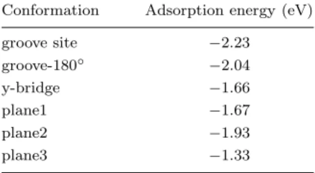
where Esurf is the total energy of the Si96H16 surface (in their optimal geometry), Emol is the total energy of alanine dipeptide, and Esurf+mol is the total energy of alanine dipeptide and Si96H16 surface together. We calculated all the energies by substituting the system to relax by having all atoms reach into their minimum en-ergy positions at temperature T = 0 K. The adsorption energies are calculated for several orientations of the molecule and for different sites. The most favorable six chemical adsorption configurations are shown in Fig. 1 and the calculated adsorp-tion energies are presented in Table 1. As it can be seen from Table 1, the groove site was found to be the most energetically favorable adsorption site for the ad-sorption of alanine dipeptide on Si(100)-2 × 1 surface. The most stable equilibrium configuration, which is obtained after geometrical optimization, is represented in Fig. 1(a). The adsorption energy of this site is (Eads = −2.23 eV), which is char-acterized by the formation of a covalent bond between Si and O atoms, and the calculated length of Si-O bond is 1.78 ˚A. These values can be compared with the tabulated one: 1.63 ˚A (Si-O, in SiO2). The Si-O bond is slightly elongated with respect to that of bulk silica, as could be expected for a surface bond. In another study dealing with the adsorption of methanol molecule on Si(100)-2 × 1 surface, the calculated Si-O bond is 1.76 ˚A.10These results are also in good agreement with our values.
The second energetically favorable state is the groove-180◦which is obtained by rotating around the CH3 axis by 180◦ [shown in Fig. 1(b)]. In this case, the other oxygen atom is bonded to the other Si atom of the considered surface site. The calculated binding energy of this configuration is −2.04 eV and the calculated Si-O bond length is 1.80 ˚A. Comparing with the groove site, the Si-O bond length is a little extended. As shown in Fig. 1(c), the system can also assume a configuration characterized by a bond between the surface and the oxygen atom. In this case, the occurrence of unsaturated dangling bonds giving rise to partially filled bands leads
Table 1. Calculated adsorption energies for the alanine dipeptide on Si(100)-2 × 1 sur-face.
Conformation Adsorption energy (eV)
groove site −2.23 groove-180◦ −2.04 y-bridge −1.66 plane1 −1.67 plane2 −1.93 plane3 −1.33
Int. J. Mod. Phys. C 2010.21:97-106. Downloaded from www.worldscientific.com
Eads =− 2.23 eV E ads =− 2.04 eV E ads =− 1.66 eV Eads =− 1.67 eV (a) (b) (c) (d) (e) Eads =− 1.93 eV (f) Eads =− 1.33 eV O H N C Si
Fig. 1. (Color online) Panels (a)–(f) show the chemical bonding configurations and corresponding binding energies for chemisorption of a alanine dipeptide on a silicon substrate. (a) Calculated alanine dipeptide adsorbed on groove, (b) groove-180◦, (c) y-bridge, (d) plane1, (e) plane2, and
(f) plane3 site of Si(100) surface.
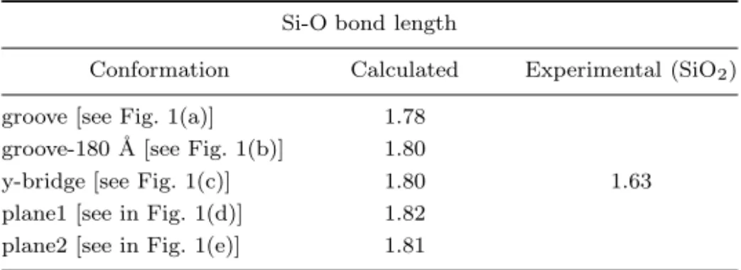
Table 2. Summary of obtained Si-O bond length and to compar with experimental value.
Si-O bond length
Conformation Calculated Experimental (SiO2)
groove [see Fig. 1(a)] 1.78 groove-180 ˚A [see Fig. 1(b)] 1.80
y-bridge [see Fig. 1(c)] 1.80 1.63 plane1 [see in Fig. 1(d)] 1.82
plane2 [see in Fig. 1(e)] 1.81
to a new reconstruction of a surface. Hence, the dangling bonds are saturated when the amino acid is adsorbed. The adsorption energy (Eads = −1.66 eV) is smaller than for the groove and groove-180◦ cases and the bond length is same as for the groove-180◦site (see Table 2).
The other two configurations which are called plane1 and plane2 are also forming Si-O bonds. The bond lengths are 1.82 and 1.81 ˚A, respectively. They are longer than the corresponding Si-O bond for the adsorption on a groove site (a difference of 0.4 and 0.3 ˚A). The adsorption energies are Eads = −1.67 eV and −1.93 eV for these configurations, respectively. One can conclude from these results, with the energy value of Eads = −2.23 eV, the groove site is the most probable adsorption site.
Int. J. Mod. Phys. C 2010.21:97-106. Downloaded from www.worldscientific.com
The last equilibrium configuration is depicted in Fig. 1(f) (plane3), which re-sults from chemisorption reactions involving the alanine dipeptide. In this case, the chemisorption energy is −1.33 eV and a Si-O bond is not formed. Consequently, all the Si-O bond lengths obtained for different adsorption sites are listed in Table 2. The results for the Si-O bond lengths are in good agreement with the previous ex-perimental and theoretical results for other systems which also form Si-O bonds.10 To support our results given above that the interaction of alanine dipeptide with Si(100)-2 × 1 surface cause to bonding between Si and O atoms, we have depicted the electron density map for one of the adsorption sites (for the most probable adsorption site) in Fig. 2. The accumulation of the charge in the Si-O bonding region can be seen from the charge density difference
∆n(r) = npeptide/surface(r) − [nsurface(r) + npeptide(r)] , (2) where npeptide/surface(r), nsurface(r) and npeptideare the electron charge density dis-tributions of the peptide on surface, clean surface and peptide, respectively. In the small insert picture of Fig. 2, the isolines at the middle correspond to the region where the difference is negative, indicating there is lowering in the electron den-sity. The isolines at the bottom part of the picture are positive showing the regions where there is an increment of electron density. The analysis leads us to realize that there is an electron density increment in the Si-O interatomic region indicating the formation of a covalent bond.
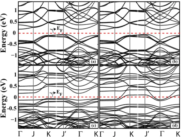
In order to see the influence of dipeptide adsorption on surface, we have calculated the surface electronic band structure for the surface and for the dipeptide-surface system for different adsorption sites. In Fig. 3 we show the re-sults in reciprocal space. The band structure of the asymmetric dimer structures of the surface are depicted in Fig. 3(a). The asymmetric surface is semiconduct-ing, and our results are in good agreement with previous calculations.34–36 The separation of the occupied and unoccupied bands associated with the asymmet-ric dimer is found as 0.17 eV. Further, we focus on the electronic band structures of the dipeptide/Si(100)-2 × 1 system given in Fig. 3(b) for groove site, (c) for groove-180◦ and (d) for y-bridge site. As seen in Fig. 3, the adsorption of the dipeptide on different sites gives rise to dramatic changes on the electronic struc-ture. Therefore, the adsorption can be conformation-selective. Firstly, when the adsorption of dipeptide on the surface occurred at groove site, because of the localization of band within the bulk valence band, the electron transferred from surface to molecule. As a result of this, the electron transfer decreases the ion-icity and increases the covalency of the asymmetric dimer. Therefore, the band structure of groove site is similar to clean surface. Secondly, the calculated band structures of the dipeptide adsorption on semiconductor surface in the groove-180◦ site are depicted in Fig. 3(c). The surface of this structure includes many quan-tum confinement states or localized states resulting from occupied surface states. On the other hand, the rotation of dipeptide molecule also reduces the molec-ular interaction and, consequently, dispersion is developed in the surface band
Int. J. Mod. Phys. C 2010.21:97-106. Downloaded from www.worldscientific.com
∆
n (r)
+
∆
n (r)
-(a)
(b)
Fig. 2. (Color online) (a) Isosurfaces of the different electron charge density. ∆n+
(r) with pink and ∆n−(r) blue isosurface showing charge accumulation and charge depletion regions
respec-tively. Isosurface charge density is taken to be 0.003 electrons/˚A3
. (b) The electron density maps for alanine dipeptide adsorbed on groove of Si surface atoms obtained from periodic LDA DFT calculation.
when compared with the clean surface band structure [Fig. 3(a)]. When we com-pare the adsorption energies, the difference between the energies associated with groove and groove-180◦ is relatively small, although the electronic structures are very different from each other. In groove-180◦ case, a distortion occurs and con-sequently the energy level of surface state shows a splitting due to Jahn–Teller effect. In groove case the π dimer states interactions are not strong enough to cause a splitting of surface states and to open up a surface gap. The difference in electronic structure is also explained by using Chadi’s37 predictions that the bonding and antibonding π dimer states have separated in energy when dimer buckles occur at groove-180◦. Although the bare surface has indirect band gap, the surface band gap was reduced by 0.1 eV at groove-180◦ and system be-comes direct band gap. In addition, the formation of antisymmetric dimer at the
Int. J. Mod. Phys. C 2010.21:97-106. Downloaded from www.worldscientific.com
−1 0
E
n
er
g
y
(
eV
)
Γ
J
K
J’
Γ
K Γ
J
K
J’
Γ
K
1 0.5 -0.5 −1 0 1 0.5 -0.5 E F (a) (b) (c) (d)E
n
er
g
y
(
eV
)
EFFig. 3. (a) The energy band structure of bare Si surface folded to the (4 × 4) unit cell, (b) energy band structure of groove, (c) groove-180◦and y-bridge site. The zero band energy is set to Fermi
Energy of EF. The points Γ, J, K, and J0are described as in Ref. 34.
interaction region results in a charge transfer from down atom to up atom at groove-180◦.
Finally, the band structure of the dipeptide adsorption in the y-bridge site are calculated and presented in Fig. 3(d). An interesting situation comes out when one examines the band structure. The O-terminated dimer is tilded due to the interac-tion between the molecules and topmost atoms of surface and thus dimer becomes asymmetric due to the charge transfer from down to up dimer atom. As a result of this, the surface bands differ from the bulk bands at the middle of the band gap, π and π∗bands overlap which in turn makes the surface gain metallic property. These electronic band structure changes are also reflected in structural modifications of the Si surface dimers. In short, the adsorption of dipeptide on semiconductor surface in y-bridge site changes the electronic properties of the surface upon adsorption.
4. Conclusions
A theoretical investigation using the DFT method has been carried out in the study of alanine dipeptide adsorption process on Si(100)-2 × 1 asymmetric surface. All probable adsorption sites over the surface and all probable molecule orientations are searched in detail. The geometrical optimization of the alanine dipeptide adsorbed on various sites was found to lead different adsorption energies (being measures of the stability of the adsorbate-substrate system). There are different favorable
Int. J. Mod. Phys. C 2010.21:97-106. Downloaded from www.worldscientific.com
adsorption sites, of which the groove site is found to be the energetically most fa-vorable. Our results have shown that the adsorption of dipeptide is undissociative and new Si-O bond is created upon adsorption. Previous experimental and theoret-ical studies of methanol adsorption on the same surface also shows that after the process new Si-O bonds are formed. Our calculated Si-O bond lengths for differ-ent sites are also in good agreemdiffer-ent with previous works.10Finally, an interesting situation comes out when we examine the band structure. The adsorption of the dipeptide on Si(100)-2 × 1 asymmetric surface in y-bridge site changes the surface electronic properties, where the surface bands overlap with the bulk conduction bands. This in turn gives the surface metallic character. This is an important re-sult, since it is generally expected that selective synthetic peptides may play an essential role in future nanotechnological applications.
Acknowledgments
Tarik C¸ elik acknowledges The Turkish Academy of Sciences. Handan Arkin ac-knowledges support by The Scientific and Technological Research Council of Turkey under the project number 104T150 and The Turkish Academy of Sciences under the program to Reward Successful Young Scientists.
References
1. S. R. Whaley et al., Nature (London) 405, 665 (2000).
2. K. Goede, P. Busch and M. Grundmann, Nano Lett. 4, 2215 (2004). 3. K. Goede et al., AIP Conf. Proc. 893, 611 (2007).
4. M. Bachmann and W. Janke, Phys. Rev. Lett. 95, 058102 (2005). 5. M. Bachmann and W. Janke, Phys. Particles Nucl. Lett. 5, 243 (2008). 6. A. J. Dyson and P. V. Smith, Surf. Sci. 194, 55 (1997).
7. C. Huang, W. Widdra and W. H. Weinberg, Surf. Sci. 312, L953 (1994). 8. B. Craig, Surf. Sci. 298, 87 (1993).
9. R. Miotto, A. C. Ferraz and G. P. Srivastava, Phys. Rev. B 65, 075401 (2002). 10. M. Carbone and K. Larsson, J. Phys.: Condens. Matter 17, 1289 (2005). 11. P. L. Silvestrelli et al., Phys. Rev. B 68, 235306 (2003).
12. J.-H. Cho and L. Kleinman, Phys. Rev. B 64, 235420 (2001).
13. X. Lu, Q. Zhang and M. C. Lin, Phys. Chem. Chem. Phys. 3, 2156 (2001). 14. P. L. Silvestrelli, Surf. Sci. 552, 17 (2004).
15. V. Efstathiou and D. P. Woodruff, Surf. Sci. 531, 304 (2003). 16. Y.-Q. Qu et al., Surf. Sci. 569, 12 (2004).
17. L. M. Ghiringhelli, P. Schravendijk and L. DelleSite, Phys. Rev. B 74, 035437 (2006). 18. A. C. Ferraz and R. Miotto, Brazilian J. Phys. 36, 2A (2006).
19. G. Jones et al., Surf. Sci. 600, 1924 (2006).
20. R. B. Rankin and D. S. Sholl, J. Chem. Phys. 124, 074703 (2006). 21. M. Nydberg et al., J. Chem. Phys. 112, 5420 (2000).
22. S. M. Barlow et al., Surf. Sci. 401, 322 (1998). 23. E. Soria et al., Surf. Sci. 451, 188 (2000). 24. A. Lopez et al., Chem. Phys. 277, 1 (2002). 25. X. Y. Xiao et al., Langmuir 18, 6274 (2002). 26. T. Roman et al., Thin Solid Films 509, 218 (2006).
Int. J. Mod. Phys. C 2010.21:97-106. Downloaded from www.worldscientific.com
27. C. S. Cucinotta et al., Phys. Rev. B 72, 245310 (2005). 28. A. Kuhnle et al., Phys. Rev. Lett. 93, 086101 (2004). 29. S. Baroni et al., http://www.pwscf.org (2001). 30. D. Vanderbilt, Phys. Rev. B 41, R7892 (1990).
31. B. A. Berg and T. C¸ elik, Phys. Rev. Lett. 69, 2292 (1992).
32. J. Dabrowski and H.-J. M¨ussig, Silicon Surfaces and Formation of Interfaces (World Scientific, Singapore, 2000).
33. M. D. Ellison and R. J. Hamers, J. Phys. Chem. B 103, 6243 (1999). 34. A. Ramstad, G. Brocks and P. J. Kelly, Phys. Rev. B 51, 14 (1995). 35. J. Ihm, M. L. Cohen and D. J. Chadi, Phys. Rev. B 21, 4592 (1980). 36. M. Rohlfing, P. Kr¨uger and J. Pollmann, Phys. Rev. B 52, 13753 (1995). 37. D. J. Chadi, Phys. Rev. Lett. 43, 47 (1979).
Int. J. Mod. Phys. C 2010.21:97-106. Downloaded from www.worldscientific.com