DO I: 10.4274/uob.215
Introduction
Renal sarcomas are rare tumors. They constitute only %1-2 of malignant renal tumors in adulthood. Though leiomyosarcoma is the most common histologic type of renal sarcoma (%50-60) (1). Renal leiomyosarcomas are aggressive tumors arising from the renal capsule, renal vein, pelvic musculature or the renal parenchyma. Diagnosis is usually post-operative and requires a thorough sampling of the tumor to rule out an epithelial component (2).
We report new four cases of primary renal leiomyosarcomas.
Case Reports
Case 1
Our first patient was a 61-year-old female. She was undergone radical nephrectomy. In the macroscopic examination, a well-circumscribed, gray-white, swirl cut surface tumoral mass was seen in the lower pole of the kidney which causing renal pelvic and ureteral dilatation. The tumor sizes were measured 9.5x7.5x6 cm. In our microscopic examination, the tumor originated from
1Akdeniz University Faculty of Medicine, Department of Medical Pathology, Antalya, Turkey 2Mardin Maternity and Pediatrics Hospital, Clinic of Medical Pathology, Mardin, Turkey 3Başkent University Alanya Hospital, Clinic of Medical Pathology, Antalya, Turkey
4Akdeniz University Faculty of Medicine, Department of Urology, Antalya, Turkey
Bahar Akkaya MD
1, Saniye Sevim Tuncer MD
2, Hampar Akkaya MD
3, Elif İnanç Gürer MD
1, Mehmet Baykara MD
4Böbreğin Primer Leiyomyosarkomu: Dört Olgu Sunumu
Primary Leiomyosarcoma of the Kidney: Four Cases
Ad dress for Cor res pon den ce/Ya z›fl ma Ad re si: Saniye Sevim Tuncer MD, Mardin Maternity and Pediatrics Hospital, Clinic of Medical Pathology, Mardin, Turkey E-mail: [email protected]
Re cei ved/Ge liş Ta ri hi: 09.12.2014 Ac cep ted/Ka bul Ta ri hi: 12.12.2014
© Bulletin of Urooncology, Pub lis hed by Ga le nos Pub lis hing. / © Üroonkoloji Bülteni, Ga le nos Ya yı ne vi ta ra fın dan ba sıl mış tır.
Renal sarcomas are rare tumors. They constitute only 1-2% of malignant renal tumors in adulthood. Leiomyosarcoma is the most common histological type of renal sarcoma (50-60%). Renal leiomyosarcomas are aggressive tumors arising from the renal capsule, renal vein, pelvic musculature or the renal parenchyma. Diagnosis is usually post operative and requires a thorough sampling of the tumor to rule out an epithelial component. We report 4 new cases of primary renal leiomyosarcomas. Typical morphologic pattern shows alternating fascicles of spindle cells with blunt-ended, non-tapering nuclei and eosinophilic cytoplasm. Nuclear pleomprphisim atypia, mitotic figures and necrosis are seen in different ratios. Immunohistochemically the tumor cells of leiomyosarcoma are positive for SMA, desmin, calponin and h-caldesmon and negative for CK, S-100, HMB-45 and CD117. These tumors are classified using the French Federation of Cancer Centers System. To make a diagnosis of a primary renal sarcoma the following criteria should be met: 1) The patient must not have a sarcoma elsewhere to rule out metastasis. 2) Gross must be compatible with origin in the kidney rather than involvement due to retroperitoneal sarcoma. 3) Sarcomatoid renal cell carcinoma must be excluded. They usually have poor prognosis. But small size (<5 cm), low histological grade, absence of lymph node metastases and radical operations are all associated with better prognosis. Despite radical nephrectomy the tumors can run an aggressive clinical course and early local and distant recurrences are common.
Key Words: Primary renal leiomyosarcoma, leiomyosarcoma, kidney
Summary
Renal sarkomlar nadir tümörlerdir. Yetişkin malign renal tümörlerin yaklaşık %1-2’sini oluştururlar. Leiomyosarkom renal sarkomların ise en sık görülen histolojik tipidir (%50-60). Renal kapsül, renal ven, pelvik kas dokusu veya renal parankimden köken alan agresif tümördür. Tanı genellikle post-operatif dönemde, tümörün epitelyal komponent içeriğini dışlayan doğru bir makroskopik örnekleme ile konur. Biz 4 primer leiomyosarkom rapor etmek istedik. Tipik morfolojik paterni eozinofilik sitoplazmalı, künt sonlanan iğsi hücrelerden oluşan alterne fasiküllerden oluşur. Nükleer pleomorfizm, atipi, mitotik figürler ve nekroz değişik oranlarda görülebilir. İmmunohistokimyasal olarak tümör hücreleri SMA, desmin, kalponin ve h-kaşdesmon pozitif, CK, S100, HMB-45 ve CD117 negatiftir. French Federation of Cancer Centers System’e göre sınıflandırılırlar. Primer renal leiomyosarkom tanısı koyabilmek için şu kriterler karşılanmalıdır: 1) Metastazı dışlamak için hastanın başka bir bölgede sarkom öyküsü olmamalıdır. 2) Makroskopik olarak tümörün retroperitoneal bir tümörün uzantısı olmadığı, primer böbrek kaynaklı olduğu görülmelidir. 3) Sarkomatoid renal hücreli karsinom ekarte edilmelidir. Genellikle kötü prognoza sahiptirler ancak küçük boyutlu (<5 cm), düşük histolojik dereceli ve lenf nodu metastazı göstermeyen ayrıca radikal nefrektomi yapılan olgular daha iyi prognoz gösterebilirler. Bazı olgular radikal nefrektomiye rağmen agresif klinik seyir gösterebilirler. Lokal rekürrens ve uzak metastaz sık görülür.
Anah tar Ke li me ler: Primer renal leiomyosarkom, leiomyosarkom, böbrek
Özet
Case Report
/ Olgu Sunumu
168
Üroonkoloji Bülteni 2015;14:168-170 Bulletin of Urooncology 2015;14:168-170
169
renal capsule and included necrosis and atypia. A diagnosis of leiomyosarcoma of kidney was made which was confirmed with positive immunstaining for actin, desmin, caldesmon and negative immunstaining for S100, Pan-CK, CD34 and CD 117 (Figures 1, 2). The Ki-67 LI was detected 30%.
Case 2
The second patient was an 81-year-old female. According to macroscopic examination of nephrectomy specimen was 17x15x11 cm and lobulated. A 5x4x3 cm tumoral lesion was seen in the lower pole near the renal capsule. The cut surface of the tumor was mucoid and whorled. The tumor included >5 mitotic figures in the 50 high power fields, necrosis and atypia. Tumor cells were immunoreactive for SMA, EMA, and vimentin. They were negative myoglobulin, desmin and S-100.
Case 3
The third patient was a 36 year-old female. Paraffin blocks which belong to her nephrectomy specimen were sent to our department for consultation. In our microscopic section we saw interlacing spindle cells. Tumor cells were positive with SMA, desmin, vimentin and negative with Pan-CK and S-100. The tumor included marked pleomorphism, mitosis and necrosis. The Ki-67 LI was detected 30%.
Case 4
The fourth patient was a 44 year-old female. She has asmptomatic gross hematuria and imaging findings was tumor of the left kidney. After a left radical nephro-ureterectomy, histology confirmed a leiomyosarcoma of the renal pelvis which was confirmed with positive positive immunstaining for actin, desmin and negative with Pan-CK and S-100.
Discussion
Sarcomas of the kidney are extremely rare. Primary leiomyosarcomas of the kidney constitute only 0.1% of all invasive renal tumors. The common signs and symptoms of renal sarcomas are abdominal or flank pain, hematuria and a palpable mass in adults. These similar symptoms were seen with large, rapidly growing renal cell carcinoma, too. These neoplasms exhibit an aggressive biological behavior and an unfavorable prognosis. Renal sarcomas are more lethal than any other genitourinary sites sarcomas (3). Renal leiomyosarcomas have been usually reported in female patients such as our patients.
Leiomyosarcomas usually have an irreguler shape, and CT or MR imaging often reveals a heterogeneously enhanced, soft tissue mass without calcification or a fat component. Sonography or anjiography has been useful in previous studies for defining the vascular structure and invasion when a mass lesion was found. It is difficult to make diagnose of leiomyosarcoma purely based upon physical and radiological examination (4).
Macroscopically, leiomyosarcomas are large, solid, grey-white, softy to firm, focally necrotic tumors (2). They may cause hydroureteronephrosis, like as our first and last patients. So the leiomyosarcomas and other sarcomas should be kept in mind among the reasons of hydroureteronephrosis.
Typical morphologic pattern shows alternating fascicles of spindle cells with blunt-ended, non-tapering nuclei and eosinophilic cytoplasm. Nuclear pleomorphism atypia, mitotic figures and necrosis are variably seen (2). Immunohistochemically the tumor cells of leiomyosarcoma are positive for SMA, desmin, calponin and h-caldesmon and negative for CK, S-100, HMB-45 and CD117 (2). These tumors are classified using the French Federation of Cancer Centers System (5).
To make a diagnosis of a primary renal sarcoma the following criteria should be met: 1) The patient must not have or have had a sarcoma elsewhere to rule out metastasis. 2) Gross must be compatible with origin in the kidney rather than involvement due to retroperitoneal sarcoma. 3) Sarcomatoid renal cell carcinoma must be excluded (2).
They usually have poor prognosis. But small size (<5 cm), low histologic grade, absence of lymph node metastases and radical operations are all associated with better prognosis. Despite radical nephrectomy the tumors can run an aggressive clinical course and early local and distant recurrences are common (5). Renal leiomyosarcomas must be differentiated from epitheloid anjiomyolipoma, the sarcomatoid variant of renal cell carcinoma, fibrosarcoma and a malignant peripheral nerve sheath tumor (5). Retroperitoneal leiomyosarcoma secondarily involving the kidney must be ruled out before the diagnosing primary renal leiomyosarcoma (3). The imaging studies and macroscopic Akkaya et al.
Primary Leiomyosarcoma of the Kidney: Four Cases
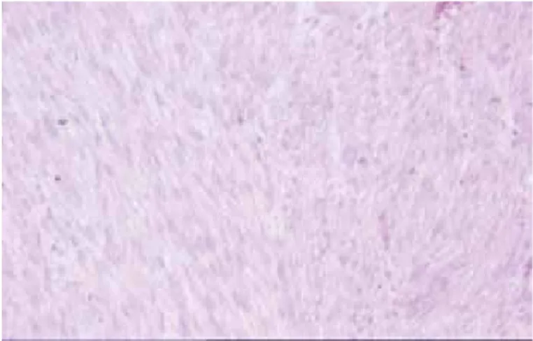
Figure 1. Tumor composed of elonged, plump cells with blunt ended, sometimes hyperchromatic nuclei (H&E; x200)
Figure 2. Tumor cells in well-differentied areas were strongly positive for desmin (Desmin)
170
examination may be useful for the differential diagnosis. Epithelioid angiomyolipoma, a variant of angiomyolipoma, can be mistaken with a leiomyosarcoma. Occasionally, the smooth muscle cells are epitheloid and exhibit nuclear atypia. They are negative for epithelial markers but positive for smooth muscle and melanocytic markers (1).
Sarcomatoid variant of renal cell carcinoma lacks the alternating fascicles, is more pleomorfic, and usually has foci of typical renal cell carcinoma. Absence of smooth muscle markers and CK positivity are supportive of a diagnosis of carcinoma (1). Radical nephrectomy is the treatment choice for leiomyosarcoma of the kidney. Many urologists and oncologists have advocated radical nephrectomy followed by either chemotherapy or radiotherapy (5).
Conclusion
Renal leiomyosarcomas are extremely rare entity with poor prognosis. The radiologic features are non-spesific, and the majority of the primary renal leimyosarcomas exhibit high-grade morphologic features and have a high metastatic potential.
Concept: Bahar Akkaya, Elif İnanç Gürer, Mehmet Baykara Design: Bahar Akkaya, Hampar Akkaya
Data Collection or Processing: Saniye Sevim Tuncer, Hampar Akkaya
Analysis Interpretation: Bahar Akkaya, Elif İnanç Gürer Literature Search: Saniye Sevim Tuncer, Hampar Akkaya Writing: Saniye Sevim Tuncer, Hampar Akkaya
Conflict of Interest: No conflict of interest was declared by the authors.
Financial Disclosure: The authors declared that this study has received no financial support.
References
1. Venkatesh K, Lamba Saini M, Niveditha SR, et al. Primary leiomyosarcoma of kidney. Patholog Res Int 2010;2010:652398. 2. Dhamne SA, Gadzil NM, Padmanabhan A. Leiomyosarcoma of the
renal pelvis. Indian J Pathol Microbiol 2009;52:549-551.
3. Miller JS, Zhou M, Brimo F, et al. Primary leiomyosarcoma of kidney: A Clinicopathologic Study of 27 Cases. Am J Surg Pathol 2010;34:238-242.
4. Chung YG, Kang SC, Yoon SM, et al. Leiomyosarcoma Arising from the Blind End of a Bifid Renal Pelvis. Yonsei Med J 2007;48:557-560. 5. Ellouze S, Abid N, Kossentini M, et al. Leiomyosarcoma of the Kidney.
Clin Genitourin Cancer 2011;9:68-69. Akkaya et al.