I
NTRODUCTIONMitral valve replacement (MVR) in patients with severe calcifi cation of mitral annulus or annular destruction (due to endocarditis or previous mitral valve operations) is associated with high rates of mortality and morbidity.1-4 In these cases, severe complications such as atrioventricular rupture, coronary artery injury, thromboembolic incidents and paravalvular leakage may occur.1,2 To prevent these complications, alternative patch materials and operative techniques may be used to reconstruct the posterior mitral annulus.1-8 The purpose of this study was to discuss the use of autologous pericardial patch to repair a mitral annulus during mitral valve replacement.
P
ATIENTS ANDM
ETHODSMitral valve replacement and annular reconstruction was performed on eight patients between 2000–2002. The demographics and clinical characteristics of the patients are shown in Table 1. All patients were assessed using preoperative transthoracic echocardiography and perioperative transesophageal echocardiography (TEE). The mitral valve pathology is shown in Table 2. Non-endocarditis patients were assessed with coronary arteriography and right and left heart catheterization. Endocarditis patients were not assessed with catheterization because they were young and preoperatively, in a poor
O
RIGINAL
C
ONTRIBUTION
The Use of Autologous Pericardium for
Complicated Mitral Valve Annulus
Mustafa Güden, MD, Kamran Kazımoğlu, MD
1, İlhan Sanisoğlu, MD,
Ertan Sağbaș, MD, Reșit Yaman, MD, Belhhan Akpınar, MD
Department of Cardiovascular Surgery Florence Nightingale Hospital, Kadir Has University, Istanbul, Turkey
1Department of Cardiovascular Surgery
Cerrahpașa Medical Faculty Istanbul University, Istanbul, Turkey
For reprint information contact:
Mustafa Güden, MD Tel: 90 212 224 4950 Fax: 90 212 239 8791 Email: [email protected] Department of Cardiovascular Surgery, Florence Nightingale Hospital, Çağlayan, İstanbul, Turkey
2004, VOL. 12, NO. 1 7 ASIAN CARDIOVASCULAR & THORACIC ANNALS
A
BSTRACTThe study aims to discuss annular repair using the autologous pericardial patch in cases with severe mitral ring calcifi cation and endocarditis during mitral valve replacement. In the study, annular reconstruction was applied, during mitral valve replacement, to 8 patients who had extensive annular calcifi cation or annular destruction due to endocarditis. After annular resection, a two-centimeter wide autologous pericardial patch was sutured continuously to the left ventricular wall close to the posterior annulus. After the valve sutures with pledgets were placed at the back of the pericardial patch, the other edge of the patch was sutured continuously to the left atrial posterior wall. Suturing was complete after the whole annulus was encircled. Thereafter, an appropriately sized mechanical prosthesis valve was used. One patient died postoperatively due to low cardiac output (early mortality 12.5%). Echocardiographical paravalvular leakage was not detected in any of the cases during follow-up. Annular dehiscence and other annular pathologies were also not detected. This reconstructive approach may positively infl uence mortality and morbidity in cases with complicated mitral pathologies.
ASIAN CARDIOVASCULAR & THORACIC ANNALS 8 2004, VOL. 12, NO. 1
Mitral Valve Replacement with Annular Repair Güden
clinical condition. Patients who had endocarditis suffered from severe mitral valve 4 (+) insuffi ciency. Both of these patients had ruptures of the posterior leafl et, and one patient had an abscess formation in the posterior commissure. Valve motions revealed a Carpentier type 2 disorder. These patients also presented with pulmonary hypertension (Pulmonary artery pressure: 62 mmHg, 75 mmHg) together with moderate pericardial effusion.
Non-endocarditis patients had mitral insuffi ciency [2–3 (+)] together with mitral valve stenosis (transvalvular average gradient 14.0 ± 4.5 mmHg, average mitral valve area 1.03 ± 0.23 cm2). A restrictive type of valve motion (Carpentier type 3) was detected in this group of patients. Four patients (66.7%) had coronary artery disease, 2 patients (33.3%) had aortic valve disease and 2 patients (33.3%) had tricuspid insufficiency while one patient had (16.7%) hypertrophic obstructive cardiomyopathy.
The procedures were performed using median sternotomy with bicaval cannulation and moderate hypothermic cardiopulmonary bypass (CPB). Antegrade and retrograde blood cardioplegia was applied for myocardial protection. Left atriotomy through the interatrial groove was preferred in 7 patients (87.5%) for exposition of the mitral valve. A superior transseptal approach was applied in the patient who had an organized thrombus
in the giant left atrium (systolic diameter, 8.8 × 7.4 cm) with tricuspid insuffi ciency. In this patient, calcifi cation had spread to the left atrium as well as mitral annulus. Endocarditis patients had ruptures of the posterior leafl et. One of the patients had abscess formation extending over posteromedial commissure while another patient had a structural disorder in posterior annulus associated with valvular rupture. Non-endocarditis patients demonstrated severe calcifi cation, particularly in the posterior annulus. Calcifi cation was localized to 1/3 of the posterior annulus in 4 cases (66.7%). In one case (16.7%), almost all of the posterior annulus was calcifi ed. In another case (16.7%), calcifi cation of one half of the posterior annulus was connected to the calcifi cation of posterior leafl et, invading the left atrial posterior wall.
These findings prompted us to perform annular reconstruction in addition to mitral valve replacement. We prefer annular resection and pericardial patch with annuloplasty technique in cases requiring annular reconstruction. Over the last two years, 8 cases had annular resection prior to annular reconstruction. Distorted annuli in endocarditis cases have been partially resected. In non-endocarditis cases, calcifi ed annuli have been resected en bloc.
The fi rst phase of reconstruction was fi nalized upon the resection of the leafl ets. At the second phase, a 2 cm wide autologous pericardial patch was continuously sutured to the left ventricular posterior wall right below the posterior annulus using 3/0 polypropylene (Propylen®; Doğsan, Turkey) (Figure 1). The epicardial surface of the pericardial patch was intentionally left outside. After the 2/0 valve Ti-Cron® sutures with pledgets (Davis & Geck; Wayne, NJ, USA) were placed at the back of the pericardial patch, the other edge of the patch was sutured continuously to the left atrial posterior wall (Figure 2). Suturing was complete after the whole annulus was encircled. Thereafter, a mechanical St. Jude® No. 27
Table 1. Demographic and clinical characteristics
Number of patients 8 Age (years) 50.7 ± 16.2 Female/Male 5/3 Atrial fi brillation 5 (62.5%) LVEF 0.53 ± 0.14 Mean LVES (cm) 3.3 ± 0.3 Mean LVED (cm) 5.0 ± 0.2 Mean LA (cm) 5.6 ± 2.1 Functional Class NYHA Class 3–4 4 (50%) NYHA Class 2 4 (50%)
LA = left atrial diameter;
LVED = left ventricular enddiastolic diameter; LVES = left ventricular endsistolic diameter.
Table 2. Mitral valve pathology
Mitral valve pathology Number of patients
Rheumatic 4 (50%)
Degenerative 2 (25%)
Endocarditis 2 (25%)
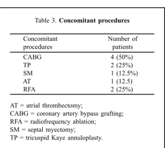
Table 3. Concomitant procedures
Concomitant Number of procedures patients CABG 4 (50%) TP 2 (25%) SM 1 (12.5%) AT 1 (12.5) RFA 2 (25%) AT = atrial thrombectomy;
CABG = coronary artery bypass grafting; RFA = radiofrequency ablation;
SM = septal myectomy;
Güden Mitral Valve Replacement with Annular Repair
2004, VOL. 12, NO. 1 9 ASIAN CARDIOVASCULAR & THORACIC ANNALS
prosthesis valve (St. Paul, Minnesota, USA) was used in 6 patients and mechanical Medtronic® No. 29 prosthesis valve (Medtronic Inc., Minneapolis, MN, USA) in 2 patients. Atriotomy closure, deairing and decannulation were carried out in the standard manner. After weaning from the cardiopulmonary bypass, transesophageal echocardiography was used to assess the prosthesis mitral valve function and paravalvular leakage.
R
ESULTSConcomitant procedures are shown in Table 3. Mean CPB time was 161.7 ± 63.2 min and aortic crossclamp time was 133.7 ± 56.2 min. An intraaortic balloon pump (IABP) was used postoperatively in one patient (12.5%). Postoperative intensive care unit stay was 2.0 ± 1.3 days and the hospital stay was 8.0 ± 3.6 days. One patient (12.5%) was re-explored because of bleeding. Another patient who suffered from chronic renal failure, hepatitis C, carotid artery stenosis and required IABP support, died on the 4th postoperative day due to low cardiac output (early mortality rate, 12.5%). All of the patients were given warfarin sodium postoperatively for anticoagulation. Paravalvular leakage was not detected during preoperative TEE. In addition, these patients were assessed by transthoracic echocardiography postoperatively during the follow-up. The follow-up period was 6.5 ± 1.7 months. During this period, patients were in NYHA functional class 1–2. No paravalvular leakage was detected in any of the cases. Neither annular dehiscence nor pathology related to new formation of calcifi cation was observed. There were no major thromboembolic events detected during follow-up. Patients who had radiofrequency ablation were free of atrial fi brillation during the 6-month follow-up period.
D
ISCUSSIONSurgical approaches in mitral valve pathologies associated with annular calcifi cation or infective endocarditis are currently under discussion.1-6 Calcifi cation of the mitral annulus appears as a semilunar calcium deposit and is often located at the posterior annulus fi brosis.2 The incidence of calcification, calculated over a series of autopsies, has been reported as 8.5%.2 In another echocardiography study, female to male proportion was reported as 3:2 with an incidence of 2.8%.2 Among the 226 mitral valve patients we operated on over the last two years, 6 patients (2.6%) were detected with severe annular calcifi cation. Severe mitral calcifi cation can be observed among the younger age group suffering from Barlow’s disease, Marfan or Hurler syndromes, though it is mostly prevalent among older patients (induced by degenerative valve disease).1-5 Additionally, people with no primary cardiac valve diseases, but suffering from systemic hypertension, diabetes or hypercalcemia may have annular calcifi cation. The type of annular calcification mentioned here is distinct from the calcifi cation in rheumatic valve disease. Calcifi cation in rheumatic valve disease is more often located over the commissures and leafl ets, and may lead to late phase annular calcifi cation.1,2 Annular calcifi cation is predominant in degenerative valve disorder and the spread into the valves is less severe than to atrium and ventricle.1,2 While our patients with rheumatic origin had calcifi cation in both leafl et and annulus, there was no calcifi cation in the leafl ets in cases of degenerative origin.
In patients with active infective endocarditis, surgery is performed to help control the infection with hemodynamic stabilization. If the infection has already led to annular destruction, radical resection of the infection focus and
Figure 1. The pericardial patch is shown sutured to the ventricle side of the annulus.
Figure 2. The pericardial patch being sutured to the left atrial side. Valve sutures passing through the pericardial patch can be seen.
ASIAN CARDIOVASCULAR & THORACIC ANNALS 10 2004, VOL. 12, NO. 1
Mitral Valve Replacement with Annular Repair Güden
annular reconstruction is considered the best approach to attain the above purpose.1
In cases with considerable annular calcifi cation, the annulus is initially decalcifi ed. En bloc annular resection is advised instead of using rongeur or ultrasound to debride calcium.1,2,7 Some surgeons may prefer prosthetic valve fi xation with big pledget sutures following partial debridement.1 However, this approach is observed to increase both mortality (particularly post-MVR left ventricular rupture rate) and morbidity (prosthetic valve dysfunction, paravalvular regurgitation and embolic incidences).1,3 Pathological studies have shown that calcifi cation in annulus is often encapsulated, covered by the endocardium; hence it can be easily resected en bloc.1 It is also thought that debridement of all the calcium and precalcium structures with annular resection can stop calcifi cation process.1 Annular resection should commence on the atrial edge of the calcifi cation, applying diligent care to the soft tissue over the circumfl ex artery and the vein.
In our case, we preferred reconstruction with an autologous fresh pericardial patch after en bloc resection of the annulus. Our reasons for this were: the availability and affordability of autologous pericardium, a fast recovery time and, resistance against infection. Autologous pericardium processed with glutaraldehyde can be used instead of fresh pericardium. This is thought to prevent progressive thickening and early degeneration.6,7 Despite discussions on this matter, the use of fresh autologous pericardium is becoming more popular in cases with surrounding support tissue (for instance, left ventricle and left atrium posterior wall) and endocarditic cases. The use of glutaraldehyde-processed pericardium in the repair of unsupported areas such as fi brous tissue between aortic and mitral annulus is therefore preferred. If autologous pericardium is not readily available (due to the previous operations of the patient, etc.), glutaraldehyde-processed bovine pericardium or Dacron material can be used for this purpose.1,2,6,7
Carpentier and colleagues suggest that Tefl on pledgets may lead to local myocardial ischemia and the use of “live vascularized” atrium tissue instead of pericardial patch accelerates recovery.2 We have not observed any postoperative ischemic events, though we used pericardial patch with Tefl on pledgets. Additionally, other fi ndings such as annular dehiscence or paravalvular leakage were not detected in postoperative echocardiography indicating late annular recovery. As can be seen from these fi ndings, a prosthesis mitral valve can be safely placed into an annulus reconstructed with autologous pericardium in cases of complex mitral pathology.1,2 Moreover, an annular reconstructive approach is thought to have a positive infl uence over the high rates of mortality and morbidity of these cases.1,3 Neither early mortality
related to the procedure, nor serious morbidity such as left ventricular rupture, thromboembolic incident or paravalvular leakage was experienced in the postoperative follow-up of our cases. We therefore advocate a reconstructive approach with annular resection. An autologous pericardial patch is easy-to-apply and may positively infl uence mortality and morbidity in cases with complex mitral pathology.
R
EFERENCES1. David TE, Feindel CM, Armstrong S, Sun Z. Reconstruction of the mitral annulus. A ten-year experience. J Thorac Cardiovasc Surg 1995;110:1323–32.
2. Carpentier AF, Pellerin M, Fuzellier JF, Relland JY. Extensive calcifi cation of the mitral valve annulus: pathology and surgical management. J Thorac Cardiovasc Surg 1996;111:718–30. 3. El Asmar B, Acker M, Couetil JP, Perier P, Dervanian P,
Chauvaud S, et al. Mitral valve repair in the extensively calcifi ed mitral valve annulus. Ann Thorac Surg 1991;52:66–9.
4. Grossi EA, Galloway AC, Steinberg BM, LeBoutillier M 3rd, Delianides J, Baumann FG, et al. Severe calcifi cation does not affect long-term outcome of mitral valve repair. Ann Thorac Surg 1994;58:685–8.
5. Nataf P, Pavie A, Jault F, Bors V, Cabrol C, Gandjbakhch I. Intraatrial insertion of a mitral prosthesis in a destroyed or calcifi ed mitral annulus. Ann Thorac Surg 1994;58:163–7.
6. Ng CK, Nesser J, Punzengruber C, Pachinger O, Auer J, Franke H, et al. Valvuloplasty with glutaraldehyde-treated autologous pericardium in patients with complex mitral valve pathology. Ann Thorac Surg 2001;71:78–85.
7. Ng CK, Punzengruber C, Pachinger O, Nesser J, Auer H, Franke H, et al. Valve repair in mitral regurgitation complicated by severe annulus calcifi cation. Ann Thorac Surg 2000;70:53–8. 8. Ruvolo G, Speziale G, Voci P, Marino B. “Patch-glue” annular
reconstruction for mitral valve replacement in severely calcifi ed mitral annulus. Ann Thorac Surg 1997;63:570–1.
9. Lin PY, Kan CD, Luo CY, Yang YJ. Mitral valve replacement in the presence of massive posterior annular calcifi cation. J Card Surg 1999;14:266–9.