Introduction
Prostate cancer is the most common cancer and benign
prostate hyperplasia (BPH) is the most common benign neoplasm in men (Jemal et al., 2004). Prostatic intraepithelial neoplasia (PIN) is a premalignant lesion
Evaluation of the proliferating activities of normal and carcinomatous
prostatic tissues with Ki-67
Meral Koyutürk
1*, Ifl›n K›l›çarslan
2and Yener Aytekin
31 Department of Histology and Embryology, Medical Faculty of Kadir Has University, 80810
Gayrettepe,‹stanbul 2
Department of Pathology, Medical Faculty of ‹stanbul University, 34390- Çapa, ‹stanbul 3
Department of Histology and Embryology, Medical Faculty of Istanbul University, 34390-Capa, Istanbul/Turkey (*author for correspondence)
Received 25 March 2005; Accepted 12 May 2005
Abstract
In this study proliferation activities of normal prostate tissue, benign prostate hyperplasia (BPH), prostatic intraepithelial neoplasia (PIN) and prostate cancer samples were evaluated immunohistochemically by using the monoclonal anti-Ki-67 antibody. Ki-67 is a nuclear non-histone protein expressed throughout the cell cycle. The proliferation rate was found to be 0.3-0.8 % in normal prostate glandular epithelium, 0.5-2.2 % in BPH, 1.6-3.7 % in high grade PIN and 5-30 % in prostate cancer cases. BPH showed a slightly higher, whereas PIN and prostate cancer showed significantly higher proliferation activity compared to normal prostate tissue. The Ki-67 proliferation index was found to correlate well with the Gleason classification in the cancer group. It is concluded that the Ki-67 proliferation index can be used a valuable diagnostic marker in PIN and tumor tissue.
Key Words: Ki-67, proliferation, prostate cancer, benign prostate hyperplasia, prostatic intraepithelial neoplasia
Normal ve karsinomatöz prostat dokular›n›n ço¤alma aktivitelerinin Ki-67 ile
de¤erlendirilmesi
Özet
Bu çal›flmada, normal prostat dokusu, benign prostat hiperplazisi (BPH), prostatik intraepitelyal neoplazi (PIN) ve prostat kanseri doku örneklerinde proliferasyon aktivitesi immünohistokimyasal yöntemle Ki-67 monoklonal antikoru kullan›larak de¤erlendirildi. Ki-67, hücre siklusu boyunca eksprese edilen nükleer bir non-histon proteindir. Proliferasyon oran›, normal prostat glanduler epitelinde % 0.3-0.8 olarak, BPH de % 0.5-2.2, yüksek evreli PIN de % 1.6-3.7 ve prostat kanser vakalar›nda % 5-30 olarak bulundu. Proliferasyon aktiviteleri normal prostat dokusu ile karfl›laflt›r›ld›¤›nda BPH de hafif art›fl, PIN ve prostat kanserinde anlaml› yüksek art›fl gösterdi. Ki-67 proliferasyonndeksinin kanser grubunda Gleason s›n›fland›rmas›yla iliflkili oldu¤u bulundu. Ki-67 proliferasyon indeksinin de¤erli bir tan› mark›r› olarak PIN ve tümöral dokularda kullan›labilece¤i sonucuna var›ld›.
Anahtar sözcükler: Ki-67, proliferasyon, prostat kanseri, benign prostat hiperplazisi, prostatik intraepiteliyal neoplazi
of the prostate (Feneley et al., 1996). Loss of cellular proliferation control is a fundamental aspect in a number of prostate diseases ranging from hyperplasia to malignant neoplasm.
Cell proliferation antibodies can detect nuclear antigens expressed during the different phases of the cell cycle (Hall and Levison, 1990). Ki-67 is a nuclear non-histone protein related to cell proliferation expressed during the G1, S, G2 and M phases with the exception of Go (Sasaki et al., 1987; Gerdes et al., 1984). The molecular weight of the Ki-67 protein was determined to be 345 kDa and 395 kDa using Western blot analysis of proliferating cells (Key et al., 1993). The results of gene screening experiments have shown that the gene expressing the Ki-67 protein was located on the long arm of human chromosome 10 (Schonk et al., 1989). Immunohistochemical detection of the Ki-67 antigen as a proliferation marker is advantageous over flow cytometry, DNA analysis and thymidine labeling index methods because they are expensive, time-consuming and labor intensive (Brown and Gatter, 1990). One weakness of immunohistochemical staining of paraffin embedded tissues is the occasional requirement for antigen retrieval to expose the epitopes (Shi et al., 1991). Initially, Ki-67 could only be applied on fresh but not on paraffin embedded tissues, since the antigen was destroyed during tissue fixation (Gerdes et al., 1992). However, recent advances in the application of Ki-67 on paraffin
sections have increased its potential to be used in diagnostics.
In this study we aimed to investigate the potential of the Ki-67 antibody for diagnostic purposes by assessing the proliferation activity in normal prostate tissues and BPH, PIN, and prostate capatients. Materials and methods
A total of 55 prostate tissue samples were investigated. Of these 10 normal prostates were obtained from a maximum of 12 hours dead autopsy materials of third decade cases. Ten out of 15 BPH specimens were obtained from radical prostatectomy and 5 transurethral prostatectomy (TURP) operations, 15 high grades PIN from peripheral tissue of radical prostatectomy cases and 15 prostate cancer specimens from l2 radical prostatectomy and 3 TURP operations. After fixation in 10 % formalin and paraffin embedding, 3µm sections were prepared on poly-L-lysine coated glass slides. The sections were deparaffinized in an oven for one hour at 56 ºC and in toluene. After dehydration in ethanol series, slides were treated with trypsin for 10 min at 37 ºC and rinsed in phosphate-buffer saline (10mM, pH 7.5). Antigen retrieval was performed in a standard household pressure-cooker. When the pressure indicator has risen at the end of 5 min, sections were incubated for 3 min in 0.01M citrate buffer.
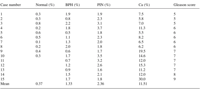
Table 1: Individual case means Ki-67 indices (%).
Case number Normal (%) BPH (%) PIN (%) Ca (%) Gleason score
1 0.3 1.9 1.9 7.5 5 2 0.3 0.8 2.3 5.8 5 3 0.8 2.2 3.1 7.0 5 4 0.2 1.8 3.7 11.3 6 5 0.6 0.5 1.8 5.5 6 6 0.5 1.1 2.3 8.2 6 7 0.1 1.3 2.0 6.5 6 8 0.2 2.0 1.8 6.2 6 9 0.4 0.6 1.7 19.5 7 10 0.3 1.7 3.5 14.6 7 11 0.7 3.2 12.0 7 12 1.2 2.6 15.3 7 13 0.9 1.6 11.2 7 14 1.5 2.1 12.0 8 15 1.7 1.8 30.0 9 Mean 0.37 1.33 2.36 11.51
Endogenous peroxidase activity was blocked with 3% hydrogen peroxide in methanol. Sections were covered with 10% normal goat serum for 20 min to prevent non-specific binding and incubated with 7B11 monoclonal antibody at 1:50 dilution (Zymed Laboratories, USA) for one hour. Then a Histostatin Plus (Zymed Laboratories, USA) broad-spectrum kit of the streptavidin-biotin system was applied. Slides were incubated with biotinylated secondary antibody then with the streptavidin-peroxidase conjugate for 20 min. The enzyme activity was developed using aminethylcarbazole (AEC) for 15 min. The sections were counterstained with haematoxylin and mounted in glycerin-gelatin. Negative control sections were prepared by substituting the Ki-67 antibody with phosphate-buffer saline.
The Ki-67 index was calculated by counting 500-1000 random nuclei under the light microscope at X400 magnification. The significance of the differences among the groups was evaluated by using the Instant 5.0 packet program with Kruskal Wallis and Dunn’s multiple comparison tests. In prostate cancer cases the significance of the difference among the stages was studied by using Spearman correlation analysis in ‘SPSS for windows 7.0’.
Results
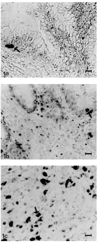
We found that in the glandular epithelium the proliferation activity of normal prostate tissue samples was low, with Ki-67 index values ranging from 0.1 to 0.8 % (mean 0.37 ± 0.21), (Table 1). Although the immunoreactivity rate in BPH gland epithelial cells was found to be 0.5-2.2 % (mean 1.33 ± 0.55) (Fig 1a, Table 1), there was no significant difference between the proliferation indices of the normal prostate tissue and BPH cases (p>0.05). The rate of cells showing positive reaction in PIN cases (Fig 1b, Table 1) was 1.6-3.7 % (mean 2.36 ± 0.70) and significantly high (p<0.01) when compared to normal prostate tissues. However there was no statistically significant difference between the Ki-67 indices of PIN and BPH cases.
According to the Gleason classification, histopathological evaluation of the 15 prostate cancer cases showed a pattern score of 5-9. The specimens of three patients, whose pattern scores were 7 and 8, showed granular cytoplasmic staining and the nuclei of these cells remained unstained. The rate of immunoreactive nuclei of the acinar epithelial cells
Figure 1. Immunohistochemical staining for Ki-67 in benign prostate hyperplasia (a), in prostatic intraepithelial neoplasia (b) and in Gleason score 9 prostate carcinoma (c), bar represents 10µm.
within the tumor (Fig 1c, Table 1) was found to be 5.5-30 % (mean 11.51 ± 6.54). In this group, Ki-67 indices were increased in parallel with Gleason pattern scores (p<0.00l). The proliferation rate of the prostate cancer cases was also significantly higher than those of normal (p<0.001), BPH (p<0.001), and PIN (p<0.05) cases.
Discussion
Immunohistochemical assessment of the proliferation rate is becoming increasingly important in the definition of neoplasm, assessment of prognosis and adjusting the effective treatment protocols (Leong and Wright, 1987, Gerdes, 1985).
Previous studies aimed to determine the proliferation rate of normal prostate tissue have found a low activity in these cells. Claus and co-workers (1993) reported a rate of 0.015 % for epithelial tissue in a series consisting of 4 cases. In a study done by Meyer (1982), where the thymidine labeling index method was adjusted according to the zones, a proliferation rate of 0.12 % in the peripheral zone and 0.11 % in the central zone has been reported. Our results are different from these previous studies, where alternative kinds of methods are applied to different age groups. Our results indicate extremely low proliferation activity in normal prostate tissues and the proliferation index in BPH is 1.3 %, slightly higher than normal prostate tissue. In previous studies higher rates, ranging from 0.4 to 3.7 %, have been reported for this group (Colombel et al., 1998; Gallee et al., 1989). Since Ki-67 is a labile antigen and can easily be destroyed during formalin fixation, standardization of appropriate antigen retrieval and quantitative assessment of immunoreactive nuclei should be of great importance in the evaluation of proliferation activity. Although our results with normal tissues samples are lower, the reading we obtain for malignant tissues are dramatically higher.
The proliferation index in high grade PIN was between that of normal prostate tissue and prostate carcinoma. Our findings, which were consistent with several previous studies (Feneley et al., 1996; Tamboli et al., 1996), showed a significantly higher Ki-67 index for PIN compared to normal prostate tissues.
Previously published data indicate different proliferation rates in prostate cancer cases. Our results are in parallel with the studies where only the Gleason classification has been shown to have a close
correlation with the Ki-67 index (Bantis et al., 2004; Stattin et al., 1997; Mc Loughlin et al., 1993). Gallee et al. (1989) reported that proliferation activity is partially related to the histologic tumor pattern. In this study, Ki-67 indices of prostate cancer were found to be significantly higher than those of normal, BPH and PIN groups, and a positive correlation with the Gleason classification was detected. In another study, the Ki-67 index of patients with Gleason score 7-10 is found to be highly correlated (Cowen et al., 2002). However proliferation indices of higher pattern scores were reported to be quite low when compared to our results. Three ductal tumors were reported a value over 50 % nuclear staining and none of the Gleason pattern scoring applied on microacinar carcinoma was reported to show a value over 25% (Oxley et al., 1998). Although we detected 30 % proliferation index in a case whose Gleason pattern score was 9, the difference was found to be insignificant. Bubendorf et al. (1998), found a correlation between the Ki-67 index and increasing Gleason score in core needle biopsies, but not in radical prostatectomy applied prostate cancer cases (Bubendorf et al., 1996). Despite these findings, the Ki-67 indices of radical prostatectomy applied prostate cancer cases were found to correlate highly with the Gleason score in our study.
Some of the investigators have reported cytoplasmic staining, which is non-specific and cannot be accepted as a positive immunoreaction (Brown et al., 1988). We also observed cytoplasmic staining in three cases of a group with 7-8 pattern score. Positive immunoreactivity was not observed within the nuclei of the epithelial cells cytoplasm where a granular form of staining was present. It has been suggested by other investigators that cytoplasmic staining encountered in both benign and malignant epithelial cells can be a cross reaction (Brown et al., 1988; Hattori, 2002).
Finally, it has been concluded that, immunohistochemical assessment of proliferation activity using the Ki-67 index can be a useful method in PIN and prostate cancer. However, before being routinely used, wide series of application and standardization of both the methods and the assessment are mandatory.
Acknowledgment
This study was supported by by the Research Fund of Istanbul University. Project No: T-442 /071197.
References
Bantis A, Giannopoulos A, Gonidi M, Liossi A, Aggelonidou E, Petrakakou E, Athanassiades P, and Athanassiadou P. Expression of p120, Ki- 67 and PCNA as proliferation biomarkers in imprint smears of prostate carcinoma and their prognostic value. Cytopathology. 15: 25-31, 2004. Brown DC, Cole D, Gatter KC, and Mason DY. Carcinoma
of the cervix uteri: an assessment of tumour proliferation using the monoclonal antibody Ki-67. Br J Cancer. 57: 178-181, 1988.
Brown DC, Gatter KC. Monoclonal antibody Ki-67: Its use in histopathology. Histopathology. 7: 489-503, 1990. Bubendorf L, Sauter G, Moch H, Schmid HP, Gasser TC,
Jordan P, and Mihatsch MJ. Ki67 labelling index: An independent predictor of progression in prostate cancer treated by radical prostatectomy. J Pathol. 178: 437-441, 1996.
Bubendorf L, Tapia C, Gasser TC, Casella R, Grunder B, Moch H, Mihatsch MJ, and Sauter G. Ki67 labelling index in core needle biopsies independently predicts tumor-specific survival in prostate cancer. Hum Pathol. 29: 949-954, 1998.
Claus S, Wrenger M, Senge T, and Schulze H. Immunohistochemical determination of age related proliferation rates in normal and benign hyperplastic human prostates. Urol Res. 21: 305-308, 1993.
Colombel M, Vacherot F, Diez SG, Fontaine E, Buttyan R, and Chopin D. Zonal variation of apoptosis and proliferation in the normal prostate and in benign prostatic hyperplasia. Br J Urol. 82: 380-385, 1998. Cowen D, Troncoso P, Khoo VS, Zagars GK, Von
Eschenbach AC, Meistrich ML, and Pollack A. Ki-67 staining is an independent correlate of Biochemical Failure in prostate cancer treated with radiotherapy. Clin Cancer Res. 8: 1148-1154, 2002.
Feneley MR, Young MPA, Chinyema C, Kirby RS, and Parkinson MC. Ki-67 expression in early prostate cancer and associated pathological lesions. J Clin Pathol. 49: 741-748, 1996.
Gallee MPW, Jong EV, Kate F JNT, Schroeder FH, and Van der Kwast TH. Monoclonal antibody Ki-67 defined growth fraction in benign prostatic hyperplasia and prostatic cancer. J Urol. 142: 1342-1346, 1989. Gerdes J, Lemke H, Baisch H, Wacker HH, Schwab U, and
Stein H. Cell cycle analysis of a cell proliferation-associated human nuclear antigen defined by the monoclonal antibody Ki-67. J Immunol. 133: 1710-1715, 1984.
Gerdes J. An immunohistological method for estimating cell growth fractions in rapid histopathological diagnosis during surgery. Int J Cancer. 35: 169-171, 1985. Gerdes J, Becker MH, Key G, and Cattoretti G.
Immunohistological detection of tumour growth fraction (Ki-67 antigen) in formalin-fixed and routinely
processed tissues. J Pathol. 168: 85-87, 1992.
Hall PA, Levison DA. Assessment of cell proliferation in histologic material. J Clin Pathol. 43: 184-192, 1990. Hattori H. Sclerosing haemangioma of the lung is positive
for MIB-1 in cell membrane and cytoplasmic staining pattern. Histopathology. 40: 291-293, 2002.
Jemal A, Tiwari RC., Murray T, Ghafoor A, Samuels A, Ward E,. Feuer EJ, and Thun MJ. Cancer Statistics, 2004. CA Cancer J Clin. 54: 8 – 29, 2004.
Key G, Becker MHG, Baron B, Duchrow M, Schluter C, Flad HD, and Gerdes J. New Ki-67 equivalent murine monoclonal antibodies (MIB 1-3) generated against bacterially expressed parts of the Ki-67 cDNA containing three 62 base pair repetitive elements encoding for the Ki-67 epitope. Lab Invest. 68: 629-636, 1993.
Leong AS, Wright J. The contribution of immunohistochemical staining in tumor diagnosis. Histopathology. 11: 1295 -1305, 1987.
Mc Loughlin J, Foster CS, Price P, Williams G, and Abel PD. Evaluation of Ki-67 monoclonal antibody as prognostic indicator for prostatic carcinoma. Br J Urol. 72: 92-97, 1993.
Meyer JS, Sufrin G, Martin SA. Proliferative activity of benign human prostate, prostatic adenocarcinoma and seminal vesicle evaluated by thymidine labeling. J Urol. 128: 1353-1356, 1982.
Oxley JD, Abbott CD, Gillatt DA, and MacIver AG. Ductal carcinomas of the prostate: a clinicopathological and immunohistochemical study. Br J Urol. 81: 109-115, 1998.
Sasaki K, Murakami T, Kawasaki M, and Takahashi M. The cell cycle associated change of the Ki-67 reactive nuclear antigen expression. J Cell Physiol. 133: 579-584, 1987.
Schonk DM, Kuijpers HJH, Van Drunen E, Van Dalen CH, Van Kessel AHG, Verheijen R, and Ramaekers FC. Assignment of the gene(s) involved in the exspression of the proliferation-related Ki-67 antigen to human chromosome 10. Hum Genet. 83: 297-299, 1989. Shi SR, Key ME, Kalra KL. Antigen retrieval in
formalin-fixed, paraffin-embedded tissues: An enhancement method for immunohistochemical staining based on microwave oven heating of tissue sections. J Histochem Cytochem. 39: 741-747, 1991.
Stattin P, Damber J, Karlberg L, Tavelin B, and Damber JE. Cell proliferation assessed by Ki-67 immunoreactivity on formalin fixed tissues is a predictive factor for survival in prostate cancer. J Urol. 157: 219-222, 1997. Tamboli P, Amin MB, Schultz DS, Linden MD, and Kubus
J. Comparative analysis of the nuclear proliferative index (Ki-67) In benign prostate, prostatic intraepithelial neoplasia, and prostatic carcinoma. Mod Pathol. 9: 1015-1019, 1996.