Pedicular stress fracture in lumbar spine
Mustafa Sirvanci*, Levent Ulusoy, Cihan Duran
Department of Diagnostic Radiology, Florence Nightingale Hospital, Kadir Has University School of Medicine, Abide-i Hurriyet caddesi No. 290, Caglayan 80220, Sisli, Istanbul, Turkey
Received 8 September 2001
Abstract
The purpose of this article is to report two cases of pedicular stress fracture of the lumbar spine, which is an uncommon cause of low back and leg pain. The relevant literature is reviewed and features of the cases that differ from those already reported are highlighted. One of our cases of pediculolysis is the first in the literature for that is caused by rotational instability induced by laminectomy. The remodeling of the fractured pedicle was striking when compared with its normal counterpart. To the authors’ knowledge, this is the first report of a pediculolysis with T2-weighted imaging findings. In addition, we report the computed tomographic (CT) and magnetic resonance (MR) imaging findings of a healed pediculolysis that has not been reported previously. D 2002 Elsevier Science Inc. All rights reserved.
Keywords: Lumbar pedicle; Stress fracture; Spondylolysis; CT; MR
1. Introduction
The lumbar pedicle is an unusual location of stress fracture, which is a common problem in specific sites. The term pediculolysis, which is first introduced by Gunzburg, describes hypertrophic fracture of the pedicle showing non-union [1,2]. This uncommon phenomenon, which histor-ically was considered to be a congenital anomaly [3], most often occurs due to contralateral spondylolysis or abnormal stress over the pedicle after spinal surgery [4 – 9]. Other causes of pedicle defects are congenital anomalies [2,10 – 12] and tumoral infiltrations [8].
This article describes two cases of unilateral pedicular stress fracture of the lumbar spine. One of the cases was associated with contralateral spondylolysis while the other was accompanied by rotational instability. Imaging features of the cases that differ from those already reported are highlighted, together with a review of the literature.
2. Case reports
2.1. Case 1
A 17-year-old man was referred to our radiology depart-ment, with a history of activity related low back and right leg pain for 1 month. His medical history was otherwise unremarkable. He denied any history of recent or remote episode of trauma. Plain lateral radiograph showed a pedi-cular cleft in L4 vertebra (Fig. 1). Magnetic resonance (MR) imaging, done for the detection of associated pathologies, revealed nonspecific pathological signal in the L4 right pedicle (Fig. 2). Computed tomographic (CT) examination was performed for differential diagnosis and demonstrated sclerosis of L4 right pedicle with a lucent fracture line with sclerotic margins, in addition to left pars interarticularis defect of the same vertebra (Fig. 3).
Trial of conservative treatment with low back muscle exercises and decrease of overall physical activities relieved his symptoms, and the imaging findings revealed the heal-ing of the pedicular fracture.
2.2. Case 2
The second patient, a 56-year-old woman presented to the Orthopedic Spine Clinic of our hospital with a
0899-7071/02/$ – see front matterD 2002 Elsevier Science Inc. All rights reserved. PII: S 0 8 9 9 - 7 0 7 1 ( 0 1 ) 0 0 3 8 9 - 8
* Corresponding author. Velioglu sokak No. 9/7 Husnufirat apartmani, 81160 Ayazma, Uskudar, Istanbul, Turkey. Tel.: +90-212-224-2273; fax: +90-212-296-5223.
E-mail address: [email protected] (M. Sirvanci).
complaint of progressive spinal deformity and low back and right leg pain. She was referred to our radiology department for CT examination in order to evaluate her symptoms. She underwent discectomy twice for extruded disc herniation with 3-month intervals, 6 years before her presentation. She felt and did well until 2 months ago when she experienced insidious onset of low back and right leg pain.
Plain radiographs demonstrated left lumbar scoliosis and L3 – L4 spondylolisthesis. L3 partial and L4 total laminec-tomy were identified. A compensatory right thoracal sco-liosis was also noted. CT scan showed that anterior displacement of L3 over L4 was on the right side suggesting rotational instability (Fig. 4a). A cleft was detected in the right L3 pedicle, which showed deformation and elongation paralleling right anterolateral displacement of L3 over L4 (Fig. 4b and c). Right neural foramen was severely nar-rowed and right L3 nerve root was under compression. Right facetectomy was detected in addition to L3 partial laminectomy. Electromyography revealed minimal, chronic right L3 nerve root denervation.
The patient was treated with anterior and left lateral fusion from L2 to S1, which relieved her symptoms sig-nificantly. Scoliosis was also treated by T4 – S1 posterior instrumentation with pedicle screws.
3. Discussion
Two types of stress fractures are known. Fatigue fractures occur in normal bone that is subject to repetitive abnormal stress. Abnormal stress placed on the pedicles may be
intensive sportive activity or may reflect an increased stress at the first mobile level above or below the rigid segment after posterior spinal fusion [5,8,13]. Unusual stress fracture of the pedicle within solid thoracolumbar fusion mass has also been reported [6]. Insufficiency fractures are due to normal stress on abnormal bone. Pediculolysis is a fatigue-type stress fracture.
Stress fractures in vertebral neural arch are located mostly in pars interarticularis. The pars interarticularis is the weakest part of the neural arch [8,14]. The majority of the neural arch fractures were reported to occur in pars interarticularis in an experimental in vitro study by applying repetitive mechanical stress on vertebrae [15]. It has been shown that the pedicle is the second weakest part of the vertebra after pars interarticularis [8,14,15]. Pedicular fatigue fractures occurred in five lumbar vertebrae out of a series of 74 vertebrae tested in the experimental in vitro study of Cyron and Hutton [15].
The first description of callus-like bony masses on the neural arch associated with contralateral spondylolysis is reported by Hadley et al. in 1955 [1,4]. In the literature, most of the cases of pediculolysis were reported to present with sclerosis in the pedicle (with or without a fracture) and associated with contralateral spondylolysis [2,16]. Bilateral pediculolysis has been reported rarely [2,5,8,17]. Gunzburg, by first using the term pediculolysis, describes reactive sclerosis and hypertrophy that develops after pedicular stress fracture [2]. Maldague used the term vertebral ani-socoria for the unilateral pedicle hypertrophy seen on plain AP radiography [18].
It has been suggested that the cause of contralateral pedicular sclerosis associated with unilateral spondylolysis
Fig. 1. Case 1: Lateral radiographs of lumbar spine at initial presentation (a) and after conservative treatment (b). The cleft still persists after treatment, however, with considerable decrease in thickness (arrow).
is the changes in the distribution of forces and abnormal stress placed upon contralateral bony structures resulting from the defect in the pars interarticularis [2,9,19,20]. Stress fracture of the contralateral lamina is also another but rarer complication of spondylolysis [21]. Response of the pedicle to abnormal stress due to instability of the neural arch is physiological hypertrophy and is well known as sclerotic pedicle in the literature [2,17,19,20]. This theory is sup-ported by decrease of the pedicular sclerosis after fusion of contralateral pars interarticularis [1]. Continued repetitive stress may lead to a fracture in the sclerosed pedicle. However, it is not clear in the literature that sclerotic period
before the fracture is a constant feature or not. In other words, it is not clear whether the sclerosis is a reactive hypertrophy due to increased stress on the pedicle due to instability or is a callus formation that occurs during repeated attempts at healing of a stress fracture [2,4]. Decrease of pedicular sclerosis after conservative or surgical treatment suggests that it is a compensatory reaction sec-ondary to laminar instability [1]. Guillodo et al. [13], in their case report of a pedicular stress fracture associated with contralateral lysis of the left isthmus, described condensa-tion of the pedicle evidencing hypertrophy 1 year before the fracture. They also reported that the pedicular fracture
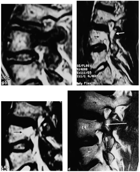
Fig. 2. Case 1: MR examination. (a) Sagittal T1-weighted image shows the nonspecific edema in the right L4 pedicle. (b) T2-weighted image reveals the hyperintense edema surrounding signal void fracture line. (c) Image just medial to (b) demonstrates the hyperintense edema in the vertebral body, adjacent to the right pedicle. After treatment, sagittal T1-weighted image (d) and axial T2-weighted image, (e) the edema is no longer seen adjacent to the signal void fracture line (arrows).
healed spontaneously despite the continuation of intensive physical activity. Contrary to this theory, Chong and Htoo [14] reported a pedicular stress fracture of recent onset with no pedicular sclerosis. They suggested that pediculo-lysis does not need to go through the stage of reactive sclerosis before the fracture takes place.
In the literature, contralateral pedicular sclerosis and stress fracture are mostly associated with unilateral spondy-lolysis [1,2,4,9,16,19,20,22]. Pedicuspondy-lolysis associated with bilateral spondylolysis has been reported rarely [3,14]. Deficiency and instability of the neural arch due to spondy-lolysis is the cause of the abnormal stress that leads to pediculolysis. Surgical laminectomy has a similar mech-anism to induce stress fractures of the pedicle [7]. In the second case of this study, right pedicular fracture was due to partial laminectomy and right facetectomy. To the authors’ knowledge, this is the first report of a pedicular stress fracture associated with rotational instability. The remodel-ing of the fractured pedicle with elongation and thinnremodel-ing was striking when compared with the contralateral normal pedicle (Fig. 4b and c).
Rarely, pedicular fracture may occur without a neural arch defect such as spondylolysis. Traughber and Havlina [17] reported bilateral pedicular stress fractures in a young female patient without any pathological process that can cause instability in the neural arch, and suggested a causa-tive relationship with the patient’s hyperactivity.
Most of the pedicular stress fractures have been described in young, athletic individuals. In the study of Sherman et al. [19] with 11 cases of pedicular sclerosis, the mean age at diagnosis was 15 years. Pediculolysis generally occurs at L2 – L5 levels and more commonly on the right side while pars interarticularis defect is often seen on the left [1,16,19]. Right-sided involvement in the majority of cases
may also be due to right-sided dominance of physical activities. Pedicular fractures were right-sided in both of our patients.
Symptoms are low back pain, leg pain due to nerve root irritation, limitation of straight leg raising, scoliosis, and local tenderness. Neurological deficits are rare. It is known that spondylolysis leads to disc degeneration, and associ-ated contralateral pediculolysis causes deterioration of dis-cal degeneration, which results in worsening of the instability [2].
The diagnosis of pediculolysis is possible by plain radiography; however, its sensitivity is limited similar to other well-known stress fractures. Plain films may dem-onstrate hypertrophy and sclerosis of the pedicle in some of these patients. Difficulty in diagnosing this lesion by plain radiography together with the unawareness of the lesion may lead to unnecessary diagnostic tests and treatment plans. Pedicular stress fracture could be seen on lateral radiograph in our first case (Fig. 1a). However, in the second case only on postoperative plain films could we identify the lucent fracture line, probably resulting from the change of orientation of the cleft postoperatively.
Aland et al. [4] and Garber and Wright [22] reported fractures of the pedicle that were not seen on the plain radiographs but were detected by CT. CT is the method of choice in the evaluation of the lesion, treatment planning, follow-up, and differentiation from other possible etiologies such as arthritis, infection, or neoplasia [17,20]. Pediculo-lytic lesion can be nicely demonstrated as a linear lucency or defect at the base of the sclerotic pedicle [2,4,22]. Contrala-teral spondylolysis, if it exists, can also be depicted. In the second case of this study, the fractured pedicle showed remodeling with elongation at CT scan, probably due to long-lasting rotational instability (Fig. 4b).
In the literature, bone scintigraphy has been reported to show normal or increased uptake [4,16,22].
MR imaging features of stress fractures are well docu-mented [23]. However, we have seen little in the literature concerning the MR appearance of pediculolysis. MR imaging may be the study of choice for discal and neuro-logical complications of pedicular stress fractures; however, it is limited in the primary evaluation of this entity. In one of the cases in Gunzburg and Fraser’s [2] series, the lesion showed hypointensity on T1-weighted images due to edema and sclerosis of the pedicle. In our first case, T2-weighted images revealed hypointense fracture line within the hyper-intense marrow edema, while T1-weighted images showed nonspecific hypointensity (Fig. 2). Awareness of the MR
features of the entity will overcome the difficulty in inter-preting the MR examinations that are the second-line imaging study for low back pain in most countries.
Osteoid osteoma and osteoblastoma are the major differ-ential diagnostic considerations. This is due to severe back pain in a healthy individual together with dense sclerosis of a pedicle on plain films. Correct diagnosis of pediculolysis with differentiation from osteoid osteoma or osteoblastoma is important because excision of the pedicle can increase the already existing instability due to pars interarticularis defect [9]. Diagnosis of a pedicular stress fracture can be made by the absence of a nidus, association of contralateral spondylolysis, and the pedicular linear defect. Congenital anomalies can also cause pedicular defects due to ossification failure. However,
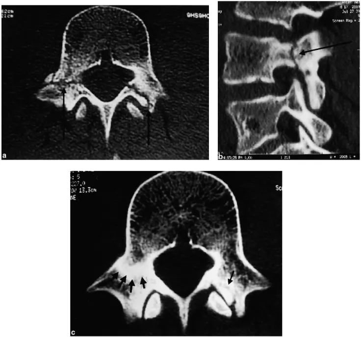
Fig. 3. Case 1: (a) Axial CT scan reveals both right pedicular and left pars interarticularis fractures (arrows). (b) Sagittal reformatted image nicely shows the pediculolysis (arrow). (c) Axial CT scan after treatment demonstrates healing in both fractures (arrows).
congenital pedicular defects do not show hypertrophic and irregular sclerotic pedicular margins as pediculolysis [2].
Surgical intervention is indicated if conservative treat-ment (rest and bracing) fails. Pediculolysis associated with contralateral pars interarticularis defect is known to cause a high degree of instability [13]. Various surgical methods such as compression fixation [16], decompression of bony over-growth and excision of the pedicle with lateral fusion [4], bone grafting of the spondylolytic defect, drilling and grafting the pedicle, and pedicular screw fixation [2] are used.
In conclusion, radiologists should be aware of pedicular stress fractures and should consider in the differential diagnosis of focal pathologic signal changes of the pedicle
at MR imaging. This is especially true for radiologists who perform MR imaging as a second-line imaging method for low back pain.
References
[1] Araki T, Harata S, Nakano K, Satoh T. Reactive sclerosis of the pedicle associated with contralateral spondylolysis. Spine 1992; 17(11):1424 – 6.
[2] Gunzburg R, Fraser RD. Stress fracture of the lumbar pedicle. Spine 1991;16(2):185 – 9.
[3] Kalideen JM, Satyapal KS, Bayat F. Case report: pedicular cleft associated with bilateral pars interarticularis defects and transverse Fig. 4. Case 2: (a) Axial CT scan shows rotational anterior translation of L3 over L4. (b) Axial image of an upper level demonstrates elongation and deformation of right L3 vertebra. Left pedicle is completely normal. (c) Oblique sagittal reformatted image reveals the fracture line. Note marginal sclerosis and, narrowing of L3 – L4 intervertebral joint.
process hypoplasia of a fifth lumbar vertebra. Br J Radiol 1994; 67:1136 – 8.
[4] Aland C, Rineberg BA, Malberg M, Fried SH. Fracture of the pedicle of the fourth lumbar vertebra associated with contralateral spondylo-lysis. J Bone Jt Surg, Am Vol 1986;68(9):1454 – 5.
[5] Tribus CB, Bradford DS. Bilateral pedicular stress fractures after suc-cessful posterior spinal fusion for adult idiopathic scoliosis. Spine 1993;18(9):1222 – 5.
[6] Knight RQ, Chan DPK. Idiopathic scoliosis with unusual stress frac-ture of the pedicle within solid fusion mass. Spine 1992;17(7):849 – 51. [7] Stanley D, Smith TWD. Contralateral pedicle stress fracture. Spine
1990;15(6):598 – 9.
[8] Robertson PA, Grobler LJ. Stress fracture of the pedicle. Spine 1993;18(7):930 – 2.
[9] Maurer SG, Wright KE, Bendo JA. Iatrogenic spondylolysis leading to contralateral pedicular stress fracture and unstable spondylolisthe-sis. Spine 2000;25(7):895 – 8.
[10] Bensaid AH, Dietemann JL, Kastler B, Gangi A, Jeung MY, Wack-enheim A. Neurofibromatosis with dural ectasia and bilateral sym-metrical pedicular clefts: report of two cases. Neuroradiology 1992; 34:107 – 9.
[11] Morin ME, Palacios E. The aplastic hypoplastic lumbar pedicle. Radiology 1974;122:639 – 42.
[12] Tomsick TA, Lebowitz ME, Campbell C. The congenital absence of pedicles in the thoracic spine. Radiology 1974;111:587 – 9.
[13] Guillodo Y, Botton E, Saraux A, Le Goff P. Contralateral spondylo-lysis and fracture of the lumbar pedicle in an elite female gymnast. Spine 2000;25(19):2541 – 3.
[14] Chong VHF, Htoo MM. Pedicular stress fracture in the lumbar spine. Australas Radiol 1997;41:306 – 7.
[15] Cyron BM, Hutton WC. The fatigue strength of the lumbar neural arch in spondylolysis. J Bone Jt Surg, Br Vol 1978;60(2):234 – 8. [16] Weatherley CR, Mehdian H, Berghe LV. Low back pain with fracture
of the pedicle and contralateral spondylolysis. J Bone Jt Surg, Br Vol 1991;73(6):990 – 3.
[17] Traughber PD, Havlina JM. Bilateral pedicle stress fractures: SPECT and CT features. J Comput Assist Tomogr 1991;15(2):338 – 40. [18] Maldague B, Malghem J. La spondylolyse en activite. J Radiol 1985;
66:263 – 74.
[19] Sherman FC, Wilkinson RH, Hall JE. Reactive sclerosis of a pedicle and spondylolysis in the lumbar spine. J Bone Jt Surg 1997;59A: 49 – 54.
[20] Wilkinson RH, Hall JE. The sclerotic pedicle: tumor or pseudotumor? Radiology 1974;111:683 – 8.
[21] O’beirne JG, Horgan JG. Stress fracture of the lamina associated with unilateral spondylolysis. Spine 1988;13(2):220 – 2.
[22] Garber JE, Wright AM. Unilateral spondylolysis and contralateral pedicle fracture. Spine 1986;11(1):63 – 6.
[23] Lee JK, Yao L. Stress fractures: MR imaging. Radiology 1988; 169:217 – 20.