doi:10.3944/AOTT.2013.2934
Correspondence: Selami Çakmak, M.D., Ass. Prof. GATA Haydarpafla E¤itim Hastanesi, Ortopedi ve Travmatoloji Klini¤i, Üsküdar, 34668, ‹stanbul, Turkey.
Tel: +90 216 - 542 20 20 e-mail: [email protected] Submitted: June 6, 2012 Accepted: March 30, 2013 ©2013 Turkish Association of Orthopaedics and Traumatology
Available online at www.aott.org.tr doi:10.3944/AOTT.2013.2934 QR (Quick Response) Code: Objective: The aim of this study was to evaluate the demographic characteristics of patients with
bilateral bisphosphonate-related low-energy femoral shaft fractures.
Methods: The clinical registry was reviewed for patients with bisphosphonate-related low-energy
fractures localized at femoral shaft between January 2008 and January 2012. Patients with a diagnosis of postmenopausal osteoporosis, bisphosphonate usage of at least 5 years and prodromal pain prior to fracture were included the study.
Results: Five women met the inclusion criteria. All patients had bilateral low-energy sequential
femoral shaft fractures. Fracture patterns were similar and atypical (transverse-short oblique fractures with lateral cortical thickening). Mean period of bisphosphonate treatment was 8.6 years. Mean patient age was 76.2 years. Union time of three patients was between 20 and 28 weeks. The remain-ing two fractures were revised for delayed union or nonunion.
Conclusion: Long-term (over 5 years) use of bisphosphonates may cause insufficiency fractures due to
increased fragility and brittleness which have a close relationship with depressed bone remodeling. While there is still no causal relationship between bisphosphonates and atypical, low-energy femoral shaft frac-tures, we have some concerns about the optimal usage time and long-term safety of bisphosphonate drugs.
Key words: Atypical femoral shaft fracture; bisphosphonate; subtrochanteric fracture.
Osteoporosis is a skeletal disease in which a low-densi-ty and micro-architectural defects in bone tissue
increase susceptibility to fractures.[1]
Currently it is estimated that more than 10 million patients have been
diagnosed with osteoporosis in the US alone.[2]
Considering a lifetime fracture risk of 40% for white females, these cases represent an approximately 9
mil-lion new osteoporotic fractures per annum.[3]
Prevention of further bone resorption and fractures is the backbone of treatment. Following current evi-dence-based guidelines, bisphosphonates are often first
considered for the treatment of osteoporosis.[4]
This class of medication may account more than 80% of total prescriptions given for osteoporosis in some countries and their efficiency in treatment of post-menopausal osteoporosis was reported to reduce verte-bral fractures by nearly 50% and hip fractures by 20 to
50%.[5]
Bisphosphonates were shown to be well
tolerat-ed and safe in large-scale clinical trials.[6]
Several rare and potentially serious adverse events have been reported to be associated with long-term bisphospho-nate use from post-marketing reports and
epidemio-Bilateral low-energy sequential femoral shaft fractures
in patients on long-term bisphosphonate therapy
Selami ÇAKMAK1, Mahir MAH‹RO⁄ULLARI2, Kenan KEKL‹KÇ‹1, Enes SARI1, Baran ERD‹K3, Osman RODOP1
1
Department of Orthopaedics and Traumatology, GATA Haydarpafla Training Hospital, ‹stanbul, Turkey;
2
Department of Orthopaedics and Traumatology, Faculty of Medicine, Medipol University, ‹stanbul, Turkey;
3
logical studies. These adverse events include dyspepsia, nausea, muscular pain, osteonecrosis of the jaw (ONJ),
and atrial fibrillation.[7]
In recent years, however, there have been increasing numbers of cases or case series about atypical subtrochanteric/femoral shaft fractures
related to bisphosphonate treatment.[8-19]
The aim of this study was to evaluate the demo-graphic characteristics of patients with bilateral bispho-sphonate-related low-energy femoral shaft fractures. Patients and methods
In this retrospective observational study, the clinical reg-istry of GATA Haydarpafla Training Hospital (Istanbul, Turkey) was reviewed for patients with femoral fractures between January 2008 and January 2012. Patients with a
diagnosis of low-energy fractures at the femoral shaft were sorted out and along with the radiological appear-ance of these fractures, patients’ demographics were recorded. Fractures occurring from a fall from standing height without any significant trauma were assessed as ‘low-energy fractures’. Patients with a diagnosis of osteoporosis using bisphosphonate drugs for a minimum of 5 years and who had prodromal pain prior to fracture were included in this study. Included patients’ fractures were labeled ‘bisphosphonate-related low-energy frac-ture’. Local ethical committee approval was obtained. Results
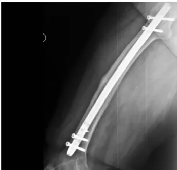
Fifty-two patients had femoral shaft fractures. Patient histories were reviewed for low-energy fractures and Fig. 1. Radiographies of patient no. 5. (a) The patient had left femur shaft fracture without any significant trauma and was treated with inter-locked intramedullary nailing. (b) After 5 months, she had thigh pain at right leg and admitted to our clinic. (c) There was an impend-ing fracture site at right femoral shaft showimpend-ing unique pattern; transverse, unicortical and showimpend-ing beakimpend-ing and cortical thickenimpend-ing at anterolateral cortex.
Fig. 2. MR images of patient no. 5. MRI sections of impending fracture localized at the right femoral shaft. (a, b) Unicortical fracture line localized at lateral cortex of femoral shaft, medullary and soft-tissue edema seen around fracture site and periosteal reaction with cortical thickening can be seen.
(a) (b)
(a) (b)
osteoporosis diagnosis. All patients used alendronate. Five patients with bilateral atypical femoral shaft frac-tures treated surgically were included in our study. Radiographic findings for atypical femoral shaft frac-tures were short-oblique-transverse fracture, transverse fracture with medial spike, cortical thickening or hyper-trophy at the lateral cortex, and stress fracture line. Patients’ mean age was 76.2 (range: 70 to 87) and mean period of bisphosphonate treatment was 8.6 (range: 5 to 14) years. All five patients were female. All patients com-plained of prodromal pain and general discomfort in the affected thigh days to weeks before the impending frac-ture. Union time of three patients was between 20 and 28 weeks. Patient number 2 had delayed union at 5 months after initial surgery and the fracture revised using an exchange nail with interlocked intramedullary nail. Patient number 4 had no union at 8 months after intramedullary nailing and the fracture was revised using open reduction, plate fixation and bone autografting. Union time was 26 weeks for both patients. Radiographs and MRI appearance of impending fracture of Patient number 5 are shown in Figures 1-3. Demographic data is given in Table 1.
Discussion
Osteoporosis is a common health problem in the elder-ly population. Increased risk of fracture can result in
disability, morbidity, decreased life quality, higher costs, and mortality. In postmenopausal osteoporotic patients, bisphosphonates have been shown to decrease
the risk of vertebral and femoral neck fractures.[20-23]
Bisphosphonates are potent inhibitors of bone resorp-Fig. 3. This impending fracture localized at the right femur diaphysis
was also treated with interlocked intramedullary nailing.
Patient 1 Patient 2 Patient 3 Patient 4 Patient 5
Age 74 72 70 78 87
Sex Female Female Female Female Female
Localization Femoral shaft Femoral distal Femoral shaft Femoral proximal Femoral shaft (Bilateral) shaft (Left) (Bilateral) shaft (Left) (Bilateral)
Femoral shaft (Right) Femoral shaft (Right)
Alendronate therapy 7 10 14 5 7
duration (year) years years years years years
Alendronate 10 mg/day 10 mg/day 10 mg/day 10 mg/day 10 mg/day
dosage for 8 years;
70 mg/week for 6 years
Fracture pattern Short oblique-transverse Transverse fracture Transverse short oblique Transverse fracture Transverse stress fracture (Both sides) with medial spike, fracture with thickening with medial spike fracture line at the
lateral cortical at the lateral cortex (Both sides) lateral cortex and mild thickening (Left) (Right) and short oblique cortical hypertrophy fracture with medial (Right) and transverse
spike (Left) fracture (Left)
Prodromal pain + + + + +
Treatment Interlocked Interlocked intramedullary Expandable Expandable intramedullary Interlocked intramedullary nailing (Right) and intramedullary nailing (revised with plate intramedullary
nailing Expandable intramedullary nailing (Both sides) fixation and autografting) nailing (Both sides) nailing (revised with interlocked (Right) and plate (Both sides)
intramedullary nailing) (Left) fixation (Left)
Time to union Right (24) Right (24) Right (28) Right (26) Right (20) (week) Left (22) Left (26) Left (26) (after revision surgery) (impending fracture)
(after revision surgery) Left (27) Left (26)
tion that inhibit osteoclast function and induce
osteo-clast apoptosis.[13,24]
Alendronate was approved by the FDA in 1995 for the treatment of osteoporosis. The pathophysiology of bisphosphonate associated fractures is thought to be associated with the inhibition of bone turnover and repair of microscopic trauma. In recent decades, human biopsy and experimental animal studies have reported suppressed bone turnover with
bisphos-phonate use.[17,25-28]
As a result, a cycle of defective repair and continual micro-trauma compounded over time gradually weakens and creates more mineralized and brittle bones, an architectural conduit for transverse or
insufficiency fractures.[13,29-31]
It has been advocated that some osteoclastic activity is necessary to repair
micro-damage and continuity of remodelization.[17,32]
In a recent study, Bala et al. reported that long-term (6 to 10 years) alendronate use compromises the micro-mechanical properties of bone and problems were relat-ed with lower crystallinity, associatrelat-ed with elastic
mod-ulus and contact hardness.[33]
Our cases also had a mini-mum of 5 years of alendronate usage (range: 5 to 14 years). Extended alendronate use may diminish mechanical properties of bone and may result in more brittle bone which in turn can result in insufficiency
fractures. In a detailed review, Ott[34]
demonstrated the mechanism of action of bisphosphonates. First, the author emphasized the common misunderstanding that ‘bisphosphonates build bone’. Second, she cited an arti-cle on fluoride treatment for osteoporosis treatment and underlined that despite increased bone density, the bone becomes more fragile. This can be example for this clinical picture of what bisphosphonate drugs do. It was also reported that overall fracture risk is similar in patients with more than 5 years of bisphosphonate use and individuals who stopped therapy.
Recently, atraumatic, low-energy or insufficiency femoral shaft/subtrochanteric site fractures have been reported in patients on prolonged bisphosphonate ther-apy.[8-19]
These studies are evaluated in detail in Table 2. Similar to our cases, some reports were of bilateral
sequential femoral shaft fractures.[10,11]
Most of the alen-dronate-related fractures in literature reported differ-ences from usual osteoporotic fractures, high-energy fractures and periprosthetic fractures, including: 1. Minor or no trauma
2. Alendronate use history for postmenopausal period 3. Prodromal (thigh) pain prior to fracture
4. Different localization from those commonly seen in osteoporotic fractures (spine, hip, wrist…etc.) 5. Bilaterality (sequential or simultaneous or
impend-ing)
6. Cortical hypertrophy or thickening at fracture site on radiographs
7. Unusual fracture pattern (transverse or short oblique; medial spike/beak)
8. Delayed fracture union time
All 5 of our cases showed all the features mentioned above. These characteristics may be useful in the diag-nosis of ‘alendronate-related low-energy fractures’.
The subtrochanteric site of the femur is subjected to maximal bending forces and is known as its strongest
region.[13,18,35]
Low-energy stress fractures usually occur in
athletes or military recruits.[36]
Bilateral femoral fractures are also usually seen as pathological fractures or follow-ing high-energy trauma such as motor-vehicle accidents. Subtrochanteric fractures (especially bilateral) occurring after low-energy events are rare and are resultant of an underlying cause that weakens the bone. With inhibition of osteoclasts and impairment of the remodelization cycle, microarchitectural damage at the site of highest stress may occur. Gaeta et al. analyzed the CT scans of tibial stress fractures and found some resorption areas
inside the typical cortical thickness site.[37]
Our radiolog-ical findings on the contralateral impending fracture can be postulated to result from chronic suppression of bone remodeling by long-term bisphosphonate treatment with accumulation of old, highly-mineralized osteons and increased brittleness of bone (especially caused by
increased Young’s modulus).[38,39]
Bisphosphonates bind the bone tightly and the skele-tal half-life of alendronate has been estimated at over 5
or 10 years.[4,20,24,40-43]Therefore, nonunion rates for such
insufficiency fractures may be higher and union may be slower or incomplete even following the discontinuation of bisphosphonates. Weil et al. studied the surgical out-comes of bisphosphonate-related fractures and reported a much higher failure rate with intramedullary nailing
which requires revision procedures.[44]
In our cases, we also detected longer union time after surgical treatment and one patient required revision due to nonunion after 8 months (with additional autografting). We now believe that these bisphosphonate-related fractures must be thoroughly evaluated and treated using different and augmented approaches, such as autografting or recom-binant bone morphogenetic proteins. Treatment modality should be chosen individually.
Questions and concerns for the long-term safety of bisphosphonates have arisen from reports of atypical femoral fractures, with studies reporting both increased
or no increased risk available in the literature.[15,45-52]
A meta-analysis based on database of three large random-ized studies found that the occurrence of sub-trochanteric or diaphyseal femur fracture (i.e. insuffi-ciency fracture of the femur) was very rare, although there were insufficient numbers of events to reach
defin-No Study, Patients Age Sex Duration of Used Additional Prodromal Fracture Bilateral Fracture Biochemical Bone BMD Time to year, no. drug use drug drugs pain site pattern on marker biopsy -2.5 union journal (years) X-ray (months) 1 Odvina CV, 2005, 9 6 0 F 3-8 Alendronate Estrogen (3), NM ST (4), sacrum, 2/9 NM N-terminal + N M Delayed
J Clin Endocrinol Metab
prednisone (2) rib, ischium, telopeptide (6) pubic rami, was low (7/9) lumbar spine 2 Schneider JP, 2006, 1 5 9 F 7 Alendronate Estrogen + S T -Cortical NM NM NM -Geriatrics
thickness, transverse with spike
3 Cheung RK, 2007, 1 8 2 F 10 NM NM NM FS + N M High + N M N M
Hong Kong Med J
OH-proline 4 Demiralp B, 2007, 1 6 5 F 7 Alendronate Steroid, + FS + Fracture line, NM NM NM NM
Arch Orthop Trauma Surg
thyroxine cortical thickening, bowing deformity 5 Goh SK, 2007, 9 66.9 F 4.2 (2.5-5) Alendronate NM + ST -Simple, NM NM + (normal) Delayed JBJS-Br 5/9 transverse, short oblique;
cortical thickening on the lateral
6 Lee P, 2007, 1 7 3 F 1,5 Alendronate NM + FS + NM NM NM + (-2.8) NM J Endocrinol Invest 7 Kwek EB, 2008, 17 66 F 4.4 (2-8) Alendronate Calcium + 13/17 ST; FS + Cortical NM NM 10/17 NM Injury (9*) (10/17) thickening; transverse or short oblique; medial cortical
spike 8 Lenart BA, 2008, 15 NM NM 5,4 Alendronate NM NM ST/FS NM Simple NM NM NM NM NEJM transverse or
oblique fracture with beaking of the cortex and diffuse cortical
thickening 9 Neviaser AS, 2008, 19 69.4 F 6.9 Alendronate None NM ST; FS NM Simple transverse NM NM NM NM JOT (min 4)
fracture, unicortical beak in area of cortical hypertrophy
10 Sayed-Noor AS, 2008, 1 7 2 F 7 Alendronate Calcium + S T + Transverse, NM NM NM 6 Acta Orthop thickening of
the lateral femoral cortex and medial spiking at the
fracture site
DS: distal shaft, FS: femoral shaft, ST: subtrochanteric; NM: not mentioned.
*Nine of these patients are also mentioned in the f
ifth study by Goh et al.
Table
2
.
No Study, Patients Age Sex Duration of Used Additional Prodromal Fracture Bilateral Fracture Biochemical Bone BMD Time to year, no. drug use drug drugs pain site pattern on marker biopsy -2.5 union journal (years) X-ray (months) 11 Visekruna M, 2008, 3 51-75 NM 5-10 NM Estrogen (2), NM NM 2/3 NM NM + + 22 J Clin Endocrin Metab prednisone (3) 2/3 (normal) 12 Ali T, 2009, 1 8 2 N M 8 NM NM -FS -Transverse C-terminal -+ 3 Age Aging with marked telopeptide (normal) cortical crosslinks thickening
were slightly elevated
13 Armamento-Villareal R, 15 43-75 F (12) 4-10 Alendronate NM NM FS (7) 2/15 NM NM NM NM NM
2009, Calsif Tissue Int
M (3) 14 Bush LA, 2009, 1 8 5 F 6 Risedronate Steroid + S T
-Mild, diffuse cortical
NM
NM
NM
NM
Radiol Case Rep
thickening and a
focal, domed, conical,
projection along the lateral cortex
15 Capeci CM, 2009, 7 6 1 F 8.6 Alendronate None + 4/7 ST (6); FS (1) + Cortical thickening, NM NM NM NM JBJS Am (5-13)
transverse, cortical spiking or beaking
16 Glennon DA, 2009, 6 60-87 F 1.5-16 Alendronate (5), NM NM ST +(1) Transverse, NM NM NM NM Bone risedronate (1)
unicortical beaking, cortical thickening
17 Sayed-Noor AS, 2009, 2 78; 55 F 9; 8 Alendronate Vit D + ST 1/2 Lateral cortical NM NM NM 5; 9 CORR (Periprosthetic) reaction, transvers fracture 18 Goddard MS, 2009, 1 6 7 F 16 Alendronate NM NM FS + Cortical thickening, NM NM NM NM Orthopedics unicortical beaking, transverse 19 Grasko JM, 2009, 1 N M N M N M N M Steroid NM ST NM NM NM NM NM NM
J Oral Maxillofac Surg
20 Ing-Lorenzini K, 2009, 8 6 7 F 16 months Alendronate Proton pump +(2) ST 4/8 Cortical thickening NM NM NM Delayed Drug Safety - 8 years inhibitor (7), at lateral cortex (2/8) prednisone (4) with a horizontal fracture line 21 Lee JK, 2009, 1 8 2 F 8 A lendronate N M N M N M + Horizontal fracture N M N M N M N M
Int J Rheum Dis
line involving the thick lateral cortex with short oblique fracture pattern
22 Leung F, 2009, 10 55-92 F 0.5-10 Alendronate N M N M ST, FS NM Femoral diaphyseal NM NM NM NM BMJ Case Rep
cortical thickening and lateral cortex
beaking 23 Schilcher J, 2009, 5 >75 F 5.8 NM NM NM FS 1/5 NM NM NM NM NM Acta Orthop
DS: distal shaft, FS: femoral shaft, ST: subtrochanteric; NM: not mentioned.
*Nine of these patients are also mentioned in the f
ifth study by Goh et al.
Table
2
.
[Contunued]
No Study, Patients Age Sex Duration of Used Additional Prodromal Fracture Bilateral Fracture Biochemical Bone BMD Time to year, no. drug use drug drugs pain site pattern on marker biopsy -2.5 union journal (years) X-ray (months) 24 Schneider JP, 2009, 3 59-66 F 5-9 NM NM 1/3 NM 2/3 NM NM NM NM NM Geriatrics 25 Somford MP, 2009, 1 7 6 F 8 Alendronate Prednisone + ST/FS -N M + + + NM
J Bone Miner Res
26 Atik S, 2010, 1 7 6 F 10 NM NM NM FS NM Transverse; medial NM NM + (T score NM
Eklem Hastalik Cerrahisi
spike at cortical 3.55 at hip) thickness site 27 Black DM, 2010, NEJM 7 69-83 NM >2 NM NM NM NM NM NM NM NM NM NM 28 Bunning RD, 2010, PM&R 4 49-59 F (3), 4.5-6 Alendronate (2), NM + ST/FS 1/4 Medial cortical NM NM NM NM M (1) pamidronate (1) thickening 29 Cermak K, 2010, CORR 4 59-77 F > 5 Alendronate NM -S T 1/4 Transverse fracture NM NM NM NM
with external cortical bone reaction and
medial cortical spike
30 Chan SS, 2010, 15 50-81 F 4-14 Alendronate NM NM ST NM Medial beak NM NM NM NM Am J Roentgenol 31 Das De S, 2010, 12 51-75 F 4.6 Alendronate NM NM ST 6/12 Thickening of NM NM NM Nonunion JBJS Br lateral femoral 3/12
cortex, transverse or slightly oblique
fracture 32 Edwards MH, 2010, 1 6 0 F 8 Alendronate Prednisone + FS + Minor cortical NM NM NM NM Osteoporos Int thickening 33 Girgis CM, 2010, 17 5 N M N M N M N M N M N M N M N M N M N M Med J Aus 34 Giusti A, 2010, 8 67.8 F 3-192 NM NM NM NM NM NM NM NM NM NM Bone months 35 Ha YC, 2010, 11 F 4.5 (3-10) NM NM NM NM NM NM NM NM NM 5 CORR 36 Isaacs JD, 2010, 41 73.7 F 7.1 Alendronate (40) NM 29/41 ST/MS 18/41 Tr an sv er se f ra ct u re N M N M N M N M CORR risedronate (1)
line and lateral
cortical thickening adjacent to the
fracture 37 Koh JSB, 2010, JOT 16 68 F 4.5 NM NM 7/16 ST/MS NM “dreaded black NM NM NM NM
line” within the cortical stress reaction on both anteroposterior and lateral views
38 Napoli N, 2010, 1 5 6 F 6 N M Prednisone -FS -NM NM NM NM NM Osteoporos Int
DS: distal shaft, FS: femoral shaft, ST: subtrochanteric; NM: not mentioned.
*Nine of these patients are also mentioned in the f
ifth study by Goh et al.
Table
2
.
[Contunued]
No Study, Patients Age Sex Duration of Used Additional Prodromal Fracture Bilateral Fracture Biochemical Bone BMD Time to year, no. drug use drug drugs pain site pattern on marker biopsy -2.5 union journal (years) X-ray (months) 39 Osugi K, 2010, 3 7 4 F NM Alendronate(2) NM NM FS 2/3 Spike-shaped NM NM NM NM Acta Orthop risedronate (1) cortical thickening laterally 40 Patel VC, 2010, 1 6 5 F 2 Ibandronate NM + FS + NM NM NM NM NM Orthopedics 41 Porrino JA, 2010, 4 66.5 F > 3 Alendronate NM 3/+ ST/FS 2/4 Localized NM NM NM NM Am J Roentgenol
lateral cortical thickening,
and the appearance of the fracture lucency 42 Venkatanarasimha N, 2 69.5 F 7.4 Alendronate Prednisolone + ST/FS 1/2 Beaking of NM NM NM NM 2010 cortex lateral
femur and marked cortical hypertrophy
43 Banffy MB, 2011, 34 68.5 6 N M N M N M S T 6/34 NM NM NM NM NM CORR 44 Gomberg SJ, 2011, 1 6 3 1 3 Alendronate NM + S T + NM NM NM NM NM
J Clin Endocrin Metab
45 Gudena R, 2011, 1 7 4 F 10 Alendronate None + FS + Lateral cortical NM NM NM 4 J Osteop cortical thickening of mid-diaphysis 46 Gunawardena I, 2011, 1 6 7 F 2 Alendronate Glucocorticoids + S T + Transverse NM NM T score NM Am J Geriat Pharma fracture pattern was -2 at hip
on the lateral half of the femoral cortex 47 Weil YA, 2011, 15 73 F 7.8 Alendronate NM NM FS (9), ST(4), 2/15 NM + N M + (T score NM JOT (4-13) DS(4) (5) low was -3 at normal range lumbar (carboxy-spine) terminal collagen crosslink) (1) osteocalcin was low
DS: distal shaft, FS: femoral shaft, ST: subtrochanteric; NM: not mentioned.
*Nine of these patients are also mentioned in the f
ifth study by Goh et al.
Table
2
.
[Contunued]
itive conclusions.[46]
Several controlled epidemiological studies examining the association between bisphospho-nate use and insufficiency fractures have also been pub-lished. Using a cohort created out of the Danish Hospital Discharge Registry and Prescription Database, Abrahamsen et al. found that high adherence to treat-ment was associated with a reduced insufficiency frac-ture risk, further suggesting that insufficiency fracfrac-tures were caused by the extensive underlying osteoporosis
instead of alendronate therapy.[45] Other studies have
shown that atypical fractures have not increased.[47,49]
In contrast, a notable interconnection between long-term bisphosphonate use and insufficiency fractures has been reported by controlled observational studies. A
Canadian report suggests that the long-term use (≥5
years) was associated with increased risk of insufficiency fracture of the femur (adjusted Odds ratio 2.74; 95% CI,
1.25-6.02).[50]
This association was not present in short-term users. Lenart et al. also reported significantly greater proportion of subtrochanteric or femoral shaft fractures in comparison to intertrochanteric or femoral neck fractures in patients who received long-term
bis-phosphonate therapy.[16]Another case-control study
sug-gested that prolonged use of alendronate may cause sup-pression of bone remodeling and may be associated with
insufficiency fractures of the femur.[13] We also believe
that the long-term use (>5 years) of alendronate may be associated with its related fractures.
At the beginning of 2010, the FDA announced a report regarding bisphosphonate-related atypical frac-tures and reported no clear connection. However, the FDA also advised physicians to prescribe bisphospho-nates according to guidelines and follow patients
close-ly.[53]
On the other hand, the Medicines and Healthcare products Regulatory Agency (MHRA), the drug regula-tory agency in the UK, recommended the cessation of alendronate therapy in patients with atypical bisphos-phonate-related fractures and the assessment of the
ben-efits of alendronate treatment.[54]
We believe that patients with atypical, bisphosphonate-related fractures should be individually reevaluated for risk factors with bone densitometry and biochemical bone turnover markers before making a decision on whether a drug holiday is necessary. The length of the drug holiday should be determined by close observation, bone miner-al densitometry and biochemicminer-al bone turnover markers (urine cross-linked N-telopeptides of Type 1 collagen, cross-linked C-telopeptides of Type 1 collagen; bone-specific alkaline phosphatase, osteocalcin, pro-peptide of
Type 1 collagen).[34,55]Consultation with an
endocrinol-ogist may be helpful in the evaluation process and frac-ture risk assessment may be completed using the
FRAX®, WHO Fracture Risk Assessment Tool.[56]
Teriparatide may be kept in mind for treatment
contin-uation.[19]
We did not perform any animal study or histomor-phological assessment for patients. There was also no detection of biochemical bone turnover markers. These features were the limitations of our study. Continuous assessment of bone turnover markers and their relation-ship with bone mineral densitometry measures may be helpful to determine the actual status of bone metabo-lism occurring inside the body which in turn may assist in the decision to continue bisphosphonate use.
In conclusion, long-term (over 5 years) use of bis-phosphonates may cause insufficiency fractures due to increased fragility and brittleness which have a close relationship with depressed bone remodeling. Although there is still no causal relationship between bisphospho-nates and atypical, low-energy femoral shaft fractures, we have some concerns about the optimal usage time and long-term safety of bisphosphonate drugs.
Conflicts of Interest: No conflicts declared. References
1. Consensus development conference: diagnosis, prophylaxis, and treatment of osteoporosis. Am J Med 1993;94:646-50. 2. Melton LJ. Report of the Surgeon General’s Workshop on
Osteoporosis and Bone Health: Prevalence and Burden of Illness [Internet]. Department of Health and Human Services. December 12-13, 2002 [cited 2012 Mar 12]. Available from: http://www.ncbi.nlm.nih.gov/books/NBK44687/pdf/TOC.pdf. 3. Johnell O, Kanis JA. An estimate of the worldwide preva-lence and disability associated with osteoporotic fractures. Osteoporos Int 2006;17:1726-33.
4. Watts NB, Bilezikian JP, Camacho PM, Greenspan SL, Haris ST, Hodgson SF, et al. American Association of Clinical Endocrinologists Medical Guidelines for Clinical Practice for the diagnosis and treatment of postmenopausal osteoporosis. Endocr Pract 2010;16:1-37.
5. Devold HM, Doung GM, Tverdal A, Furu K, Meyer HE, Falch JA, et al. Prescription of anti-osteoporosis drugs dur-ing 2004-2007 - a nationwide register study in Norway. Eur J Clin Pharmacol 2010;66:299-306.
6. Wells GA, Cranney A, Peterson J, Boucher M, Shea B, Robinson V, et al. Alendronate for the primary and second-ary prevention of osteoporotic fractures in postmenopausal women. Cochrane Database Syst Rev 2008;1:CD001155. 7. Schubert M, Klatte I, Linek W, Müller B, Döring K, Eckelt
U, et al. The Saxon bisphosphonate register - therapy and prevention of bisphosphonate-related osteonecrosis of the jaws. Oral Oncol 2012;48:349-54.
8. Atik OS, Suluova F, Görmeli G, Yildirim A, Ali AKh. Insufficiency femoral fractures in patients undergoing pro-longed alendronate therapy. Eklem Hastalik Cerrahisi 2010; 21:56-59.
9. Aydogan NH, Gul D, Ozturk A, Alemdaroglu KB, Kara T, Gultac E. Fractures of the lower limb following
bisphospho-nate use and their surgical treatment: five case reports. [Article in Turkish] Acta Orthop Traumatol Turc 2011;Suppl 1:12.
10. Capeci CM, Tejwani NC. Bilateral low-energy simultaneous or sequential femoral fractures in patients on long-term alen-dronate therapy. J Bone Joint Surg Am 2009;91:2556-61. 11. Cheung RK, Leung KK, Lee KC, Chow TC. Sequential
non-traumatic femoral shaft fractures in a patient on long-term alendronate. Hong Kong Med J 2007;13:485-9. 12. Demiralp B, Ilgan S, Ozgur Karacalioglu A, Cicek EI,
Yildrim D, Erler K. Bilateral femoral insuffiency fractures treated with inflatable intramedullary nails: a case report. Arch Orthop Trauma Surg 2007;127:597-601.
13. Goh SK, Yang KY, Koh JS, Wong MK, Chua SY, Chua DT, et al. Subtrochanteric insufficiency fractures in patients on alendronate therapy: a caution. J Bone Joint Surg Br 2007; 89:349-53.
14. Isaacs JD, Shidiak L, Harris IA, Szomor ZL. Femoral insuf-ficiency fractures associated with prolonged bisphosphonate therapy. Clin Orthop Relat Res 2010;468:3384-92.
15. Lenart BA, Lorich DG, Lane JM. Atypical fractures of the femoral diaphysis in postmenopausal women taking alen-dronate. N Engl J Med 2008;358:1304-6.
16. Lenart BA, Neviaser AS, Lyman S, Chang CC, Edobor-Osula F, Steele B, et al. Association of low-energy femoral fractures with prolonged bisphosphonate use: a case control study. Osteoporos Int 2009;20:1353-62.
17. Odvina CV, Zerwekh JE, Rao DS, Maalouf N, Gottschalk FA, Pak CY. Severely suppressed bone turnover: a potential com-plication of alendronate therapy. J Clin Endocrinol Metab 2005;90:1294-301.
18. Sayed-Noor AS, Sjödén GO. Case reports: two femoral insufficiency fractures after long-term alendronate therapy. Clin Orthop Relat Res 2009;467:1921-6.
19. Visekruna M, Wilson D, McKiernan FE. Severely sup-pressed bone turnover and atypical skeletal fragility. J Clin Endocrinol Metab 2008;93:2948-52.
20. Bone HG, Hosking D, Devogelaer JP, Tucci JR, Emkey RD, Tonino RP, et al. Ten years’ experience with alendronate for osteoporosis in postmenopausal women. N Engl J Med 2004; 350:1189-99.
21. Eastell R, Barton I, Hannon RA, Chines A, Garnero P, Delmas PD. Relationship of early changes in bone resorp-tion to the reducresorp-tion in fracture risk with risedronate. J Bone Miner Res 2003;18:1051-6.
22. Bilezikian JP. Efficacy of bisphosphonates in reducing fracture risk in postmenopausal osteoporosis. Am J Med 2009;122: S14-21.
23. Tonino RP, Meunier PJ, Emkey R, Rodriguez-Portales JA, Menkes CJ, Wasnich RD, et al. Skeletal benefits of alen-dronate: 7-year treatment of postmenopausal osteoporotic women. Phase III Osteoporosis Treatment Study Group. J Clin Endocrinol Metab 2000;85:3109-15.
24. Drake MT, Clarke BL, Khosla S. Bisphosphonates: mecha-nism of action and role in clinical practice. Mayo Clin Proc 2008;83:1032-45.
25. Mashiba T, Hirano T, Turner CH, Forwood MR, Johnston CC, Burr DB. Suppressed bone turnover by bisphosphonates increases microdamage accumulation and reduces some bio-mechanical properties in dog rib. J Bone Miner Res 2000; 15:613-20.
26. Li J, Mashiba T, Burr DB. Bisphosphonate treatment sup-presses not only stochastic remodeling but also the targeted repair of microdamage. Calcif Tissue Int 2001;69:281-6. 27. Cao Y, Mori S, Mashiba T, Westmore MS, Ma L, Sato M, et
al. Raloxifene, estrogen, and alendronate affect the processes of fracture repair differently in ovariectomized rats. J Bone Miner Res 2002;17:2237-46.
28. Armamento-Villareal R, Napoli N, Diemer K, Watkins M, Civitelli R, Teitelbaum S, et al. Bone turnover in bone biop-sies of patients with low-energy cortical fractures receiving bisphosphonates: a case series. Calcif Tissue Int 2009;85:37-44.
29. Stepan JJ, Burr DB, Pavo I, Sipos A, Michalska D, Li J, et al. Low bone mineral density is associated with bone micro-damage accumulation in postmenopausal women with osteo-porosis. Bone 2007;41:378-85.
30. Sellmeyer DE. Atypical fractures as a potential complication of long-term bisphosphonate therapy. JAMA 2010;304:1480-4.
31. Burr DB, Forwood MR, Fyhrie DP, Martin RB, Schaffler MB, Turner CH. Bone microdamage and skeletal fragility in osteoporotic and stress fractures. J Bone Miner Res 1997;12:6-15.
32. Mashiba T, Turner CH, Hirano T, Forwood MR, Jacob DS, Johnston CC, et al. Effects of high-dose etidronate treatment on microdamage accumulation and biomechanical properties in beagle bone before occurrence of spontaneous fractures. Bone 2001;29:271-8.
33. Bala Y, Depalle B, Farlay D, Douillard T, Meille S, Follet H, et al. Bone micromechanical properties are compromised during long-term alendronate therapy independently of mineralization. J Bone Miner Res 2012;27:825-34.
34. Ott SM. What is the optimal duration of bisphosphonate therapy? Cleve Clin J Med 2011;78:619-30.
35. Feldman F. Atypical diaphyseal femoral fractures – new aspects. Skeletal Radiol 2012;41:75-81.
36. Nieves JW, Cosman F. Atypical subtrochanteric and femoral shaft fractures and possible association with bisphospho-nates. Curr Osteoporos Rep 2010;8:34-9.
37. Gaeta M, Minutoli F, Vinci S, Salamone I, D’Andrea L, Bitto L, et al. High-resolution CT grading of tibial stress reactions in distance runners. AJR Am J Roentgenol 2006;187:789-93. 38. Mashiba T, Mori S, Burr DB, Komatsubara S, Cao Y,
Manabe T, et al. The effects of suppressed bone remodeling by bisphosphonates on microdamage accumulation and degree of mineralization in the cortical bone of dog rib. J Bone Miner Metab 2005;23:36-42.
39. Currey JD. Effects of differences in mineralization on the mechanical properties of bone. Philos Trans R Soc Lond B Biol Sci 1984;304:509-18.
40. Black DM, Schwartz AV, Ensrud KE, Cauley JA, Levis S, Quandt SA, et al. Effects of continuing or stopping alen-dronate after 5 years of treatment: the Fracture Intervention Trial Long-term Extension (FLEX): a randomized trial. JAMA 2006;296:2927-38.
41. Fleisch H. Bisphosphonates: mechanisms of action. Endocr Rev 1998;19:80-100.
42. Gertz BJ, Holland SD, Kline WF, Matuszewski BK, Porras AG. Clinical pharmacology of alendronate sodium. Osteo-poros Int 1993;3 Suppl 3:S13-6.
43. Ott SM. Long-term safety of bisphosphonates. J Clin Endocrinol Metab 2005;90:1897-9.
44. Weil YA, Rivkin G, Safran O, Liebergall M, Foldes AJ. The outcome of surgically treated femur fractures associated with long-term bisphosphonate use. J Trauma 2011;71:186-90. 45. Abrahamsen B, Eiken P, Eastell R. Subtrochanteric and
dia-physeal femur fractures in patients treated with alendronate: a register-based national cohort study. J Bone Miner Res 2009; 24:1095-102.
46. Black DM, Kelly MP, Genant HK, Palermo L, Eastell R, Bucci-Rechtweg C, et al. Bisphosphonates and fractures of the subtrochanteric or diaphyseal femur. N Engl J Med 2010; 362:1761-71.
47. Giusti A, Hamdy NA, Dekkers OM, Ramautar SR, Dijkstra S, Papapoulos SE. Atypical fractures and bisphosphonate therapy: a cohort study of patients with femoral fracture with radiographic adjudication of fracture site and features. Bone 2011;48:966-71.
48. Kim SY, Schneeweiss S, Katz JN, Levin R, Solomon DH. Oral bisphosphonates and risk of subtrochanteric or diaphy-seal femur fractures in a population-based cohort. J Bone Miner Res 2011;26:993-1001.
49. Nieves JW, Bilezikian JP, Lane JM, Einhorn TA, Wang Y, Steinbuch M, et al. Fragility fractures of the hip and femur: incidence and patient characteristics. Osteoporos Int 2010; 21:399-408.
50. Park-Wyllie LY, Mamdani MM, Juurlink DN, Hawker GA, Gunraj N, Austin PC, et al. Bisphosphonate use and the risk
of subtrochanteric or femoral shaft fractures in older women. JAMA 2011;305:783-9.
51. Rizzoli R, Akesson K, Bouxsein M, Kanis JA, Napoli N, Papapoulos S, et al. Subtrochanteric fractures after long-term treatment with bisphosphonates: a European Society on Clinical and Economic Aspects of Osteoporosis and Osteoarthritis, and International Osteoporosis Foundation Working Group Report. Osteoporos Int 2011;22:373-90. 52. Wang Z, Bhattacharyya T. Trends in incidence of
sub-trochanteric fragility fractures and bisphosphonate use among the US elderly, 1996-2007. J Bone Miner Res 2011; 26:553-60.
53. No authors listed. FDA Drug Safety Communication: safety update for osteoporosis drugs, bisphosphonates, and atypical fractures, 2010 [cited 2011 June 27]. Available from: http://www.fda.gov/drugs/drugsafety/ucm229009.htm. 54. Venkatanarasimha N, Miles G, Suresh P. Subtrochanteric
femoral insufficiency fractures related to the use of long-term bis-phosphonates: a pictorial review. Emerg Radiol 2010;17:511-5. 55. Iizuka T, Matsukawa M. Potential excessive suppression of
bone turnover with long-term oral bisphosphonate therapy in postmenopausal osteoporotic patients. Climacteric 2008; 11:287-95.
56. Kanis JA. FRAX: WHO Fracture Risk Assessment Tool [Internet]. World Health Organization Collaborating Centre for Metabolic Bone Diseases, University of Sheffield, UK. [Cited 2012 Feb 22] Available from: http://www.shef.ac.uk/ FRAX